The Scaling Gap: What’s Holding Back Digital Behavioral Health
April 20, 2026


Background
Hodgkin lymphoma (HL), originally named Hodgkin’s disease, is an uncommon monoclonal lymphoid tumor with high rates of recovery. Through multiple clinical and biological investigations, this disease has been classified into nodular lymphocyte-predominant Hodgkin lymphoma and classical Hodgkin lymphoma.
The clinical presentation and pathology of these two diseases are distinctly different. Almost all (95%) cases of Hodgkin lymphoma are attributed to classical Hodgkin lymphoma.
This disease is further divided into the following four subgroups:
Hodgkin lymphomas have 4 distinct features which can be used to identify the disease:
4 out of 5 patients recover from Hodgkin Lymphoma, and it has a generally favorable prognosis for most patients.
Epidemiology
Hodgkin lymphoma is an uncommon malignancy in the United States, with an estimated incidence rate of 2.6 occurrences per 100,000 individuals. The disease accounts for 11% of all lymphomas diagnosed in the United States.
It has a bimodal distribution, with most affected patients being between the ages of 20 and 40, and another peak occurring between the ages of 55 and older. It is more prevalent in males than females, particularly in children, where 85 percent of occurrences occur in boys.
Hodgkin lymphoma with nodular sclerosis is more prevalent in young persons, whereas mixed cellularity Hodgkin lymphoma is more prevalent in older adults.
Given below is the incidence rates of all the subtypes of Classical Hodgkin lymphoma:
Nodular lymphocyte-predominant Hodgkin lymphoma, accounts for around 5% of all Hodgkin lymphomas.
Anatomy
Pathophysiology
Unique neoplastic cells are present in both the classical and NLP-HL types of Hodgkin lymphoma. The Reed-Sternberg (RS) cell is a giant multinucleated neoplastic cell with two mirror-image nuclei inside a reactive cellular background.
For classical HL, the RS cell is pathognomonic. RS cells are produced from B cells with mutations in the IgH-variable region portion of the germinal center. Cytokines such as IL-5 and transforming growth factor-beta are secreted by the RS in order to recruit reactive cells. RS cells are often aneuploid, with no discernible cytogenetic abnormalities.
In the majority of isolated RS cells, conformational changes have been identified in the clonal lg gene. Immunohistochemistry stains for RS cells are positive for CD30, CD15, but typically negative for CD20 and CD45 that are positive only in neoplastic NLP-HL cells. In addition to CD15 and CD30, RS cells are usually positive for PAX5, CD25, HLA-DR, ICAM-1, Fascin, CD95 (apo-1/fas), TRAF1, CD40, and CD86. There are RS cell variants that include the Hodgkin cell, mummified cells, and lacunar cells. Hodgkin cells are mononuclear RS-cell variants.
Mummified cells show condensed cytoplasm and pyknotic reddish nuclei with smudgy chromatin. Lacunar cells have multilobulated nuclei, small nucleoli, and abundant, pale cytoplasm that often retracts during tissue fixation and sectioning, leaving the nucleus in what appears to be empty space (lacune-like space).
On the other hand, NLP-HL lacks the typical RS cells but has lymphocytic and histiocytic cells, which are characterized by larger cells with folded multilobulated nuclei (also known as “popcorn cells” or LP cells). The LP cells show a nucleus with multiple nucleoli that are basophilic and smaller than those seen in RS cells.
LP cells show clonally rearranged immunoglobulin genes that are only detected in isolated single LP cells. The LP cells are usually positive for C020, CD45, EMA, CD79a, CD75, BCL6, BOB.1, OCT2, and J chain.
Etiology
Hodgkin lymphoma’s exact cause is uncertain. However, Epstein-Barr virus (EBV) infection, autoimmune disorders, and immunosuppression all raise the chance of Hodgkin lymphoma. In Hodgkin lymphoma, there is additional evidence of a hereditary tendency. EBV was shown to be more prevalent in Hodgkin lymphoma subtypes with mixed cellularity and Hodgkin lymphomas which present lower levels of lymphocytes.
Immune deficiency has been considered as a probable cause of EBV-positive illness. No other virus has been identified as having a significant influence in the pathogenesis of Hodgkin lymphoma. Immunosuppression as a result of solid organ or hematopoietic cell transplantation, immunosuppressive medication therapy, or HIV infection all increase the chance of developing Hodgkin lymphoma.
HIV patients frequently present with an advanced stage of the infection, atypical lymph node locations, and a disappointing prognosis. Studies have discovered a tenfold increase in the risk of developing HL among same-sex siblings of Hodgkin lymphoma patients, implying that certain gene-environments are predisposed to developing Hodgkin lymphoma.
Genetics
Prognostic Factors
The prognoses of Hodgkin lymphoma cases are often decided based on 3 subgroups. The first stage, called the “Early-stage favorable”, in which the patient can be in the first or second stage of the disease without presenting any unfavorable risk factor.
The second sub-group also includes the first and second stage of Hodgkin lymphoma but with unfavorable risk factors. These include:
The last stage is the advanced stage, with the disease being in stage III or IV. The risk factors associated with this stage are:
Clinical History
Age Group:
Associated Comorbidities or Activities:
Acuity of Presentation:
Physical Examination
Lymph Node Examination:
Examination of Other Organs:
Skin Examination:
Respiratory Examination:
Neurological Examination:
B Symptoms:
Age group
Associated comorbidity
Associated activity
Acuity of presentation
Differential Diagnoses
Laboratory Studies
Imaging Studies
Procedures
Histologic Findings
Staging
Treatment Paradigm
Staging: Accurate staging is crucial to determine the extent of the disease. Staging often includes imaging studies such as CT scans and PET-CT scans, bone marrow biopsy, and other tests.
Risk Stratification: Based on the stage of the disease and other factors, patients may be stratified into different risk groups, such as low-risk, intermediate-risk, or high-risk. This helps guide treatment decisions.
Chemotherapy: The standard first-line chemotherapy for Hodgkin lymphoma is the ABVD regimen, which includes the drugs doxorubicin, bleomycin, vinblastine, and dacarbazine. This regimen is typically administered over several months.
Radiation Therapy: In some cases, especially for early-stage disease, radiation therapy may be used after chemotherapy to target specific involved lymph node regions.
Autologous Stem Cell Transplantation: This involves high-dose chemotherapy followed by the infusion of the patient’s own previously collected stem cells.
Immunotherapy:
Checkpoint Inhibitors:
Novel Agents:
by Stage
by Modality
Chemotherapy
Radiation Therapy
Surgical Interventions
Hormone Therapy
Immunotherapy
Hyperthermia
Photodynamic Therapy
Stem Cell Transplant
Targeted Therapy
Palliative Care
use-of-a-non-pharmacological-approach-for-treating-hodgkin-lymphoma
Role of Antineoplastics, Alkylating for treating Hodgkin lymphoma
Antineoplastic agents, particularly alkylating agents, play a significant role in the treatment of Hodgkin lymphoma. A family of chemotherapy medications known as alkylating agents tamper with cancer cells’ DNA to stop them from proliferating and dividing They are commonly used in combination with other chemotherapy drugs in various treatment regimens for Hodgkin lymphoma.
Role of Antineoplastics, Antibiotic for treating Hodgkin lymphoma
In the treatment of Hodgkin lymphoma, antibiotics with antineoplastic properties are sometimes used as part of combination chemotherapy regimens. One such example is bleomycin. Bleomycin is an antibiotic that has antineoplastic effects and is commonly used in combination with other chemotherapy drugs for the treatment of Hodgkin lymphoma.
Bleomycin: It functions by inducing DNA strand breaks, leading to inhibition of DNA synthesis and ultimately causing cell death. It has a specific affinity for the G-C base pairs in DNA.
Bleomycin is typically administered intravenously, and the dose and schedule are determined by the specific chemotherapy regimen being used.
Role of Antineoplastics, Vinca Alkaloid for treating Hodgkin lymphoma
Vinca alkaloids are a class of antineoplastic agents that are derived from the periwinkle plant (Vinca rosea). By preventing the development of microtubules, which are necessary for the segregation of chromosome during cell division, they obstruct cell division. Vinca alkaloids are often used in combination with other chemotherapy drugs to treat various cancers, including Hodgkin lymphoma.
Role of Antineoplastics, Podophyllotoxin Derivatives for treating Hodgkin lymphoma
Podophyllotoxin derivatives are a class of antineoplastic agents that are derived from the roots of the Mayapple plant (Podophyllum peltatum). These agents interfere with cell division by inhibiting the action of topoisomerase II, an enzyme involved in DNA replication. Podophyllotoxin derivatives are used in the treatment of various cancers, including Hodgkin lymphoma. The most notable podophyllotoxin derivative used in Hodgkin lymphoma treatment is etoposide.
Role of Antineoplastics, Alkylating, DMARDs, Immunomodulators for treating Hodgkin lymphoma
The mainstays of treatment for Hodgkin lymphoma include radiation therapy, chemotherapy, and occasionally, stem cell transplantation. Among the various classes of drugs used in the treatment of Hodgkin lymphoma, alkylating agents, disease-modifying antirheumatic drugs (DMARDs), and immunomodulators play specific roles.
Role of Immunosuppressants; Antineoplastics, Antimetabolite; DMARDs, Immunomodulators for treating Hodgkin lymphoma
Antimetabolite antineoplastic drugs disrupt cellular activities by competing for receptor or enzymes involved in DNA synthesis. The tissues most susceptible to the impacts are those with high rates of cellular metabolism.
Role of Antineoplastics, Anthracyclines for treating Hodgkin lymphoma
Anthracyclines are a class of antineoplastic agents commonly used in the treatment of Hodgkin lymphoma. They are known for their ability to interfere with DNA replication and inhibit the growth of cancer cells. Anthracyclines are often included in combination chemotherapy regimens for Hodgkin lymphoma due to their effectiveness. Anthracyclines, such as doxorubicin and epirubicin, exert their anticancer effects by intercalating with DNA, inhibiting topoisomerase II, and generating free radicals that cause DNA damage. These actions ultimately lead to cell cycle arrest and apoptosis (programmed cell death).
Role of PD-1/PD-L1 Inhibitors for treating Hodgkin lymphoma
PD-L1 is frequently overexpressed by malignant Reed-Sternberg cells, which aids in immune evasion. Inhibitors of PD-1/PD-L1 interfere with this relationship, making it easier for the immune system to find and eliminate cancer cells. PD-1/PD-L1 inhibitors block the PD-1 receptor on T cells or PD-L1 on cancer cells, preventing the suppression of the immune response. PD-1/PD-L1 inhibitors are primarily used in relapsed or refractory Hodgkin lymphoma, where traditional treatments have been less effective.
use-of-intervention-with-a-procedure-in-treating-hodgkin-lymphoma
use-of-phases-in-managing-hodgkin-lymphoma
Diagnostic Phase: The diagnostic phase involves obtaining a definitive diagnosis of Hodgkin lymphoma through various tests, including biopsies, imaging studies (such as CT scans and PET-CT scans), and laboratory tests. During this phase, healthcare providers perform a comprehensive evaluation to confirm the presence of Hodgkin lymphoma, determine the subtype, assess the extent of disease involvement (staging), and establish baseline parameters for treatment planning.
Staging Phase: Staging involves determining the extent of Hodgkin lymphoma spread throughout the body. Accurate staging helps guide treatment decisions and prognosis. Staging typically involves the combination of imaging studies (such as CT scans, PET-CT scans, and sometimes MRI scans), bone marrow biopsy, and other tests to assess lymph node involvement and detect potential spread to other organs or tissues.
Treatment Planning Phase: Healthcare professionals create a personalized treatment plan for each patient throughout the course of treatment planning phase, taking into account the patient’s general health, illness stage, treatment objectives, and other variables. Treatment planning may involve multidisciplinary discussions among oncologists, hematologists, radiation oncologists, and other specialists to determine the most appropriate treatment approach. Immunotherapy, radiation therapy, chemotherapy, or a mix of these treatments may be part of the selected course of care.
Treatment Phases:
Monitoring and Surveillance Phase: After completing active treatment, patients enter a phase of monitoring and surveillance to detect and manage any signs of disease recurrence or treatment-related complications. Regular follow-up visits, imaging studies, laboratory tests, and physical examinations are performed to monitor the patient’s response to treatment, assess for recurrence, and manage any long-term side effects or late effects of treatment.
Medication
150 - 200
mg/m^2
Intravenous (IV)
as a single dose for at least six weeks
Or divided into daily injections as a dose of 75-100 mg/m2 given IV for every two days
initial dose:
3.7
mg/m^2
Intravenous (IV)
usual dose: 5.5-7.4 mg per m2 IV once every 7 days
Max: 18.5 mg per m2 once every seven days
The patient should not take a high dose if the white cell count reduces to 3000 cells per mm3
ABVD regimen:
0.25 - 0.5
unit/kg
Intravenous (IV)
for days 1 and 15 of a 28-day cycle in combination with doxorubicin, vinblastine, and dacarbazine.
BEACOPP regimen: 0.25 to 0.5 units/kg IV on day 8 of a 28-day cycle in combination with procarbazine, etoposide, prednisone, and cyclophosphamide.
Stanford V regimen: 0.125 to 0.25 units/kg /dose in combination with mechlorethamine, vinblastine, doxorubicin, etoposide, and prednisone in weeks 2,4,6,8,10, and 12.
Single dose regimen:
60 - 75
mg/m^2
Intravenous (IV)
over 3 to 10 minutes once in 21 days.
Combination regimen: 25 mg/m^2 IV on days 1 and 15 every 28 days in combination with bleomycin, vinblastine, and dacarbazine for 2 to 4 cycles.
For untreated patients:
130
mg/m^2
Orally
as a single dose every six weeks
patients with compromised bone marrow function as a dose of 100 mg per m2 orally as a single dose every six weeks
240
mg
Intravenous (IV)
every 2 weeks
Continue the dose until unacceptable toxicity or disease progression
150
mg/m^2
Intravenous (IV)
once a day
5
days
in combination therapy; repeat every 4 weeks
OR
375 mg/m2 Intravenous (IV) on Day 1 in combination therapy; repeat every 15 days
The drug is indicated for classical Hodgkin Lymphoma (cHL) or previously untreated cHL
First-line therapy for previously untreated Stage III cHL:
1.2 mg/kg intravenously every 2 weeks; do not exceed more than 120 mg/dose
Continue until 12 doses
cHL consolidation:
Indicated for cHL
Start the treatment within 4–6 weeks after auto-HSCT
1.8 mg/kg intravenously every 3 weeks; do not exceed 180 mg/dose
Continue until a maximum of 16 cycles, unacceptable toxicity, or disease progression
Relapsed cHL:
Indicated for cHL after failure
1.8 mg/kg intravenously every 3 weeks; do not exceed the dose of more than 180 mg/dose
Continue until disease progression or unacceptable toxicity
Systemic Anaplastic Large Cell Lymphoma:
Previously untreated-
ndicated for the treatment of sALCL that is previously untreated
1.8 mg/kg intravenously every 3 weeks for 6-8 doses; do not exceed the dose of more than 180 mg/dose
Relapsed sALCL
Indicated to treat (sALCL) after the failure of multiagent chemotherapy regimen
1.8 mg/kg intravenously every 3 weeks; do not exceed the dose of more than 180 mg/dose
When vinblastine alone without combination with other drugs, the initial dose :
6.5
mg/m^2
Intravenous (IV)
When used with a combination of other drugs, the initial dose of 6.5 mg per m2 IV
ABVD (high-risk lymphoma) Children and adolescents :
0.25 - 0.5
unit/kg
Intravenous (IV)
on days 1 and 15 of a 28-day cycle for 2 to 6 cycles in combination with dacarbazine, vinblastine, and doxorubicin.
BEACOPP regimen: Children and adolescents
0.25 to 0.5 units/kg IV on day 7 of a 28-day cycle for 2 to 4 cycles in combination with procarbazine, etoposide, prednisone, and cyclophosphamide.
Stanford V regimen: Adolescents > 16 years
0.125 to 0.25 units/kg/dose IV in combination with mechlorethamine, vinblastine, doxorubicin, etoposide, and prednisone in weeks 2, 4, 6, 8, 10, and 12.
50
mg/m^2
Intravenous (IV)
per day on day 1 of a 21-day cycle in combination with vincristine, prednisone, and cyclophosphamide.
25 mg/m^2 per day on days 1 and 2 of a 21-day cycle in combination with vincristine, prednisone, and cyclophosphamide.
For advanced-stage ABVD regimen: 25 mg/m^2 per day on days 0 and 14 of a 28-day cycle in combination with bleomycin, vinblastine, and dacarbazine.
For untreated patients:
130
mg/m^2
as a single dose every six weeks
patients with compromised bone marrow function as a dose of 100 mg per m2 orally as a single dose every six weeks
150
mg/m^2
every day
5
days
, repeat every 4 Weeks OR
375 mg/m² IV on Day 1; repeat every 15 Days
150
mg/m^2
every day
5
days
, repeat every 4 Weeks OR
375 mg/m² IV on Day 1; repeat every 15 Days
Future Trends
References
Hodgkin lymphoma (HL), originally named Hodgkin’s disease, is an uncommon monoclonal lymphoid tumor with high rates of recovery. Through multiple clinical and biological investigations, this disease has been classified into nodular lymphocyte-predominant Hodgkin lymphoma and classical Hodgkin lymphoma.
The clinical presentation and pathology of these two diseases are distinctly different. Almost all (95%) cases of Hodgkin lymphoma are attributed to classical Hodgkin lymphoma.
This disease is further divided into the following four subgroups:
Hodgkin lymphomas have 4 distinct features which can be used to identify the disease:
4 out of 5 patients recover from Hodgkin Lymphoma, and it has a generally favorable prognosis for most patients.
Hodgkin lymphoma is an uncommon malignancy in the United States, with an estimated incidence rate of 2.6 occurrences per 100,000 individuals. The disease accounts for 11% of all lymphomas diagnosed in the United States.
It has a bimodal distribution, with most affected patients being between the ages of 20 and 40, and another peak occurring between the ages of 55 and older. It is more prevalent in males than females, particularly in children, where 85 percent of occurrences occur in boys.
Hodgkin lymphoma with nodular sclerosis is more prevalent in young persons, whereas mixed cellularity Hodgkin lymphoma is more prevalent in older adults.
Given below is the incidence rates of all the subtypes of Classical Hodgkin lymphoma:
Nodular lymphocyte-predominant Hodgkin lymphoma, accounts for around 5% of all Hodgkin lymphomas.
Unique neoplastic cells are present in both the classical and NLP-HL types of Hodgkin lymphoma. The Reed-Sternberg (RS) cell is a giant multinucleated neoplastic cell with two mirror-image nuclei inside a reactive cellular background.
For classical HL, the RS cell is pathognomonic. RS cells are produced from B cells with mutations in the IgH-variable region portion of the germinal center. Cytokines such as IL-5 and transforming growth factor-beta are secreted by the RS in order to recruit reactive cells. RS cells are often aneuploid, with no discernible cytogenetic abnormalities.
In the majority of isolated RS cells, conformational changes have been identified in the clonal lg gene. Immunohistochemistry stains for RS cells are positive for CD30, CD15, but typically negative for CD20 and CD45 that are positive only in neoplastic NLP-HL cells. In addition to CD15 and CD30, RS cells are usually positive for PAX5, CD25, HLA-DR, ICAM-1, Fascin, CD95 (apo-1/fas), TRAF1, CD40, and CD86. There are RS cell variants that include the Hodgkin cell, mummified cells, and lacunar cells. Hodgkin cells are mononuclear RS-cell variants.
Mummified cells show condensed cytoplasm and pyknotic reddish nuclei with smudgy chromatin. Lacunar cells have multilobulated nuclei, small nucleoli, and abundant, pale cytoplasm that often retracts during tissue fixation and sectioning, leaving the nucleus in what appears to be empty space (lacune-like space).
On the other hand, NLP-HL lacks the typical RS cells but has lymphocytic and histiocytic cells, which are characterized by larger cells with folded multilobulated nuclei (also known as “popcorn cells” or LP cells). The LP cells show a nucleus with multiple nucleoli that are basophilic and smaller than those seen in RS cells.
LP cells show clonally rearranged immunoglobulin genes that are only detected in isolated single LP cells. The LP cells are usually positive for C020, CD45, EMA, CD79a, CD75, BCL6, BOB.1, OCT2, and J chain.
Hodgkin lymphoma’s exact cause is uncertain. However, Epstein-Barr virus (EBV) infection, autoimmune disorders, and immunosuppression all raise the chance of Hodgkin lymphoma. In Hodgkin lymphoma, there is additional evidence of a hereditary tendency. EBV was shown to be more prevalent in Hodgkin lymphoma subtypes with mixed cellularity and Hodgkin lymphomas which present lower levels of lymphocytes.
Immune deficiency has been considered as a probable cause of EBV-positive illness. No other virus has been identified as having a significant influence in the pathogenesis of Hodgkin lymphoma. Immunosuppression as a result of solid organ or hematopoietic cell transplantation, immunosuppressive medication therapy, or HIV infection all increase the chance of developing Hodgkin lymphoma.
HIV patients frequently present with an advanced stage of the infection, atypical lymph node locations, and a disappointing prognosis. Studies have discovered a tenfold increase in the risk of developing HL among same-sex siblings of Hodgkin lymphoma patients, implying that certain gene-environments are predisposed to developing Hodgkin lymphoma.
The prognoses of Hodgkin lymphoma cases are often decided based on 3 subgroups. The first stage, called the “Early-stage favorable”, in which the patient can be in the first or second stage of the disease without presenting any unfavorable risk factor.
The second sub-group also includes the first and second stage of Hodgkin lymphoma but with unfavorable risk factors. These include:
The last stage is the advanced stage, with the disease being in stage III or IV. The risk factors associated with this stage are:
Age Group:
Associated Comorbidities or Activities:
Acuity of Presentation:
Lymph Node Examination:
Examination of Other Organs:
Skin Examination:
Respiratory Examination:
Neurological Examination:
B Symptoms:
Staging: Accurate staging is crucial to determine the extent of the disease. Staging often includes imaging studies such as CT scans and PET-CT scans, bone marrow biopsy, and other tests.
Risk Stratification: Based on the stage of the disease and other factors, patients may be stratified into different risk groups, such as low-risk, intermediate-risk, or high-risk. This helps guide treatment decisions.
Chemotherapy: The standard first-line chemotherapy for Hodgkin lymphoma is the ABVD regimen, which includes the drugs doxorubicin, bleomycin, vinblastine, and dacarbazine. This regimen is typically administered over several months.
Radiation Therapy: In some cases, especially for early-stage disease, radiation therapy may be used after chemotherapy to target specific involved lymph node regions.
Autologous Stem Cell Transplantation: This involves high-dose chemotherapy followed by the infusion of the patient’s own previously collected stem cells.
Immunotherapy:
Checkpoint Inhibitors:
Novel Agents:
Antineoplastic agents, particularly alkylating agents, play a significant role in the treatment of Hodgkin lymphoma. A family of chemotherapy medications known as alkylating agents tamper with cancer cells’ DNA to stop them from proliferating and dividing They are commonly used in combination with other chemotherapy drugs in various treatment regimens for Hodgkin lymphoma.
In the treatment of Hodgkin lymphoma, antibiotics with antineoplastic properties are sometimes used as part of combination chemotherapy regimens. One such example is bleomycin. Bleomycin is an antibiotic that has antineoplastic effects and is commonly used in combination with other chemotherapy drugs for the treatment of Hodgkin lymphoma.
Bleomycin: It functions by inducing DNA strand breaks, leading to inhibition of DNA synthesis and ultimately causing cell death. It has a specific affinity for the G-C base pairs in DNA.
Bleomycin is typically administered intravenously, and the dose and schedule are determined by the specific chemotherapy regimen being used.
Vinca alkaloids are a class of antineoplastic agents that are derived from the periwinkle plant (Vinca rosea). By preventing the development of microtubules, which are necessary for the segregation of chromosome during cell division, they obstruct cell division. Vinca alkaloids are often used in combination with other chemotherapy drugs to treat various cancers, including Hodgkin lymphoma.
Podophyllotoxin derivatives are a class of antineoplastic agents that are derived from the roots of the Mayapple plant (Podophyllum peltatum). These agents interfere with cell division by inhibiting the action of topoisomerase II, an enzyme involved in DNA replication. Podophyllotoxin derivatives are used in the treatment of various cancers, including Hodgkin lymphoma. The most notable podophyllotoxin derivative used in Hodgkin lymphoma treatment is etoposide.
The mainstays of treatment for Hodgkin lymphoma include radiation therapy, chemotherapy, and occasionally, stem cell transplantation. Among the various classes of drugs used in the treatment of Hodgkin lymphoma, alkylating agents, disease-modifying antirheumatic drugs (DMARDs), and immunomodulators play specific roles.
Antimetabolite antineoplastic drugs disrupt cellular activities by competing for receptor or enzymes involved in DNA synthesis. The tissues most susceptible to the impacts are those with high rates of cellular metabolism.
Anthracyclines are a class of antineoplastic agents commonly used in the treatment of Hodgkin lymphoma. They are known for their ability to interfere with DNA replication and inhibit the growth of cancer cells. Anthracyclines are often included in combination chemotherapy regimens for Hodgkin lymphoma due to their effectiveness. Anthracyclines, such as doxorubicin and epirubicin, exert their anticancer effects by intercalating with DNA, inhibiting topoisomerase II, and generating free radicals that cause DNA damage. These actions ultimately lead to cell cycle arrest and apoptosis (programmed cell death).
PD-L1 is frequently overexpressed by malignant Reed-Sternberg cells, which aids in immune evasion. Inhibitors of PD-1/PD-L1 interfere with this relationship, making it easier for the immune system to find and eliminate cancer cells. PD-1/PD-L1 inhibitors block the PD-1 receptor on T cells or PD-L1 on cancer cells, preventing the suppression of the immune response. PD-1/PD-L1 inhibitors are primarily used in relapsed or refractory Hodgkin lymphoma, where traditional treatments have been less effective.
Diagnostic Phase: The diagnostic phase involves obtaining a definitive diagnosis of Hodgkin lymphoma through various tests, including biopsies, imaging studies (such as CT scans and PET-CT scans), and laboratory tests. During this phase, healthcare providers perform a comprehensive evaluation to confirm the presence of Hodgkin lymphoma, determine the subtype, assess the extent of disease involvement (staging), and establish baseline parameters for treatment planning.
Staging Phase: Staging involves determining the extent of Hodgkin lymphoma spread throughout the body. Accurate staging helps guide treatment decisions and prognosis. Staging typically involves the combination of imaging studies (such as CT scans, PET-CT scans, and sometimes MRI scans), bone marrow biopsy, and other tests to assess lymph node involvement and detect potential spread to other organs or tissues.
Treatment Planning Phase: Healthcare professionals create a personalized treatment plan for each patient throughout the course of treatment planning phase, taking into account the patient’s general health, illness stage, treatment objectives, and other variables. Treatment planning may involve multidisciplinary discussions among oncologists, hematologists, radiation oncologists, and other specialists to determine the most appropriate treatment approach. Immunotherapy, radiation therapy, chemotherapy, or a mix of these treatments may be part of the selected course of care.
Treatment Phases:
Monitoring and Surveillance Phase: After completing active treatment, patients enter a phase of monitoring and surveillance to detect and manage any signs of disease recurrence or treatment-related complications. Regular follow-up visits, imaging studies, laboratory tests, and physical examinations are performed to monitor the patient’s response to treatment, assess for recurrence, and manage any long-term side effects or late effects of treatment.

Both our subscription plans include Free CME/CPD AMA PRA Category 1 credits.
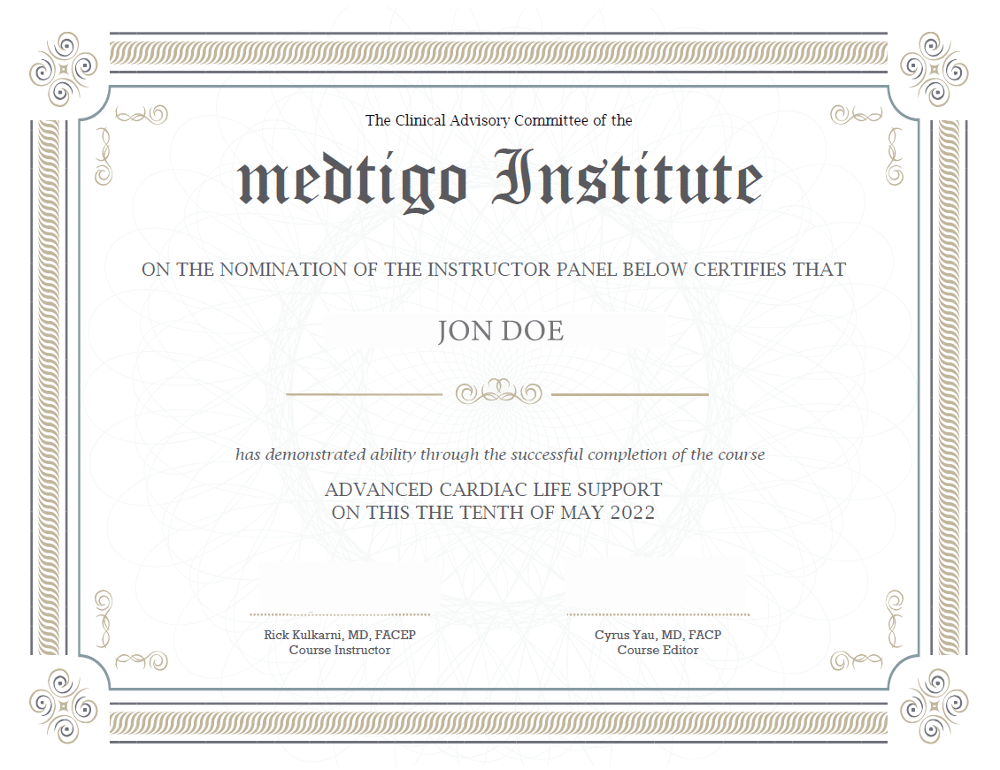
On course completion, you will receive a full-sized presentation quality digital certificate.
A dynamic medical simulation platform designed to train healthcare professionals and students to effectively run code situations through an immersive hands-on experience in a live, interactive 3D environment.
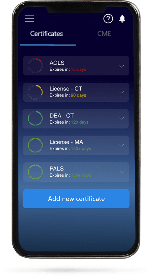
When you have your licenses, certificates and CMEs in one place, it's easier to track your career growth. You can easily share these with hospitals as well, using your medtigo app.