Scientists at the National Institutes of Health (NIH) have developed the first high-resolution digital twin of human retinal pigment epithelial (RPE) cells. This breakthrough offers new insights into how these critical eye cells function in health and how they deteriorate in age-related macular degeneration (AMD). AMD is a leading cause of vision loss in people aged 50 and older. The new modeling platform could accelerate the discovery of treatments for this and other blinding diseases.
The study led by investigators at NIH’s National Eye Institute (NEI) represents the first subcellular-resolution digital replica of a differentiated human primary cell. As per Kapil Bharti, Ph.D., NEI scientific director, this achievement shows how the eye can serve as an ideal model system to develop advanced computational tools that could be broadly applied across biomedical research. Scientists can now observe and measure cellular organization in unprecedented detail by reconstructing the intricate three-dimensional architecture of RPE cells.
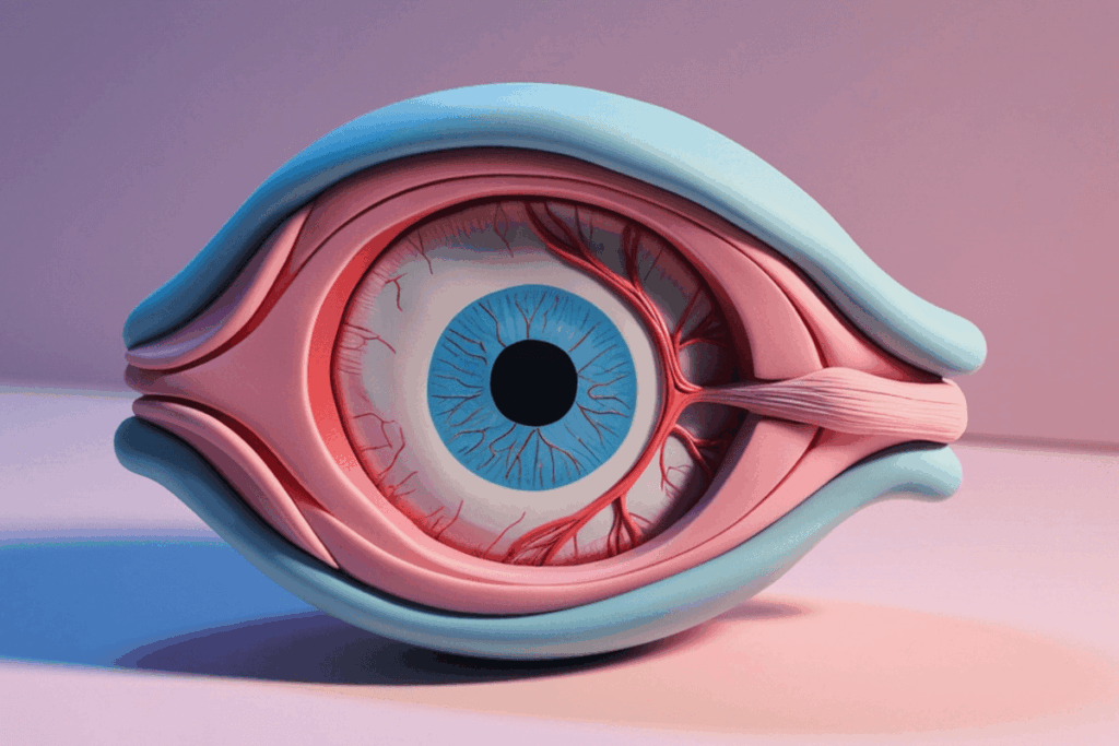
RPE cells play an important supportive role in the retina. They maintain and nourish photoreceptors, the light-sensing cells responsible for vision, by recycling worn-out photoreceptor components each day. These cells also regulate the transport of oxygen, nutrient and metabolic waste between the retina and its blood supply. When RPE cells degenerate, as occurs in AMD, photoreceptors subsequently die, which leads to progressive and irreversible vision loss.
A defining feature of healthy RPE cells is their polarity, the organized top-to-bottom orientation that allows them to perform specialised functions. The apical (top) side faces the photoreceptors and handles recycling functions, while the basal (bottom) side faces the blood supply to manage nutrient intake and waste removal. Loss of this polarity disrupts cell function and is a hallmark of disease progression.
Researchers used RPE cells derived from induced pluripotent stem cells (iPSCs), originally developed by the Allen Institute for Cell Science, to construct a digital twin. These stem cells were differentiated into RPE cells at NEI. The team collected massive amounts of imaging data using automated confocal microscopy. Three-dimensional (3D) imaging data from about 1.3 million RPE cells were gathered across nearly 4,000 fields of view. This large dataset provided the foundation for building a comprehensive and data-driven cellular model.
The scientists developed an artificial intelligence algorithm called Polarity Organization with Learning-Based Analysis for RPE Image Segmentation (POLARIS) to analyze the images. POLARIS was trained to detect and segment key cellular components, which include nuclei, mitochondria, cytoskeletal structures, and overall cell boundaries. It also measured cell shape, volume, and spatial organization at different developmental stages. The segmentation process assigned labels to individual voxels (3D pixels), which enables precise reconstruction of subcellular structures.
By applying AI-driven analysis and mathematical modeling, the team quantified how RPE cells change over time as they mature. They focused particularly on the emergence of polarity, mapping how organelles and cytoskeletal elements reorganize during development. The results showed that healthy RPE cells follow a predictable trajectory toward a fully polarized state. This discovery allowed researchers to create a detailed atlas of both polarized and non-polarized cells, which provides a reference framework for comparison.
This digital atlas serves as a baseline to detect deviations associated with disease. In AMD and other retinal disorders, RPE cells lose their structural organization before they die. With the digital twin platform, researchers can detect subtle subcellular changes that may signal early disease processes. Such insights could guide the development of therapies aimed at preserving or restoring cell polarity before irreversible damage occurs.
The digital twin approach has broad implications. Cell polarity is fundamental to many tissues in the body, and its disruption is linked to a variety of diseases, which include degenerative conditions and cancer. The researchers believe the same AI-based modeling strategy could be adapted to study other cell types and organ systems.
Davide Ortolan, Ph.D., the study’s first and senior author, emphasized that the integration of artificial intelligence with mathematical modeling offers a new window into previously hidden cellular dynamics. Rather than simply observing static images, scientists can now analyze dynamic organizational patterns and predict how cells might respond to stress or therapeutic interventions. This capability transforms the digital twin into both a diagnostic and discovery tool.
Overall, the NIH team has created a highly detailed, AI-powered digital replica of RPE cells that captures their structure and polarity at subcellular resolution. By revealing how healthy cells organize themselves and how that organization fails in the disease, the platform holds promise to understand and treat age-related macular degeneration and potentially many other diseases driven by disrupted cell polarity.
Reference: National Institutes of Health (NIH). NIH scientists develop “digital twin” of eye cells to understand and treat age-related macular degeneration. Published February 10, 2026. Accessed February 12, 2026. NIH scientists develop “digital twin” of eye cells to understand and treat age-related macular degeneration