The fovea is a specialized region of the retina essential for high-acuity vision. Its pathogenesis includes the movement of inner retinal layers and the congestion of cone photoreceptors. Arrested formation results in foveal hypoplasia (FH), a condition usually associated with albinism, nystagmus, and reduced visual acuity. Established genetic causes of FH largely involve pigmentation pathways (e.g., TYR, OCA2), but about 30% of cases are not yet resolved, suggesting additional developmental mechanisms.
Previous genome-wide association studies (GWAS) have explained characteristics associated with retinal thickness; however, no study has yet investigated the genetic basis of the foveal pit morphology itself. This study represents the first large-scale GWAS of foveal pit depth, aiming to clarify the re-establishment of the genetic environment of foveal development and foveal hypoplasia.
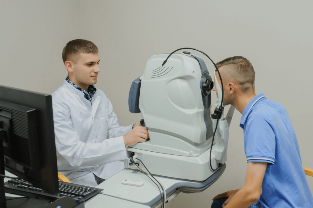
Optical coherence tomography (OCT) B-scans from 61,269 UK Biobank participants were analyzed. A deep-learning algorithm (ResNet-50) was used to measure the pit depth in terms of a vertical distance between the pit floor and rim peaks, excluding poor-quality scans.
Genotyping of participants was followed by GWAS using REGENIE (v3.4.1), adjusted for age, sex, height, and ancestry. The genome-wide significance was defined as P < 5 x 10⁻⁸. The association study of rare variants was also carried out on whole-exome sequencing data across 59,313 individuals in an exome-wide rare-variant association study (RVAS) with P < 5 × 10‾9 as a discovery threshold—variant-to-gene mapping combined 12 complementary criteria, which were positional, regulatory, and disease-informed evidence. Pathway enrichment, transferability across the ancestry, and genetic correlation with ocular traits were also evaluated.
The mean foveal pit depth among the European ancestry cohort (n = 61,269) was 116.4 μm (range 2.7-231.1 μm ). GWAS of 35.8 million variants found 126 sentinel variants, including 47 novel associations. Heritability, which was determined using SNP, was estimated to be ∼0.29 (SE = 0.03). The priority of integrative mapping was 129 putative causal genes, with 64 not previously associated with foveal biology. Enriched pathways included retinoic acid metabolism (e.g., CYP26A1), photoreceptor differentiation (e.g., VSX2), extracellular matrix organization, and pigmentation (e.g., OCA2, PMEL).
Two rare missense variants that were found and associated with a significantly shallower pits (up to 69 μm) was 5 x 10‾9: ACTN3 (11:66560171-T) and ESYT3 (3:138472579-A) (P = 5 × 10 ‾9). Carriers were confirmed to have FH-like characteristics by OCT scans.
Cross-ancestry analysis indicated that participants of African origin (median: 124.4 μm) and South Asian descent (119.5 μm) exhibited greater pit depths compared to Europeans. Polygenic scores derived from European cohorts predicted pit depth in South Asian participants (β = 4.90, P = 3.9 × 10‾30) and Africans (β = 2.62, P = 1.6 × 10⁻⁸).
Monogenic FH genes (TYR, OCA2, PAX6, AHR) and systemic disease genes (COL11A1, KIF11, TUBB4B, PHYH) were overlapping with it. The presence of FH was also established by review of OCTs among affected persons in syndromic cases, and even in individuals with recurrent TUBB4B p.(Arg390Trp) variants. Genetic correlation results indicated an association with refractive error (rg = 0.16, P = 3.17 x 10‾10), but not with glaucoma or macular degeneration.
This initial GWAS of human foveal morphology identifies over 120 sentinel variants and highlights 64 novel genes influencing foveal structure. FH is also caused by both common polygenic and rare large-effect variants (ACTN3, ESYT3). It is found that developmental processes other than pigmentation, pathways such as retinoic acid metabolism, retinal cell fate specification, vascular patterning, and extracellular matrix organization are implicated.
Systemic disease-related genes (PHYH, COL11A1, TUBB4B) also influence foveal development, supporting a continuum model where both rare mutations and polygenic variation shape foveal morphology. These findings redefine the biological pathway of FH, broaden its clinical spectrum, and offer a basis for future diagnostic and therapeutic advances in macular disorders.
References: Hunt C, Yoon HJ, Lirio A, et al. Genome-wide insights into the genes and pathways shaping human foveal development: redefining the genetic landscape of foveal hypoplasia. Invest Ophthalmol Vis Sci. 2025;66(12):22. doi:10.1167/iovs.66.12.22


