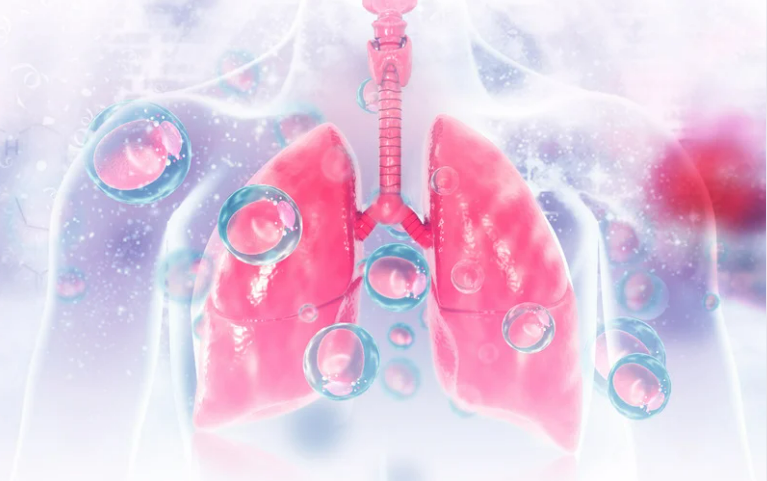
Gas exchange and immune responses to respiratory pathogens depend on the human alveolus, which limits the translational utility of animal lungs because of fundamental differences in lung structure and function across species. Organ-on-chip technologies, particularly those using human iPSC-derived cells, offer a promising alternative; however, scalable models of the distal lung remain lacking.
The researchers developed an iPSC-derived lung-on-chip (iLoC), a multicellular, autologous human model, constructed from a single iPSC line in this study. The iLoC demonstrated an important role for endothelial cells in immune homeostasis and modeled early infection in tuberculosis, with macrophage infection and death as the major phenotypes. This research also identified a cell-type-specific role for the autophagy gene ATG14 in supporting macrophage survival during Mycobacterium tuberculosis infection, using genetically engineered iPSCs.
The advent of the iLoC provides a viable alternative to animal testing, an approach strongly encouraged under the FDA Modernization Act 2.0. Organ-on-chip systems are increasingly recognized as legitimate tools for human tissue modelling and for predicting disease responses.
To build the iLoC, researchers first differentiated human iPSCs into lung progenitor cells. These cells were then directed to form two key alveolar cell types: type I alveolar cells (iAT1), which facilitate nutrient exchange, and type II alveolar cells (iAT2), which produce surfactant to keep air sacs open. The cells were cultured on a microfluidic chip that applied mild mechanical stretching, mimicking natural breathing. This was then succeeded by the addition of an air-liquid interface at day 7, which further promotes cellular maturation.
A strong and effective epithelial layer of alveolar cells had formed by day 14. Hallmark characteristics of the adult human alveoli, in the form of microvilli, tight junctions, desmosomes, and lamellar bodies, are essential for surfactant storage and secretion. While marker expression levels were lower than those observed in adult lung tissue, the cells maintained alveolar epithelial identity and retained the capacity for self-organization and proliferation.
The system was further improved by incorporating iPSC-derived vascular endothelial cells under the epithelial layer, forming a physiologically relevant epithelial-endothelial barrier. These endothelial cells persisted for at least 14 days and were essential for the development of immune responses in the chip.
Using this platform, the team simulated early TB infection. Upon introduction of Mycobacterium tuberculosis (Mtb), both alveolar epithelial cells and macrophages were infected, as was previously observed in humans. Unlike the conventional 2D cultures, however, the iLoC did not support robust bacterial replication. Rather, macrophage cell death occurred within 2 days, reflecting the sporadic and unpredictable nature of early TB infection in actual lungs.
This study presents a multicellular human lung-on-chip (iLoC) genetically engineered with four human isogenic iPSC-derived cell types within an alveoli-like microenvironment, which facilitates genetic manipulation of the system using CRISPR. Although the iLoC reproduces several critical characteristics of the human alveolus, it remains a preliminary platform that can be further optimized, including fluidic flow, cell types and phenotypes, and scalable cryopreservation.
The iLoC was found to exhibit cell-type-specific responses to mechanical stretch and to occupy a central position in adaptive responses of endothelial cells in the control of alveolar differentiation, immune signaling, and macrophage heterogeneity, as revealed by single-cell analyses. As an early model of tuberculosis, the iLoC model was used to recapitulate physiologically relevant infection events, inflammation, barrier disruption, and tissue damage that are not well modeled in 2D cultures or animal systems.
Notably, the genetic loss of ATG14 demonstrated that autophagy plays a macrophage-specific role in cell survival and immune signaling during Mycobacterium tuberculosis infection, indicating the association between the autophagy defect and the severe lung pathology. Overall, the iLoC provides a human-relevant environment for studying early mechanisms of respiratory disease and offers significant potential for drug discovery and pulmonary disease modeling.
References: Chak Hon Luk et al. Autologous human iPSC–derived alveolus-on-chip reveals early pathological events of Mycobacterium tuberculosis infection. Sci Adv. 2026;12:eaea9874. doi:10.1126/sciadv.aea9874