Understanding microcirculation at the whole-organ level is crucial for diagnosing vascular diseases and improving treatment outcomes. Nevertheless, current imaging techniques have notable limitations. Computed tomography angiography (CTA) and photon-counting CT provide excellent anatomical detail but lack temporal resolution, whereas MRI offers dynamic flow imaging at a high cost and with limited accessibility. Traditional ultrasound, though inexpensive and portable, lacks sufficient spatial resolution.
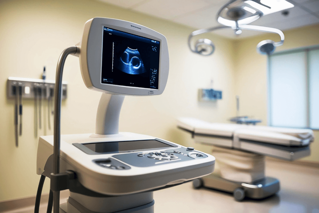
Recently, ultrasound localization microscopy (ULM) has demonstrated super-resolution imaging in small animals and limited two-dimensional imaging in humans. However, its ability to capture whole-organ views remains restricted due to complex hardware and narrow fields of view. This new study introduces a multi-lens ultrasound array that enables low-cost, high-resolution, and three-dimensional imaging of entire organs in large animals.
The research team developed a probe consisting of 252 elements (with a large aperture of 104 × 82 mm²) arranged in a multi-lens array probe. Each element is three times larger than the standard elements and uses compound diverging acoustic lenses to recover low directivity while maintaining resolution. This configuration achieved volumetric acquisition rates of 312 Hz and a spatial resolution of 125-200 µm across imaging volumes of 120 x100 x 82 mm³.
The system was validated through numerical simulation, in vitro phantom studies, and in vivo imaging of swine models (heart, kidney, and liver). A 3D-ULM was used to map blood flow with the help of microbubbles (MBs). The motion correction algorithms and flow quantification (Poiseuille flow profile and Murray law correlation) were applied. Skeletonization was used to estimate vessel radius, and Fourier Shell Correlation (FSC) was used to estimate spatial resolution.
Simulations revealed that the multi-lens array exhibited higher sensitivity and lower directivity compared to conventional or sparse arrays. Angular directivity at a depth of 35 mm was 33.17° compared to 10.27° for large elements without lenses. The volume sensitivity (1050 cm³) was significantly higher than that of conventional arrays (443-580 cm³). A lateral resolution of 1.06 mm and a flow rate ranging between 73 ± 6 mL h⁻¹ and 209 ± 5 mL h⁻¹ were validated in vitro phantom tests with a strong linear correlation to set flow rates (R² = 0.98).
Ex vivo heart imaging provided detailed coronary microcirculation of 120 × 100 × 82 mm³, showing vessels ranging in diameter between 75 and 600 µm and flow velocities of 10-300 mm s⁻¹. The flow-rate/ radius relationship followed Murray’s law (exponent = 2.3, R² = 0.82), achieving 125 µm spatial resolution.
In vivo kidney imaging (60 × 80 × 40 mm³) showed velocities up to 280 mm s⁻¹, vessel radii of 70-400 µm, and resolution of 147 µm with an exponent of flow rate to radius was 2.13 (R² = 0.70). In vivo liver imaging (65 × 100 × 82 mm³) achieved 200 µm resolution at velocities up to 300 mm s⁻¹, and a flow exponent of 2.36 (R² = 0.61) in the power-law. The profiles of the flow in all organs were consistent with laminar Poiseuille flow.
This study demonstrates a breakthrough in ultrasonic imaging, enabling whole-organ 3D microvascular mapping with microscopic precision at a fraction of MRI or CT cost (< €200k vs. ~€1M). The multi-lens design offers greater sensitivity, covers a wider area, and produces super-resolution with standard ultrasound systems. Potential applications include cardiovascular research, kidney disease screening, and early detection of liver pathologies.
Current limitations include edge sensitivity loss, limited visualization of capillaries smaller than 10 µm, and the need for optimized microbubble protocols. Nevertheless, this technology represents the future of medical imaging, aiming to scale toward human clinical use, integrate AI-guided analysis, and support digital twin modeling for personalized medicine.
Reference: Haidour N, Favre H, Mateo P, et al. Multi-lens ultrasound arrays enable large scale three-dimensional micro-vascularization characterization over whole organs. Nature Communications. 2025;16:9317. doi:10.1038/s41467-025-64911-z