Epidemiology studies the distribution and determinants of health-related states or events in specified populations. Rothia aeria is a rare bacterium that can cause infections in humans, especially in the heart valves (infective endocarditis). According to a review of 67 cases of Rothia bacteremia (blood infection) at Mayo Clinic from 2002 to 2012, only one case was caused by Rothia aeria, while the rest were caused by Rothia mucilaginosa.
The patient with Rothia aeria bacteremia had acute myeloid leukemia and was neutropenic (low white blood cell count). The source of the infection was unknown. The patient was treated with vancomycin and cefepime and survived.Another case report from Korea described a patient with Rothia aeria infective endocarditis who had a bicuspid aortic valve (a congenital heart defect). The patient presented with fever, chills, and diarrhea.
He was treated with ceftriaxone and gentamicin and underwent valve replacement surgery. He recovered without complications.A third case report from Japan described a patient with Rothia aeria pneumonia who was immunocompetent (regular immune system)—having a history of COPD (chronic obstructive pulmonary disease) present in the patient’s bronchiectasis. He presented with cough, sputum, and dyspnea.
He was treated with meropenem and levofloxacin and improved clinically and radiologically.These cases suggest that the uncommon Rothia aeria infection can affect both immunocompromised and immunocompetent hosts. The risk factors, transmission routes, and reservoirs of Rothia aeria need to be better understood. More studies are needed to determine the epidemiology and pathogenicity of this bacterium.
It is a Gram-positive, non-motile, and non-spore-forming bacterium. Rothia aeria is an aerobic microorganism that requires oxygen to grow and survive.
Domain: Bacteria
Phylum: Actinobacteria
Class: Actinobacteria
Order: Micrococcales
Family: Micrococcaceae
Genus: Rothia
Species: Rothia aeria
Morphologically, Rothia aeria appears as small, coccoid cells that typically occur in pairs or clusters. These cells are generally non-pigmented, although some strains may exhibit pink pigmentation due to the production of carotenoids. Rothia aeria is often described as being slightly larger than other species within the Rothia genus.
Rothia aeria is part of the normal microbiota in various human habitats, including the oral cavity, respiratory tract, and skin. It has been isolated from dental plaque, saliva, sputum, and lung tissues. While it is generally considered a commensal bacterium, meaning it typically coexists with its host without causing harm, Rothia aeria has also been associated with opportunistic infections in specific clinical settings.
There needs to be more information on the antigenic types of R. aeria infection, as it is a rare and recently discovered pathogen. However, some studies have reported that R. aeria can produce capsular polysaccharides that may act as antigens and contribute to its virulence and biofilm formation. Rothia antigens can also activate lymphocytes in patients with periodontal disease and induce tumor necrosis factor α (TNF-α) production in macrophages. The antibody response to R. aeria infection is poorly understood. Still, some studies have suggested that R. aeria is usually susceptible to vancomycin and most beta-lactams but resistant to oxacillin.
R. aeria is a relatively new species of bacteria first isolated from the air in the Russian space laboratory Mir in 2004. Since then, only a few strains of R. aeria have been identified and characterized from clinical specimens, such as blood, sputum, heart valve, and joint fluid. Some of the strains that have been reported are WSA-8, A1-17B, F0474, A1-22, and A1-3. Their molecular methods, such as 16S rRNA gene sequencing or MALDI-TOF mass spectrometry, can distinguish these strains. The type of strain of R. aeriais A1-17B, which has been deposited in several culture collections under different accession numbers
The process through which an infection results in illness is known as pathogenesis, part of the normal oral flora in humans. Rothia aeria can cause infectious endocarditis (IE). an infection of the heart’s inner lining or heart valves. A recent study found that the heart valves in IE can develop biofilms from Rothia aeria & another species of Rothia called Rothia dentocariosa. “Biofilms”—communities of bacteria affixed to a surface—are shielded by a slimy matrix. Antibiotic resistance in bacteria is exacerbated by biofilms and immune system assaults. The biofilms of Rothia aeria & Rothia dentocariosa in two cases of IE were identified and viewed in the study using fluorescence in situ hybridization (FISH) & PCR.
The study suggested that the potential of Rothia species for biofilms must be considered when treating IE caused by these bacteria. The study also recommended biofilm-effective antibiotics and raising knowledge about the worms Rothia aeria & Rothia dentocariosa a disease that is uncommon yet important for IE.The virulence factors of Rothia aeria, which are the molecules that help the bacterium cause disease, are still unknown. However, some studies have reported that Rothia aeria can produce enterobactin, a siderophore that helps the bacterium acquire iron from the host. Enterobactin may also interfere with the host’s immune response and promote inflammation.
Host defenses against Rothia aeria need to be better understood. Still, some studies suggest it can form biofilms on heart valves and other surfaces, which may help evade the immune system and antibiotics. It can also degrade and detoxify gluten, a protein that triggers an immune response in people with celiac disease.Rothia aeria infections are usually treated with beta-lactam antibiotics such as penicillin, ampicillin, or cephalosporins. However, some cases of resistance have been reported, so antibiotic susceptibility testing is recommended. In some cases, surgical intervention may be necessary to remove the infected tissue or device.
The symptoms of Rothia aeria infection may vary depending on the site and severity of the infection. Some possible symptoms are:
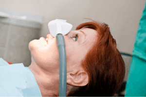
Fever, chills, malaise, and night sweats in cases of bacteremia or endocarditis.
Cough, dyspnea, sputum production, and chest pain in cases of pneumonia.
Joint pain, swelling, redness, and reduced mobility in cases of septic arthritis.
Abdominal pain, pelvic pain, vaginal discharge, and menstrual irregularities in cases of tubal-ovarian abscess.
Eye pain, swelling, redness, and tearing in cases of dacryoadenitis.
Some of these symptoms may be nonspecific or overlap with other conditions, so it is essential must consult a physician for a precise diagnosis and course of action.
Rothia aeria infection can be diagnosed by culturing the bacterium from a clinical specimen, such as blood, sputum, or heart valve tissue. However, culturing Rothia aeria can be challenging, as it may grow slowly or not on some media or need clarification with other Gram-positive bacteria. Therefore, molecular methods such as PCR and sequencing are more reliable and accurate for identifying Rothia aeria.
These methods can detect and distinguish the specific DNA sequences of Rothia aeria from other Rothia species or related bacteria. Another technique that can be used to visualize and identify Rothia aeria in situ is (fluorescence in situ hybridization) FISH. This method uses fluorescent probes that bind to the ribosomal RNA of Rothia aeria and emit a signal that can be detected by microscopy. FISH can also show the morphology and distribution of Rothia aeria in biofilms or tissues.
Rothia aeria infection is a rare condition mainly affecting immunocompromised patients or those with underlying heart disease or dental abnormalities. The prevention of R. aeria infection depends on the specific risk factors and the site of infection. Some general measures that may help prevent R. aeria infection are:
Keeping up with routine dental checkups and proper oral hygiene to
avoid dental caries, gingivitis, and periodontitis, which can be sources of R. aeria bacteremia and endocarditis.
Avoid intravenous drug use or using sterile needles and syringes to prevent R. aeria bloodstream infection and endocarditis.
Taking prophylactic antibiotics before dental or surgical procedures if there is a history of heart valve disease, prosthetic valve, congenital heart defect, or previous endocarditis, as recommended by the guidelines.
Seeking medical attention promptly if there are indications of an illness, such as fever, chills, fatigue, stomach discomfort, coughing, shortness of breath, pelvic pain, or vaginal discharge.
Following the prescribed antibiotic regimen and completing the course of treatment if diagnosed with R. aeria infection to prevent complications and relapse.
Epidemiology studies the distribution and determinants of health-related states or events in specified populations. Rothia aeria is a rare bacterium that can cause infections in humans, especially in the heart valves (infective endocarditis). According to a review of 67 cases of Rothia bacteremia (blood infection) at Mayo Clinic from 2002 to 2012, only one case was caused by Rothia aeria, while the rest were caused by Rothia mucilaginosa.
The patient with Rothia aeria bacteremia had acute myeloid leukemia and was neutropenic (low white blood cell count). The source of the infection was unknown. The patient was treated with vancomycin and cefepime and survived.Another case report from Korea described a patient with Rothia aeria infective endocarditis who had a bicuspid aortic valve (a congenital heart defect). The patient presented with fever, chills, and diarrhea.
He was treated with ceftriaxone and gentamicin and underwent valve replacement surgery. He recovered without complications.A third case report from Japan described a patient with Rothia aeria pneumonia who was immunocompetent (regular immune system)—having a history of COPD (chronic obstructive pulmonary disease) present in the patient’s bronchiectasis. He presented with cough, sputum, and dyspnea.
He was treated with meropenem and levofloxacin and improved clinically and radiologically.These cases suggest that the uncommon Rothia aeria infection can affect both immunocompromised and immunocompetent hosts. The risk factors, transmission routes, and reservoirs of Rothia aeria need to be better understood. More studies are needed to determine the epidemiology and pathogenicity of this bacterium.
It is a Gram-positive, non-motile, and non-spore-forming bacterium. Rothia aeria is an aerobic microorganism that requires oxygen to grow and survive.
Domain: Bacteria
Phylum: Actinobacteria
Class: Actinobacteria
Order: Micrococcales
Family: Micrococcaceae
Genus: Rothia
Species: Rothia aeria
Morphologically, Rothia aeria appears as small, coccoid cells that typically occur in pairs or clusters. These cells are generally non-pigmented, although some strains may exhibit pink pigmentation due to the production of carotenoids. Rothia aeria is often described as being slightly larger than other species within the Rothia genus.
Rothia aeria is part of the normal microbiota in various human habitats, including the oral cavity, respiratory tract, and skin. It has been isolated from dental plaque, saliva, sputum, and lung tissues. While it is generally considered a commensal bacterium, meaning it typically coexists with its host without causing harm, Rothia aeria has also been associated with opportunistic infections in specific clinical settings.
There needs to be more information on the antigenic types of R. aeria infection, as it is a rare and recently discovered pathogen. However, some studies have reported that R. aeria can produce capsular polysaccharides that may act as antigens and contribute to its virulence and biofilm formation. Rothia antigens can also activate lymphocytes in patients with periodontal disease and induce tumor necrosis factor α (TNF-α) production in macrophages. The antibody response to R. aeria infection is poorly understood. Still, some studies have suggested that R. aeria is usually susceptible to vancomycin and most beta-lactams but resistant to oxacillin.
R. aeria is a relatively new species of bacteria first isolated from the air in the Russian space laboratory Mir in 2004. Since then, only a few strains of R. aeria have been identified and characterized from clinical specimens, such as blood, sputum, heart valve, and joint fluid. Some of the strains that have been reported are WSA-8, A1-17B, F0474, A1-22, and A1-3. Their molecular methods, such as 16S rRNA gene sequencing or MALDI-TOF mass spectrometry, can distinguish these strains. The type of strain of R. aeriais A1-17B, which has been deposited in several culture collections under different accession numbers
The process through which an infection results in illness is known as pathogenesis, part of the normal oral flora in humans. Rothia aeria can cause infectious endocarditis (IE). an infection of the heart’s inner lining or heart valves. A recent study found that the heart valves in IE can develop biofilms from Rothia aeria & another species of Rothia called Rothia dentocariosa. “Biofilms”—communities of bacteria affixed to a surface—are shielded by a slimy matrix. Antibiotic resistance in bacteria is exacerbated by biofilms and immune system assaults. The biofilms of Rothia aeria & Rothia dentocariosa in two cases of IE were identified and viewed in the study using fluorescence in situ hybridization (FISH) & PCR.
The study suggested that the potential of Rothia species for biofilms must be considered when treating IE caused by these bacteria. The study also recommended biofilm-effective antibiotics and raising knowledge about the worms Rothia aeria & Rothia dentocariosa a disease that is uncommon yet important for IE.The virulence factors of Rothia aeria, which are the molecules that help the bacterium cause disease, are still unknown. However, some studies have reported that Rothia aeria can produce enterobactin, a siderophore that helps the bacterium acquire iron from the host. Enterobactin may also interfere with the host’s immune response and promote inflammation.
Host defenses against Rothia aeria need to be better understood. Still, some studies suggest it can form biofilms on heart valves and other surfaces, which may help evade the immune system and antibiotics. It can also degrade and detoxify gluten, a protein that triggers an immune response in people with celiac disease.Rothia aeria infections are usually treated with beta-lactam antibiotics such as penicillin, ampicillin, or cephalosporins. However, some cases of resistance have been reported, so antibiotic susceptibility testing is recommended. In some cases, surgical intervention may be necessary to remove the infected tissue or device.
The symptoms of Rothia aeria infection may vary depending on the site and severity of the infection. Some possible symptoms are:
Fever, chills, malaise, and night sweats in cases of bacteremia or endocarditis.
Cough, dyspnea, sputum production, and chest pain in cases of pneumonia.
Joint pain, swelling, redness, and reduced mobility in cases of septic arthritis.
Abdominal pain, pelvic pain, vaginal discharge, and menstrual irregularities in cases of tubal-ovarian abscess.
Eye pain, swelling, redness, and tearing in cases of dacryoadenitis.
Some of these symptoms may be nonspecific or overlap with other conditions, so it is essential must consult a physician for a precise diagnosis and course of action.
Rothia aeria infection can be diagnosed by culturing the bacterium from a clinical specimen, such as blood, sputum, or heart valve tissue. However, culturing Rothia aeria can be challenging, as it may grow slowly or not on some media or need clarification with other Gram-positive bacteria. Therefore, molecular methods such as PCR and sequencing are more reliable and accurate for identifying Rothia aeria.
These methods can detect and distinguish the specific DNA sequences of Rothia aeria from other Rothia species or related bacteria. Another technique that can be used to visualize and identify Rothia aeria in situ is (fluorescence in situ hybridization) FISH. This method uses fluorescent probes that bind to the ribosomal RNA of Rothia aeria and emit a signal that can be detected by microscopy. FISH can also show the morphology and distribution of Rothia aeria in biofilms or tissues.
Rothia aeria infection is a rare condition mainly affecting immunocompromised patients or those with underlying heart disease or dental abnormalities. The prevention of R. aeria infection depends on the specific risk factors and the site of infection. Some general measures that may help prevent R. aeria infection are:
Keeping up with routine dental checkups and proper oral hygiene to
avoid dental caries, gingivitis, and periodontitis, which can be sources of R. aeria bacteremia and endocarditis.
Avoid intravenous drug use or using sterile needles and syringes to prevent R. aeria bloodstream infection and endocarditis.
Taking prophylactic antibiotics before dental or surgical procedures if there is a history of heart valve disease, prosthetic valve, congenital heart defect, or previous endocarditis, as recommended by the guidelines.
Seeking medical attention promptly if there are indications of an illness, such as fever, chills, fatigue, stomach discomfort, coughing, shortness of breath, pelvic pain, or vaginal discharge.
Following the prescribed antibiotic regimen and completing the course of treatment if diagnosed with R. aeria infection to prevent complications and relapse.
Both our subscription plans include Free CME/CPD AMA PRA Category 1 credits.
Digital Certificate PDF
On course completion, you will receive a full-sized presentation quality digital certificate.
medtigo Simulation
A dynamic medical simulation platform designed to train healthcare professionals and students to effectively run code situations through an immersive hands-on experience in a live, interactive 3D environment.
medtigo Points
medtigo points is our unique point redemption system created to award users for interacting on our site. These points can be redeemed for special discounts on the medtigo marketplace as well as towards the membership cost itself.
Community Forum post/reply = 5 points
*Redemption of points can occur only through the medtigo marketplace, courses, or simulation system. Money will not be credited to your bank account. 10 points = $1.
All Your Certificates in One Place
When you have your licenses, certificates and CMEs in one place, it's easier to track your career growth. You can easily share these with hospitals as well, using your medtigo app.