Facial nerve repair is a micro surgical process done to repair the facial nerve to function properly.
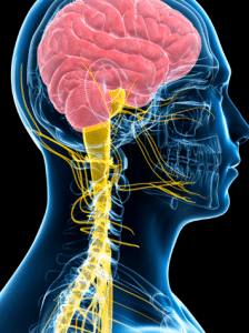
Anatomy of facial nerve
This nerve oversees all muscles that are for the purposes of smiling, frowning, blinking as well as chewing. The facial nerve can get damaged and may cause loss of the ability to close one or both sides of the face and may cause physical weakness and emotional discomfort. If facial nerve repair is needed, this is usually done by an otolaryngologist (ear, nose, and throat doctor), a neurosurgeon or a Plastic surgeon. Nerve repair is dependent on the degree of injury one has received and the type of procedure to be undertaken.
Indications
Bell’s palsy: This is one of the most frequent causes of facial paralysis which affects the facial muscles and leads to their weakness or complete paralysis. It causes sudden weakness or paralysis of the muscles of the face on one side of the face.
Trauma: Some other common conditions that may lead to facial paralysis include exposure to extreme cold, brain tumors, strokes, nervous system disorders, and injury to the face through an accident or a slip.
Incomplete facial paralysis: If facial functions are paresthetic but not completely paralytic and there is no improvement recorded in the first 6 months then facial nerve repair might be carried out.
Contraindications
Extensive Nerve Damage:There are cases when the facial nerve has been damaged beyond any possibility of efficient repair or when the gap in the nerve is too large to be bridged by a graft and the surgical intervention should be avoided. Severe Comorbidities: There are certain conditions where facial nerve repair is not advisable for children or patients with uncontrolled diabetes or severe cardiovascular diseases, immunosuppression, and other chronic ailments that can complicate surgery, facial nerve repair may not be advisable. Active Infection: Any wound infection or systemic infection is a contraindication that can affect outcomes and needs to be treated before surgery is performed for the repair.
Outcomes
Equipment
Operating microscope
Microsurgical Instruments
Jeweler’s forceps
Straight and curved micro scissors
Non-locking micro needle driver
Sutures and Needles
10-0 thin nylon sutures
Suction Tip
Bipolar Jeweler’s Forceps
Nerve Sheath Materials
Retractors
Fibrin Glue
Nerve Stimulator
Patient preparation
Pre-operative Consultation During the initial consultation, it is also important for the surgeon to find out the medical history, all medications, and even allergies of the patient. Some physical tests are important to evaluate the severity of the facial paralysis and the root cause. Preparing for Surgery To minimize the chances of complication or adverse interaction during surgery, the surgeon will advise on when and how to stop certain medicines or supplements that may result in clotting or otherwise cause difficulties in operations under anesthesia.
You may be told to avoid eating any food or only to take specific fluids for a period of time prior to the surgery. Medical Conditions It may include surgical risks associated with certain medical conditions like diabetes, heart diseases, or other chronic diseases, and which need to be well managed before having surgery.
Direct End-to-End Coaptation technique
Step 1: Preparation: Anesthesia: This procedure is done under general anesthesia.
Exposure: The facial nerve is then exposed after careful incision. Specifically, the surgeon exposes the whole region of nerve damage and makes sure this area is visible and accessible.
Step 2: Debridement:This step helps eliminate areas of poor-quality nerve tissue that may not be suitable for procedure.
Step 3: Alignment: Microscopic Assistance: Surgical procedure to repair peripheral nerves is done while using an operating microscope.
Fascicular Alignment: The nerve’s fascicles, or collections of nerve fibres, are precisely positioned. The precise alignment of motor and sensory fibers is essential to restore the integrity of functioning as well as lead to functional improvement.
Step 4: Coaptation: Suturing: The distal and proximal ends of the nerve are then joined in fashion with use of fine, non-absorbing suture material commonly nylon (8-0 or 10-0 nylon). To reduce damage to the nerve fibres, the sutures are inserted via the epineurium, or outer layer of the nerve.
Number of Sutures: Typically, 3-4 are enough to interconnect the nerve endings without the need for significantly applying much tension.
Step 5: Tension-Free Repair: It is significant to ensure that the nerve ends are properly aligned or coapted without unwanted tension. Excessive tension reduces blood flow and chance of nerve regeneration thus must be avoided. In some cases it might be required to free the nerve or the tissues surrounding it from tension to decrease the pressure on it.
Step 6: Wound Closure: Saline is then used to wash the surgical site in order to eliminate any debris and loose adherent tissue. Close the wound in a stepwise manner and minimize tissue trauma to adjacent structures.
Step 7 Postoperative Care Monitoring: Regular visits are conducted to check on nerve continuity and to address any limitations in motor function. Other modalities that may be used to determine the status of the nerves include electrophysiological tests.
Nerve substitution techniques
Hypoglossal-Facial Nerve Transfer: The hypoglossal nerve, which is the XII cranial nerve involved in the movements of the tongue, should be transferred partially or entirely to the facial nerve.
Procedure: Aberration of the hypoglossal nerve can be seen, and the muscle is separated out from the hypoglossal nerve. The hypoglossal nerve is used as donor, a portion of which is excised and end-to-end sutured to the distal part of the facial nerve. In the cases of a complete transaction, the hypoglossal nerve is preserved to avoid considerable reduction of tongue mobility.
Masseteric-Facial Nerve Transfer: Masseteric nerve belongs to the trigeminal nerve (cranial nerve V), to the facial nerve and utilized in chewing.
Procedure: It is identified through the infratemporal fossa and divides at the mental foramen into two branches, buccinator nerve and mental nerve. It is then incised and anastomosed to the end of the facial nerve stump distally.
Cross-Facial Nerve Grafting (CFNG) Cross-facial nerve grafting is a surgical intervention performed when the facial nerve of a patient is damaged and it is not possible to repair it directly, and involves the use of a healthy facial nerve from the opposite, non-affected side of the face, to the unaffected musculature on the paralyzed face.
Procedure The recipient nerve is cut and a section of nerve, usually the sural nerve, is taken from the leg. The graft is then passed underneath the skin in a careful manner across the face all the way to the paralyzed side. Simultaneously, one extremity of the graft is anastomosed with a branch of the normal facial nerve.
Aftercare:
The area, which is grafted must be closely observed after surgery, as well as nerve function and the reinnervation of muscles. Motor control is central in the recovery process as muscles must be retrained through an optimal dose of physical therapy.
Laboratory tests Electromyography (EMG): This test helps in recording the electricity generated by muscles in the body. It may assist in ascertaining whether there was any facial nerve damage and, if there was, the extent of the damage.
Electroneurography (ENoG): This test compares the speed / strength with which electricity signals travelling through a nerve. It can be used in diagnosing disorders in the facial nerves, particularly in establishing the extent of the injury that has occurred. Additional diagnostic tests may include MRI scan or CT scan to necessitate out other probable causes of the facial paralysis such as a tumor or a skull fracture.
Complications
Incomplete return of function: This is the common condition that happens when the nerve fibers do not regenerate efficiently again. The amount of recovery one will experience after the surgery depends on the degree of the first injury, and with how much time has elapsed from the accident to the surgery.
Synkinesis: This can happen when the nerve fibers that were repaired grow back incorrectly; thus, making two muscles do the work that was previously done by one. For instance, you might smile when you try to blink.
Infection: Like with any operation, there is a likelihood of developing an infection which can be minimized by following all precautions. This is usually responsive to antibiotics.
Facial numbness: It is normal to loose sensation in part of the surgery area during operation and is often temporary. Although, in some cases, the condition may be irreversible in that the individual affected cannot feel sensations anywhere on their body.
Change in facial contour: The surgery may affect the face and lead to some level of asymmetry, distortion, or any similar thing.
Facial nerve repair is a micro surgical process done to repair the facial nerve to function properly.
Anatomy of facial nerve
This nerve oversees all muscles that are for the purposes of smiling, frowning, blinking as well as chewing. The facial nerve can get damaged and may cause loss of the ability to close one or both sides of the face and may cause physical weakness and emotional discomfort. If facial nerve repair is needed, this is usually done by an otolaryngologist (ear, nose, and throat doctor), a neurosurgeon or a Plastic surgeon. Nerve repair is dependent on the degree of injury one has received and the type of procedure to be undertaken.
Bell’s palsy: This is one of the most frequent causes of facial paralysis which affects the facial muscles and leads to their weakness or complete paralysis. It causes sudden weakness or paralysis of the muscles of the face on one side of the face.
Trauma: Some other common conditions that may lead to facial paralysis include exposure to extreme cold, brain tumors, strokes, nervous system disorders, and injury to the face through an accident or a slip.
Incomplete facial paralysis: If facial functions are paresthetic but not completely paralytic and there is no improvement recorded in the first 6 months then facial nerve repair might be carried out.
Extensive Nerve Damage:There are cases when the facial nerve has been damaged beyond any possibility of efficient repair or when the gap in the nerve is too large to be bridged by a graft and the surgical intervention should be avoided. Severe Comorbidities: There are certain conditions where facial nerve repair is not advisable for children or patients with uncontrolled diabetes or severe cardiovascular diseases, immunosuppression, and other chronic ailments that can complicate surgery, facial nerve repair may not be advisable. Active Infection: Any wound infection or systemic infection is a contraindication that can affect outcomes and needs to be treated before surgery is performed for the repair.
Operating microscope
Microsurgical Instruments
Jeweler’s forceps
Straight and curved micro scissors
Non-locking micro needle driver
Sutures and Needles
10-0 thin nylon sutures
Suction Tip
Bipolar Jeweler’s Forceps
Nerve Sheath Materials
Retractors
Fibrin Glue
Nerve Stimulator
Pre-operative Consultation During the initial consultation, it is also important for the surgeon to find out the medical history, all medications, and even allergies of the patient. Some physical tests are important to evaluate the severity of the facial paralysis and the root cause. Preparing for Surgery To minimize the chances of complication or adverse interaction during surgery, the surgeon will advise on when and how to stop certain medicines or supplements that may result in clotting or otherwise cause difficulties in operations under anesthesia.
You may be told to avoid eating any food or only to take specific fluids for a period of time prior to the surgery. Medical Conditions It may include surgical risks associated with certain medical conditions like diabetes, heart diseases, or other chronic diseases, and which need to be well managed before having surgery.
Step 1: Preparation: Anesthesia: This procedure is done under general anesthesia.
Exposure: The facial nerve is then exposed after careful incision. Specifically, the surgeon exposes the whole region of nerve damage and makes sure this area is visible and accessible.
Step 2: Debridement:This step helps eliminate areas of poor-quality nerve tissue that may not be suitable for procedure.
Step 3: Alignment: Microscopic Assistance: Surgical procedure to repair peripheral nerves is done while using an operating microscope.
Fascicular Alignment: The nerve’s fascicles, or collections of nerve fibres, are precisely positioned. The precise alignment of motor and sensory fibers is essential to restore the integrity of functioning as well as lead to functional improvement.
Step 4: Coaptation: Suturing: The distal and proximal ends of the nerve are then joined in fashion with use of fine, non-absorbing suture material commonly nylon (8-0 or 10-0 nylon). To reduce damage to the nerve fibres, the sutures are inserted via the epineurium, or outer layer of the nerve.
Number of Sutures: Typically, 3-4 are enough to interconnect the nerve endings without the need for significantly applying much tension.
Step 5: Tension-Free Repair: It is significant to ensure that the nerve ends are properly aligned or coapted without unwanted tension. Excessive tension reduces blood flow and chance of nerve regeneration thus must be avoided. In some cases it might be required to free the nerve or the tissues surrounding it from tension to decrease the pressure on it.
Step 6: Wound Closure: Saline is then used to wash the surgical site in order to eliminate any debris and loose adherent tissue. Close the wound in a stepwise manner and minimize tissue trauma to adjacent structures.
Step 7 Postoperative Care Monitoring: Regular visits are conducted to check on nerve continuity and to address any limitations in motor function. Other modalities that may be used to determine the status of the nerves include electrophysiological tests.
Hypoglossal-Facial Nerve Transfer: The hypoglossal nerve, which is the XII cranial nerve involved in the movements of the tongue, should be transferred partially or entirely to the facial nerve.
Procedure: Aberration of the hypoglossal nerve can be seen, and the muscle is separated out from the hypoglossal nerve. The hypoglossal nerve is used as donor, a portion of which is excised and end-to-end sutured to the distal part of the facial nerve. In the cases of a complete transaction, the hypoglossal nerve is preserved to avoid considerable reduction of tongue mobility.
Masseteric-Facial Nerve Transfer: Masseteric nerve belongs to the trigeminal nerve (cranial nerve V), to the facial nerve and utilized in chewing.
Procedure: It is identified through the infratemporal fossa and divides at the mental foramen into two branches, buccinator nerve and mental nerve. It is then incised and anastomosed to the end of the facial nerve stump distally.
Cross-Facial Nerve Grafting (CFNG) Cross-facial nerve grafting is a surgical intervention performed when the facial nerve of a patient is damaged and it is not possible to repair it directly, and involves the use of a healthy facial nerve from the opposite, non-affected side of the face, to the unaffected musculature on the paralyzed face.
Procedure The recipient nerve is cut and a section of nerve, usually the sural nerve, is taken from the leg. The graft is then passed underneath the skin in a careful manner across the face all the way to the paralyzed side. Simultaneously, one extremity of the graft is anastomosed with a branch of the normal facial nerve.
Aftercare:
The area, which is grafted must be closely observed after surgery, as well as nerve function and the reinnervation of muscles. Motor control is central in the recovery process as muscles must be retrained through an optimal dose of physical therapy.
Laboratory tests Electromyography (EMG): This test helps in recording the electricity generated by muscles in the body. It may assist in ascertaining whether there was any facial nerve damage and, if there was, the extent of the damage.
Electroneurography (ENoG): This test compares the speed / strength with which electricity signals travelling through a nerve. It can be used in diagnosing disorders in the facial nerves, particularly in establishing the extent of the injury that has occurred. Additional diagnostic tests may include MRI scan or CT scan to necessitate out other probable causes of the facial paralysis such as a tumor or a skull fracture.
Incomplete return of function: This is the common condition that happens when the nerve fibers do not regenerate efficiently again. The amount of recovery one will experience after the surgery depends on the degree of the first injury, and with how much time has elapsed from the accident to the surgery.
Synkinesis: This can happen when the nerve fibers that were repaired grow back incorrectly; thus, making two muscles do the work that was previously done by one. For instance, you might smile when you try to blink.
Infection: Like with any operation, there is a likelihood of developing an infection which can be minimized by following all precautions. This is usually responsive to antibiotics.
Facial numbness: It is normal to loose sensation in part of the surgery area during operation and is often temporary. Although, in some cases, the condition may be irreversible in that the individual affected cannot feel sensations anywhere on their body.
Change in facial contour: The surgery may affect the face and lead to some level of asymmetry, distortion, or any similar thing.
Both our subscription plans include Free CME/CPD AMA PRA Category 1 credits.
Digital Certificate PDF
On course completion, you will receive a full-sized presentation quality digital certificate.
medtigo Simulation
A dynamic medical simulation platform designed to train healthcare professionals and students to effectively run code situations through an immersive hands-on experience in a live, interactive 3D environment.
medtigo Points
medtigo points is our unique point redemption system created to award users for interacting on our site. These points can be redeemed for special discounts on the medtigo marketplace as well as towards the membership cost itself.
Community Forum post/reply = 5 points
*Redemption of points can occur only through the medtigo marketplace, courses, or simulation system. Money will not be credited to your bank account. 10 points = $1.
All Your Certificates in One Place
When you have your licenses, certificates and CMEs in one place, it's easier to track your career growth. You can easily share these with hospitals as well, using your medtigo app.