
Rising Adoption of Continuous Glucose Monitors in Medicare Advantage: Trends from 2021–2023
April 13, 2026


Background
Actinic keratoses, commonly known as senile keratoses or sun keratoses, are benign intraepithelial neoplasms and one of the most prevalent skin conditions examined by dermatologists.
AKs are frequently connected with prolonged sun exposure and may manifest as irregular, red, scaly papules or plaques on sun-exposed areas of the body.
AKs have the potential to develop into invasive SCC if left untreated, highlighting the significance of early identification and the establishment of a treatment plan. Several management strategies are available for AKs.
Epidemiology
Actinic keratoses are most prevalent on persistently sun-exposed regions on the body. Older individuals have a higher risk of developing these due to the longer accumulated exposure to the sun.
Regions which are more susceptible to developing actinic keratoses include the scalp, face, dorsal surface of the hands, and the back of the arms.
Risk factors which are linked to actinic keratoses are:
Anatomy
Pathophysiology
The pathophysiology underlying the development of actinic keratoses is complicated. Through the breakdown of regulatory mechanisms involved in cell development and differentiation, excessive and accumulative UV radiation exposure can produce a multitude of pathologic alterations in epidermal keratinocytes. Inflammation and immunosuppression leads to the growth of dysplastic keratinocytes within the epidermis, which gives birth to actinic keratosis.
Etiology
Most cases of actinic keratoses develop due to the damage caused to skin because of UV radiation through chronic sun exposure.
Genetics
Prognostic Factors
Most cases of actinic keratoses regress spontaneously, due to some mechanism not completely understood. Other cases have a risk of evolving into squamous cell carcinoma.
Actinic keratoses is the most comma risk factor for squamous cell carcinoma, as most cases develop from former AKs or between a region affected by actinic keratoses. For a favorable outcome, patients are advised regular skin exams to spot new AKs and diagnose SCC in the early stages.
Clinical History
Physical Examination
Age group
Associated comorbidity
Associated activity
Acuity of presentation
Differential Diagnoses
Laboratory Studies
Imaging Studies
Procedures
Histologic Findings
Staging
Treatment Paradigm
by Stage
by Modality
Chemotherapy
Radiation Therapy
Surgical Interventions
Hormone Therapy
Immunotherapy
Hyperthermia
Photodynamic Therapy
Stem Cell Transplant
Targeted Therapy
Palliative Care
non-pharmacological-treatment-of-actinic-keratosis
Lifestyle modifications:
Use of Topical Antineoplastic Agents in the treatment of Actinic Keratosis
Fluorouracil Topical (Fluoroplex, Efudex, Carac, Tolak):
Ingenol Mebutate Topical (Picato):
Use of Topical Immunomodulator in the treatment of Actinic Keratosis
Imiquimod Topical (Aldara and Zyclara):
Use of Topical Photosensitizing Agents in the treatment of Actinic Keratosis
Aminolevulinic acid (Levulan Kerastick, Ameluz)
Use of Topical Nonsteroidal Anti-inflammatory agents in the treatment of Actinic Keratosis
Diclofenac Transdermal Gel (3%):
Use of Microtubule inhibitor in the treatment of Actinic Keratosis
Tirbanibulin Topical (Klisyri):
cryosurgery-in-the-treatment-of-actinic-keratosis
Cryosurgery, also known as cryotherapy, is a common and effective treatment for actinic keratosis (AK), a precancerous skin condition caused by sun exposure. Here’s how cryosurgery is used in the treatment of AK:
cosmetic-resurfacing-procedures-involved-in-actinic-keratosis
Cosmetic resurfacing procedures can be considered for the treatment of actinic keratosis (AK) when AK lesions are limited, and the patient has a desire to improve appearance and texture of skin. These procedures aim to remove the affected skin layers, promote healthy skin growth, and provide a more youthful and rejuvenated appearance.
management-of-actinic-keratosis
Acute Phase:
Treatment Phase:
Chronic Phase:
Medication
Apply 0.5% cream 4 weeks; and continue up to 4 weeks for greater lesion
Apply 2.5%/3.75% cream to the affected area once a day
Extremities or Trunk: Apply 0.05% of gel to the affected area every day for two days.
Scalp or Face: Apply 0.015% of gel to the affected area daily for three days; avoid applying the gel in or close to the eyes, the mouth, or the lips.
Applying gel in more than one area at once is not advised.
should be applied to one contiguous skin area that is no more than 25 cm² (5×5)
Not to be used as a spot therapy for actinic keratosis upon more than 25 cm2 of areas simultaneously.
Apply thin layer to affected area of skin for every 12 hours up to 2 to 3 months
Apply illumination to specific areas, possibly for therapeutic purposes
The treatment session lasts for 8 weeks
If the treated lesions have not entirely resolved after the initial 8-week treatment session, a second treatment may be administered
Apply to the affected area on the face or scalp for five days in a row daily
Apply only one single-dose package at a time
methyl aminolevulinate topical
Apply the topical cream on the lesion using gauze prepared by a curette
Apply a thin layer of 1 mm thickness
Do not keep more than 1 gm of cream on the skin
Let the cream on the skin for 2.5-4 hours only
Apply topically onto affected area one time in a day until lesions are cleared
Minimum 10 lesions at a time may be treated
Dosing modification
Renal impairment
Not suggested
Hepatic impairment
Dose modification not required
Apply to affected areas twice daily for 28 days
One illumination dose and one application at the treatment site for eight weeks of treatment session
Duration: May be repeated for a second time if the lesions are not entirely resolved with treatment for eight weeks
Future Trends
References
https://www.cedars-sinai.org/health-library/diseases-and-conditions/a/actinic-keratosis.html
https://www.ncbi.nlm.nih.gov/books/NBK557401/
Actinic keratoses, commonly known as senile keratoses or sun keratoses, are benign intraepithelial neoplasms and one of the most prevalent skin conditions examined by dermatologists.
AKs are frequently connected with prolonged sun exposure and may manifest as irregular, red, scaly papules or plaques on sun-exposed areas of the body.
AKs have the potential to develop into invasive SCC if left untreated, highlighting the significance of early identification and the establishment of a treatment plan. Several management strategies are available for AKs.
Actinic keratoses are most prevalent on persistently sun-exposed regions on the body. Older individuals have a higher risk of developing these due to the longer accumulated exposure to the sun.
Regions which are more susceptible to developing actinic keratoses include the scalp, face, dorsal surface of the hands, and the back of the arms.
Risk factors which are linked to actinic keratoses are:
The pathophysiology underlying the development of actinic keratoses is complicated. Through the breakdown of regulatory mechanisms involved in cell development and differentiation, excessive and accumulative UV radiation exposure can produce a multitude of pathologic alterations in epidermal keratinocytes. Inflammation and immunosuppression leads to the growth of dysplastic keratinocytes within the epidermis, which gives birth to actinic keratosis.
Most cases of actinic keratoses develop due to the damage caused to skin because of UV radiation through chronic sun exposure.
Most cases of actinic keratoses regress spontaneously, due to some mechanism not completely understood. Other cases have a risk of evolving into squamous cell carcinoma.
Actinic keratoses is the most comma risk factor for squamous cell carcinoma, as most cases develop from former AKs or between a region affected by actinic keratoses. For a favorable outcome, patients are advised regular skin exams to spot new AKs and diagnose SCC in the early stages.
Lifestyle modifications:
Dermatology, General
Fluorouracil Topical (Fluoroplex, Efudex, Carac, Tolak):
Ingenol Mebutate Topical (Picato):
Imiquimod Topical (Aldara and Zyclara):
Aminolevulinic acid (Levulan Kerastick, Ameluz)
Diclofenac Transdermal Gel (3%):
Tirbanibulin Topical (Klisyri):
Cryosurgery, also known as cryotherapy, is a common and effective treatment for actinic keratosis (AK), a precancerous skin condition caused by sun exposure. Here’s how cryosurgery is used in the treatment of AK:
Cosmetic resurfacing procedures can be considered for the treatment of actinic keratosis (AK) when AK lesions are limited, and the patient has a desire to improve appearance and texture of skin. These procedures aim to remove the affected skin layers, promote healthy skin growth, and provide a more youthful and rejuvenated appearance.
Acute Phase:
Treatment Phase:
Chronic Phase:
https://www.cedars-sinai.org/health-library/diseases-and-conditions/a/actinic-keratosis.html
https://www.ncbi.nlm.nih.gov/books/NBK557401/

Both our subscription plans include Free CME/CPD AMA PRA Category 1 credits.
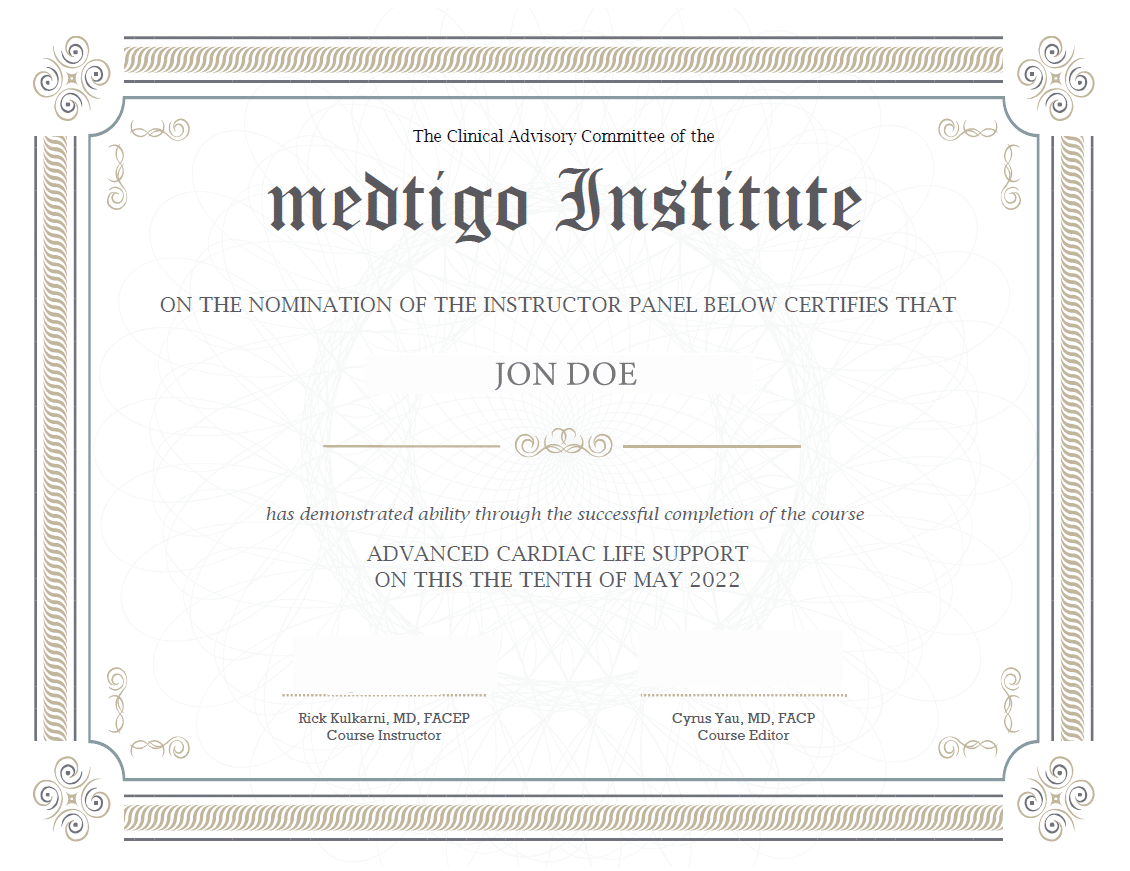
On course completion, you will receive a full-sized presentation quality digital certificate.
A dynamic medical simulation platform designed to train healthcare professionals and students to effectively run code situations through an immersive hands-on experience in a live, interactive 3D environment.
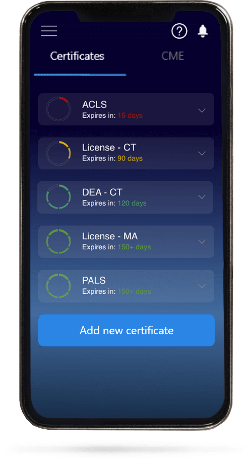
When you have your licenses, certificates and CMEs in one place, it's easier to track your career growth. You can easily share these with hospitals as well, using your medtigo app.