The symptoms of brain abscess vary based on the stage of infection. The early phase is known as focal cerebritis. It exhibits acute inflammatory responses like localized edema and vascular congestion. The starting stage is cerebritis. After 2 to 3 weeks, the abscess develops liquefaction and necrosis. It is a form of capsule. This capsule contains an inner layer of granulation tissue, an outer layer of astroglia tissue, and a middle layer of the collagenous layer. The parenchyma in brain is edematous.
Epidemiology
Brain abscess is common in developing countries, around 8% of the intracranial masses. About 1500 to 2500 new cases are noted in the U.S. every year. Fungal brain abscess is associated with the use of broad-spectrum antibiotics and immunosuppressive drugs. This disease is more common in adult men under the age of 30 years and is seen in children in the age of 4 to 7 years. Vaccination has decreased the cases in young children. Men are affected than women. The female-to-male ratio is 1:2 to 1:3. There are no differences in cases because of the season and geographical variations.
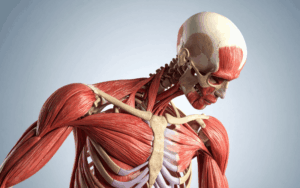
Anatomy
Pathophysiology
The symptoms of brain abscess vary based on the stage of infection. The early phase is known as focal cerebritis. It exhibits acute inflammatory responses like localized edema and vascular congestion. The starting stage is cerebritis. After 2 to 3 weeks, the abscess develops liquefaction and necrosis. It is a form of capsule. This capsule contains an inner layer of granulation tissue, an outer layer of astroglia tissue, and a middle layer of the collagenous layer. The parenchyma in brain is edematous.
Etiology
Abscesses can develop in localized areas and spread to other parts of the body. They originate from the infections in the areas like sinuses or ear but can go to more distant areas like lung or heart.
Direct local spread
Brain abscess can develop from conditions and infection like otitis media, mastoiditis, infections of frontal or ethmoid sinus, facial trauma, presence of foreign body or mental fragments and paranasal sinus infection in the brain. Paranasal sinus infections are about 30 to 50% of the cases. Dental infections lead to frontal lobe abscesses.
Hematogenous spread and Generalized Septicemia
Diseases which may lead to hematogenous seeding in the brain are cystic fibrosis, bronchopleural fistula, pulmonary arteriovenous malformation, lung infections, and pneumonia. About 60% of cases in children occur in cyanotic congenital heart disease. Other factors like thrombosis. Ventricular aneurysms and bacterial endocarditis. Intra-abdominal. Skin infections and pelvic infections are common risks. About 10% of patients who have pulmonary arteriovenous malformations may lead to a brain abscess. Brain abscesses linked with bacterial infections lead to multiple abscesses. The common microbial pathogens are streptococcal and staphylococcal. The common pathogens are Staphylococcus aureus and Viridian streptococci.
Causative pathogens of brain abscesses are:
pneumoniae
Streptococcus species
Staphylococcus species
Viridians streptococci
epidermidis
Gram-negative enteric
aureus
Klebsielle pneumoniae
Nocardia
Escherichia coli
Actinomycetales
Hemophilus species
Corynebacterium
Pseudomonas species
Fusobacterium species
Fungi
Parasites
Prognosis:
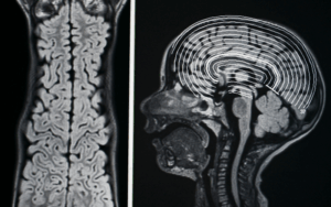
MRI and CT scans have reduced the mortality rate from 10 to 5%. The rupture of brain abscess is still a serious and fatal problem. Long-term neurological results from infection are based on the time of diagnosis and administration of antibiotics.
Genetics
Prognostic Factors
Clinical History
About 2 out of 3 patients are suffering from this condition. The symptoms can last for 2 weeks or less, varying from indolent to fulminant. Symptoms occur because of the size and location of lesions. < half of the patients have symptoms like headache, neurologic deficit, and fever.
Temporal love abscess: visual defects, headache, ipsilateral aphasia
Occipital abscess: neck rigidity
Frontal abscess: Deterioration of mental status, inattention, grand mal seizures, drowsiness, motor speech disease, headache
Brain abscess can lead to papilledema, vomiting, encephalitis, and purulent meningitis in the starting stage of infection. Severe headache can be caused in older children and adults. Younger infants may have bulging fontanels. Severe cerebral edema can lead to coma and lethargy. Some pathogens have particular clinical symptoms.
Age group
Associated comorbidity
Seizures
Brain herniation
Septicemia
Ventriculitis
Meningitis
Thrombosis of intracranial blood vessels
Neurological deficits
Death
Elevated intracranial pressure
Associated activity
Acuity of presentation
Differential Diagnoses
Cryptococcosis
Cysticercosis
Focal encephalitis
Mycotic aneurysm
Septic dural sinus thrombosis
Brain cancer
Bacterial meningitis
Laboratory Studies
Imaging Studies
Procedures
Histologic Findings
Staging
Treatment Paradigm
Medical care:
Brain abscess is a life-threatening disease. It needs antimicrobial treatment to control intracranial pressure. Treatment includes surgical excision or drainage with extended administration of antibiotics for 4 to 8 weeks. Early microbiological diagnosis is necessary to plan for proper treatment. This information can be given by CT-guided needle aspiration. The use of Glucocorticosteroids is controversial because of the immunosuppressive effects and negative results. Dexamethasone is given to treat cerebral edema with brain abscess. The duration of treatment is depending on the condition of the patient. Etiologic pathogen and no. od abscess present and response to treatment. The antimicrobial course is about 6 to 8 weeks long to repair the brain tissue. Empiric treatment must be active against oral streptococci, methicillin-susceptible staphylococci, anaerobes, and Enterobacteriaceae. Empiric antimicrobial treatment must be against the etiologic agents depending on predispose conditions, primary infection source and presumed pathogenesis of abscess formation.
by Stage
by Modality
Chemotherapy
Radiation Therapy
Surgical Interventions
Hormone Therapy
Immunotherapy
Hyperthermia
Photodynamic Therapy
Stem Cell Transplant
Targeted Therapy
Palliative Care
Administration of pharmaceutical agents with drugs Use of antibiotics
Ampicillin: It has a bactericidal activity on susceptible pathogens.
Cefotaxime: It is a 3rd generation cephalosporin. It has a broad-spectrum activity against gram-negative bacteria. It binds to PBPs by inhibiting the synthesis of cell wall and bacterial growth.
Ceftriaxone: It is a 3rd generation cephalosporin. It has a broad-spectrum activity against gram-negative bacteria. It binds to PBPs by inhibiting the synthesis of cell wall and bacterial growth.
Chloramphenicol: It binds to 50s ribosomal subunits by bacteria. It inhibits the growth of the bacteria by blocking the synthesis of protein.
Imipenem: It is used to treat infections because of multiple organisms.
Metronidazole: It contains an imidazole ring. It is active against protozoa and different anaerobic bacteria.
Vancomycin: It is an antibiotic which is active against Enterococcus species.
Use of corticosteroids
Dexamethasone: It is a corticosteroid which is used to decrease intracranial pressure.
The symptoms of brain abscess vary based on the stage of infection. The early phase is known as focal cerebritis. It exhibits acute inflammatory responses like localized edema and vascular congestion. The starting stage is cerebritis. After 2 to 3 weeks, the abscess develops liquefaction and necrosis. It is a form of capsule. This capsule contains an inner layer of granulation tissue, an outer layer of astroglia tissue, and a middle layer of the collagenous layer. The parenchyma in brain is edematous.
Brain abscess is common in developing countries, around 8% of the intracranial masses. About 1500 to 2500 new cases are noted in the U.S. every year. Fungal brain abscess is associated with the use of broad-spectrum antibiotics and immunosuppressive drugs. This disease is more common in adult men under the age of 30 years and is seen in children in the age of 4 to 7 years. Vaccination has decreased the cases in young children. Men are affected than women. The female-to-male ratio is 1:2 to 1:3. There are no differences in cases because of the season and geographical variations.
The symptoms of brain abscess vary based on the stage of infection. The early phase is known as focal cerebritis. It exhibits acute inflammatory responses like localized edema and vascular congestion. The starting stage is cerebritis. After 2 to 3 weeks, the abscess develops liquefaction and necrosis. It is a form of capsule. This capsule contains an inner layer of granulation tissue, an outer layer of astroglia tissue, and a middle layer of the collagenous layer. The parenchyma in brain is edematous.
Abscesses can develop in localized areas and spread to other parts of the body. They originate from the infections in the areas like sinuses or ear but can go to more distant areas like lung or heart.
Direct local spread
Brain abscess can develop from conditions and infection like otitis media, mastoiditis, infections of frontal or ethmoid sinus, facial trauma, presence of foreign body or mental fragments and paranasal sinus infection in the brain. Paranasal sinus infections are about 30 to 50% of the cases. Dental infections lead to frontal lobe abscesses.
Hematogenous spread and Generalized Septicemia
Diseases which may lead to hematogenous seeding in the brain are cystic fibrosis, bronchopleural fistula, pulmonary arteriovenous malformation, lung infections, and pneumonia. About 60% of cases in children occur in cyanotic congenital heart disease. Other factors like thrombosis. Ventricular aneurysms and bacterial endocarditis. Intra-abdominal. Skin infections and pelvic infections are common risks. About 10% of patients who have pulmonary arteriovenous malformations may lead to a brain abscess. Brain abscesses linked with bacterial infections lead to multiple abscesses. The common microbial pathogens are streptococcal and staphylococcal. The common pathogens are Staphylococcus aureus and Viridian streptococci.
Causative pathogens of brain abscesses are:
pneumoniae
Streptococcus species
Staphylococcus species
Viridians streptococci
epidermidis
Gram-negative enteric
aureus
Klebsielle pneumoniae
Nocardia
Escherichia coli
Actinomycetales
Hemophilus species
Corynebacterium
Pseudomonas species
Fusobacterium species
Fungi
Parasites
Prognosis:
MRI and CT scans have reduced the mortality rate from 10 to 5%. The rupture of brain abscess is still a serious and fatal problem. Long-term neurological results from infection are based on the time of diagnosis and administration of antibiotics.
About 2 out of 3 patients are suffering from this condition. The symptoms can last for 2 weeks or less, varying from indolent to fulminant. Symptoms occur because of the size and location of lesions. < half of the patients have symptoms like headache, neurologic deficit, and fever.
Temporal love abscess: visual defects, headache, ipsilateral aphasia
Occipital abscess: neck rigidity
Frontal abscess: Deterioration of mental status, inattention, grand mal seizures, drowsiness, motor speech disease, headache
Brain abscess can lead to papilledema, vomiting, encephalitis, and purulent meningitis in the starting stage of infection. Severe headache can be caused in older children and adults. Younger infants may have bulging fontanels. Severe cerebral edema can lead to coma and lethargy. Some pathogens have particular clinical symptoms.
Seizures
Brain herniation
Septicemia
Ventriculitis
Meningitis
Thrombosis of intracranial blood vessels
Neurological deficits
Death
Elevated intracranial pressure
Cryptococcosis
Cysticercosis
Focal encephalitis
Mycotic aneurysm
Septic dural sinus thrombosis
Brain cancer
Bacterial meningitis
Medical care:
Brain abscess is a life-threatening disease. It needs antimicrobial treatment to control intracranial pressure. Treatment includes surgical excision or drainage with extended administration of antibiotics for 4 to 8 weeks. Early microbiological diagnosis is necessary to plan for proper treatment. This information can be given by CT-guided needle aspiration. The use of Glucocorticosteroids is controversial because of the immunosuppressive effects and negative results. Dexamethasone is given to treat cerebral edema with brain abscess. The duration of treatment is depending on the condition of the patient. Etiologic pathogen and no. od abscess present and response to treatment. The antimicrobial course is about 6 to 8 weeks long to repair the brain tissue. Empiric treatment must be active against oral streptococci, methicillin-susceptible staphylococci, anaerobes, and Enterobacteriaceae. Empiric antimicrobial treatment must be against the etiologic agents depending on predispose conditions, primary infection source and presumed pathogenesis of abscess formation.
Infectious Disease
Ampicillin: It has a bactericidal activity on susceptible pathogens.
Cefotaxime: It is a 3rd generation cephalosporin. It has a broad-spectrum activity against gram-negative bacteria. It binds to PBPs by inhibiting the synthesis of cell wall and bacterial growth.
Ceftriaxone: It is a 3rd generation cephalosporin. It has a broad-spectrum activity against gram-negative bacteria. It binds to PBPs by inhibiting the synthesis of cell wall and bacterial growth.
Chloramphenicol: It binds to 50s ribosomal subunits by bacteria. It inhibits the growth of the bacteria by blocking the synthesis of protein.
Imipenem: It is used to treat infections because of multiple organisms.
Metronidazole: It contains an imidazole ring. It is active against protozoa and different anaerobic bacteria.
Vancomycin: It is an antibiotic which is active against Enterococcus species.
Infectious Disease
Dexamethasone: It is a corticosteroid which is used to decrease intracranial pressure.
Infectious Disease
medtigo
Brain Abscess
Updated :
September 23, 2024
The symptoms of brain abscess vary based on the stage of infection. The early phase is known as focal cerebritis. It exhibits acute inflammatory responses like localized edema and vascular congestion. The starting stage is cerebritis. After 2 to 3 weeks, the abscess develops liquefaction and necrosis. It is a form of capsule. This capsule contains an inner layer of granulation tissue, an outer layer of astroglia tissue, and a middle layer of the collagenous layer. The parenchyma in brain is edematous.
Brain abscess is common in developing countries, around 8% of the intracranial masses. About 1500 to 2500 new cases are noted in the U.S. every year. Fungal brain abscess is associated with the use of broad-spectrum antibiotics and immunosuppressive drugs. This disease is more common in adult men under the age of 30 years and is seen in children in the age of 4 to 7 years. Vaccination has decreased the cases in young children. Men are affected than women. The female-to-male ratio is 1:2 to 1:3. There are no differences in cases because of the season and geographical variations.
The symptoms of brain abscess vary based on the stage of infection. The early phase is known as focal cerebritis. It exhibits acute inflammatory responses like localized edema and vascular congestion. The starting stage is cerebritis. After 2 to 3 weeks, the abscess develops liquefaction and necrosis. It is a form of capsule. This capsule contains an inner layer of granulation tissue, an outer layer of astroglia tissue, and a middle layer of the collagenous layer. The parenchyma in brain is edematous.
Abscesses can develop in localized areas and spread to other parts of the body. They originate from the infections in the areas like sinuses or ear but can go to more distant areas like lung or heart.
Direct local spread
Brain abscess can develop from conditions and infection like otitis media, mastoiditis, infections of frontal or ethmoid sinus, facial trauma, presence of foreign body or mental fragments and paranasal sinus infection in the brain. Paranasal sinus infections are about 30 to 50% of the cases. Dental infections lead to frontal lobe abscesses.
Hematogenous spread and Generalized Septicemia
Diseases which may lead to hematogenous seeding in the brain are cystic fibrosis, bronchopleural fistula, pulmonary arteriovenous malformation, lung infections, and pneumonia. About 60% of cases in children occur in cyanotic congenital heart disease. Other factors like thrombosis. Ventricular aneurysms and bacterial endocarditis. Intra-abdominal. Skin infections and pelvic infections are common risks. About 10% of patients who have pulmonary arteriovenous malformations may lead to a brain abscess. Brain abscesses linked with bacterial infections lead to multiple abscesses. The common microbial pathogens are streptococcal and staphylococcal. The common pathogens are Staphylococcus aureus and Viridian streptococci.
Causative pathogens of brain abscesses are:
pneumoniae
Streptococcus species
Staphylococcus species
Viridians streptococci
epidermidis
Gram-negative enteric
aureus
Klebsielle pneumoniae
Nocardia
Escherichia coli
Actinomycetales
Hemophilus species
Corynebacterium
Pseudomonas species
Fusobacterium species
Fungi
Parasites
Prognosis:
MRI and CT scans have reduced the mortality rate from 10 to 5%. The rupture of brain abscess is still a serious and fatal problem. Long-term neurological results from infection are based on the time of diagnosis and administration of antibiotics.
About 2 out of 3 patients are suffering from this condition. The symptoms can last for 2 weeks or less, varying from indolent to fulminant. Symptoms occur because of the size and location of lesions. < half of the patients have symptoms like headache, neurologic deficit, and fever.
Temporal love abscess: visual defects, headache, ipsilateral aphasia
Occipital abscess: neck rigidity
Frontal abscess: Deterioration of mental status, inattention, grand mal seizures, drowsiness, motor speech disease, headache
Brain abscess can lead to papilledema, vomiting, encephalitis, and purulent meningitis in the starting stage of infection. Severe headache can be caused in older children and adults. Younger infants may have bulging fontanels. Severe cerebral edema can lead to coma and lethargy. Some pathogens have particular clinical symptoms.
Seizures
Brain herniation
Septicemia
Ventriculitis
Meningitis
Thrombosis of intracranial blood vessels
Neurological deficits
Death
Elevated intracranial pressure
Cryptococcosis
Cysticercosis
Focal encephalitis
Mycotic aneurysm
Septic dural sinus thrombosis
Brain cancer
Bacterial meningitis
Medical care:
Brain abscess is a life-threatening disease. It needs antimicrobial treatment to control intracranial pressure. Treatment includes surgical excision or drainage with extended administration of antibiotics for 4 to 8 weeks. Early microbiological diagnosis is necessary to plan for proper treatment. This information can be given by CT-guided needle aspiration. The use of Glucocorticosteroids is controversial because of the immunosuppressive effects and negative results. Dexamethasone is given to treat cerebral edema with brain abscess. The duration of treatment is depending on the condition of the patient. Etiologic pathogen and no. od abscess present and response to treatment. The antimicrobial course is about 6 to 8 weeks long to repair the brain tissue. Empiric treatment must be active against oral streptococci, methicillin-susceptible staphylococci, anaerobes, and Enterobacteriaceae. Empiric antimicrobial treatment must be against the etiologic agents depending on predispose conditions, primary infection source and presumed pathogenesis of abscess formation.
Infectious Disease
Ampicillin: It has a bactericidal activity on susceptible pathogens.
Cefotaxime: It is a 3rd generation cephalosporin. It has a broad-spectrum activity against gram-negative bacteria. It binds to PBPs by inhibiting the synthesis of cell wall and bacterial growth.
Ceftriaxone: It is a 3rd generation cephalosporin. It has a broad-spectrum activity against gram-negative bacteria. It binds to PBPs by inhibiting the synthesis of cell wall and bacterial growth.
Chloramphenicol: It binds to 50s ribosomal subunits by bacteria. It inhibits the growth of the bacteria by blocking the synthesis of protein.
Imipenem: It is used to treat infections because of multiple organisms.
Metronidazole: It contains an imidazole ring. It is active against protozoa and different anaerobic bacteria.
Vancomycin: It is an antibiotic which is active against Enterococcus species.
Infectious Disease
Dexamethasone: It is a corticosteroid which is used to decrease intracranial pressure.
Infectious Disease
Loading...
Free CME credits
Both our subscription plans include Free CME/CPD AMA PRA Category 1 credits.
Digital Certificate PDF
On course completion, you will receive a full-sized presentation quality digital certificate.
medtigo Simulation
A dynamic medical simulation platform designed to train healthcare professionals and students to effectively run code situations through an immersive hands-on experience in a live, interactive 3D environment.
medtigo Points
medtigo points is our unique point redemption system created to award users for interacting on our site. These points can be redeemed for special discounts on the medtigo marketplace as well as towards the membership cost itself.
Community Forum post/reply = 5 points
*Redemption of points can occur only through the medtigo marketplace, courses, or simulation system. Money will not be credited to your bank account. 10 points = $1.
All Your Certificates in One Place
When you have your licenses, certificates and CMEs in one place, it's easier to track your career growth. You can easily share these with hospitals as well, using your medtigo app.