RyR1 Structural Alterations Explain Statin-Associated Muscle Dysfunction
December 16, 2025


Background
Eumycetoma, also known as fungal mycetoma or maduromycosis,
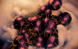
is a chronic infectious disease that affects the skin, subcutaneous tissues, and sometimes bones. It is one of the two main types of mycetoma, the other being actinomycetoma, caused by bacteria. Eumycetoma is explicitly caused by certain fungi and is characterized by the formation of granules or grains within the affected tissues.
The primary causative agents of eumycetoma are fungi belonging to various genera, including Madurella, Scedosporium, Pseudallescheria, and Fusarium, among others. These fungi are typically found in soil, decaying vegetation, or organic matter. Infection occurs when fungal spores enter the body through a wound, a cut, or even a small puncture, commonly on the foot. Once inside, the fungi begin to grow and increase, leading to the development of mycetoma.
Eumycetoma typically presents as a painless, slowly progressive swelling or mass in the affected body part, most commonly the foot. The infection can spread to the surrounding tissues, leading to the formation of abscesses and draining sinuses. Over time, the fungi can invade the bones, causing bone destruction and deformities.
Epidemiology
Anatomy
Pathophysiology
The pathogenesis of eumycetoma is not fully understood, but it involves several key steps:
Etiology
The main etiological agents associated with eumycetoma:
Genetics
Prognostic Factors
Clinical History
Physical Examination
Age group
Associated comorbidity
Associated activity
Acuity of presentation
Differential Diagnoses
The differential diagnosis of eumycetoma (fungal mycetoma) includes various conditions with chronic subcutaneous swellings, abscesses, or draining sinuses. Distinguishing eumycetoma from these other conditions can be challenging due to overlapping clinical features. Some of the key differential diagnoses to consider are:
Laboratory Studies
Imaging Studies
Procedures
Histologic Findings
Staging
Treatment Paradigm
Medical Therapy:
Surgical Interventions:
Supportive Care:
by Stage
by Modality
Chemotherapy
Radiation Therapy
Surgical Interventions
Hormone Therapy
Immunotherapy
Hyperthermia
Photodynamic Therapy
Stem Cell Transplant
Targeted Therapy
Palliative Care
use-of-a-non-pharmacological-approach-for-treating-eumycetoma
Non-pharmacological approaches can be used as adjuncts to the medical and surgical treatment of eumycetoma (fungal mycetoma). While antifungal medications and surgical interventions are the mainstay of treatment, non-pharmacological approaches can support managing the condition and improving the patient’s quality of life. Some non-pharmacological approaches that can be considered include:
Patient Education and Self-Care: Educating patients about their condition, treatment plan, and the importance of adherence to medical advice can enhance treatment outcomes. Encouraging self-care practices, such as keeping the affected area clean and dry, can improve wound healing.
Psychological Support: Dealing with a chronic condition like eumycetoma can be emotionally challenging. Psychological support, counseling, and support groups can help patients cope with the psychological impact of the disease and improve their overall well-being.
Prosthetics and Orthotics: In cases where eumycetoma has led to limb amputation or significant deformities, prosthetics or orthotics can aid in restoring function and mobility.
Role of Antifungal therapy for treating Eumycetoma
Antifungal therapy plays a central and critical role in treating eumycetoma (fungal mycetoma). It is the primary mode of medical treatment aimed at targeting the causative fungi responsible for the infection. Antifungal therapy eliminates or controls fungal growth, resolves infection, and prevents disease progression and complications. The treatment of eumycetoma (fungal mycetoma) is primarily based on azole antifungal therapy, guided by reported in-vitro susceptibility data. Azole antifungals, such as itraconazole and voriconazole, are the mainstay of medical treatment for eumycetoma caused by specific fungal species. These antifungal agents have demonstrated promising activity against certain causative fungi, such as Madurella mycetomatis.
Itraconazole:
Itraconazole, an antifungal medication, exerts its mechanism of action by inhibiting the synthesis of ergosterol, a vital component of the fungal cell membrane. This disruption of the cell membrane integrity leads to the loss of fungal cell viability, ultimately resulting in the death of the fungus. The mechanism of action of itraconazole involves several steps:
Voriconazole:
Voriconazole is an antifungal medication used to treat eumycetoma (fungal mycetoma). It involves inhibiting a specific fungal enzyme called cytochrome P450 14-alpha demethylase. This enzyme is crucial for synthesizing ergosterol, a vital component of the fungal cell membrane. By inhibiting this enzyme, voriconazole disrupts the production of ergosterol, destabilizing the fungal cell membrane. This disruption compromises the fungal cell’s integrity, causing cellular damage and ultimately resulting in the death of the fungus. Voriconazole’s ability to interfere with ergosterol synthesis makes it effective against a wide range of fungal species, including those responsible for causing eumycetoma.
use-of-intervention-with-a-procedure-in-treating-eumycetoma
The main objective of these procedures is to remove infected tissue, reduce the fungal burden, and promote wound healing. Surgical interventions are typically considered in cases with extensive involvement, large masses, bone invasion, or when antifungal therapy alone cannot control the infection. Some common procedures used in the management of eumycetoma include:
use-of-phases-in-managing-eumycetoma
The typical phases involved in managing eumycetoma:
Medication
Future Trends
References
Mycetoma:pubmed.ncbi.nlm.nih
Molecular Detection and Identification: ncbi.nlm.nih
Global Burden of Human: ncbi.nlm.nih
Mycetoma:ncbi.nlm.nih
Eumycetoma, also known as fungal mycetoma or maduromycosis,
is a chronic infectious disease that affects the skin, subcutaneous tissues, and sometimes bones. It is one of the two main types of mycetoma, the other being actinomycetoma, caused by bacteria. Eumycetoma is explicitly caused by certain fungi and is characterized by the formation of granules or grains within the affected tissues.
The primary causative agents of eumycetoma are fungi belonging to various genera, including Madurella, Scedosporium, Pseudallescheria, and Fusarium, among others. These fungi are typically found in soil, decaying vegetation, or organic matter. Infection occurs when fungal spores enter the body through a wound, a cut, or even a small puncture, commonly on the foot. Once inside, the fungi begin to grow and increase, leading to the development of mycetoma.
Eumycetoma typically presents as a painless, slowly progressive swelling or mass in the affected body part, most commonly the foot. The infection can spread to the surrounding tissues, leading to the formation of abscesses and draining sinuses. Over time, the fungi can invade the bones, causing bone destruction and deformities.
The pathogenesis of eumycetoma is not fully understood, but it involves several key steps:
The main etiological agents associated with eumycetoma:
The differential diagnosis of eumycetoma (fungal mycetoma) includes various conditions with chronic subcutaneous swellings, abscesses, or draining sinuses. Distinguishing eumycetoma from these other conditions can be challenging due to overlapping clinical features. Some of the key differential diagnoses to consider are:
Medical Therapy:
Surgical Interventions:
Supportive Care:
Dermatology, General
Pain Management
Non-pharmacological approaches can be used as adjuncts to the medical and surgical treatment of eumycetoma (fungal mycetoma). While antifungal medications and surgical interventions are the mainstay of treatment, non-pharmacological approaches can support managing the condition and improving the patient’s quality of life. Some non-pharmacological approaches that can be considered include:
Patient Education and Self-Care: Educating patients about their condition, treatment plan, and the importance of adherence to medical advice can enhance treatment outcomes. Encouraging self-care practices, such as keeping the affected area clean and dry, can improve wound healing.
Psychological Support: Dealing with a chronic condition like eumycetoma can be emotionally challenging. Psychological support, counseling, and support groups can help patients cope with the psychological impact of the disease and improve their overall well-being.
Prosthetics and Orthotics: In cases where eumycetoma has led to limb amputation or significant deformities, prosthetics or orthotics can aid in restoring function and mobility.
Dermatology, General
Antifungal therapy plays a central and critical role in treating eumycetoma (fungal mycetoma). It is the primary mode of medical treatment aimed at targeting the causative fungi responsible for the infection. Antifungal therapy eliminates or controls fungal growth, resolves infection, and prevents disease progression and complications. The treatment of eumycetoma (fungal mycetoma) is primarily based on azole antifungal therapy, guided by reported in-vitro susceptibility data. Azole antifungals, such as itraconazole and voriconazole, are the mainstay of medical treatment for eumycetoma caused by specific fungal species. These antifungal agents have demonstrated promising activity against certain causative fungi, such as Madurella mycetomatis.
Itraconazole:
Itraconazole, an antifungal medication, exerts its mechanism of action by inhibiting the synthesis of ergosterol, a vital component of the fungal cell membrane. This disruption of the cell membrane integrity leads to the loss of fungal cell viability, ultimately resulting in the death of the fungus. The mechanism of action of itraconazole involves several steps:
Voriconazole:
Voriconazole is an antifungal medication used to treat eumycetoma (fungal mycetoma). It involves inhibiting a specific fungal enzyme called cytochrome P450 14-alpha demethylase. This enzyme is crucial for synthesizing ergosterol, a vital component of the fungal cell membrane. By inhibiting this enzyme, voriconazole disrupts the production of ergosterol, destabilizing the fungal cell membrane. This disruption compromises the fungal cell’s integrity, causing cellular damage and ultimately resulting in the death of the fungus. Voriconazole’s ability to interfere with ergosterol synthesis makes it effective against a wide range of fungal species, including those responsible for causing eumycetoma.
Dermatology, General
The main objective of these procedures is to remove infected tissue, reduce the fungal burden, and promote wound healing. Surgical interventions are typically considered in cases with extensive involvement, large masses, bone invasion, or when antifungal therapy alone cannot control the infection. Some common procedures used in the management of eumycetoma include:
Dermatology, General
The typical phases involved in managing eumycetoma:
Mycetoma:pubmed.ncbi.nlm.nih
Molecular Detection and Identification: ncbi.nlm.nih
Global Burden of Human: ncbi.nlm.nih
Mycetoma:ncbi.nlm.nih

Both our subscription plans include Free CME/CPD AMA PRA Category 1 credits.

On course completion, you will receive a full-sized presentation quality digital certificate.
A dynamic medical simulation platform designed to train healthcare professionals and students to effectively run code situations through an immersive hands-on experience in a live, interactive 3D environment.

When you have your licenses, certificates and CMEs in one place, it's easier to track your career growth. You can easily share these with hospitals as well, using your medtigo app.