RyR1 Structural Alterations Explain Statin-Associated Muscle Dysfunction
December 16, 2025


Background
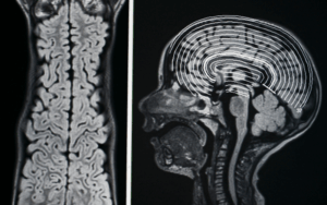
Foster Kennedy Syndrome is a medical condition characterized by a specific pattern of findings observed in patients with certain types of brain tumors, typically involving the frontal lobes or olfactory groove.
It is named after the American neurologist Robert Foster Kennedy, who first described the syndrome in the 1910s. The syndrome manifests as a combination of optic atrophy (damage to the optic nerve) in one eye and papilledema (swelling of the optic disc) in the other eye.
Epidemiology
Foster Kennedy Syndrome is a relatively rare condition, and specific epidemiological data regarding its prevalence and incidence are limited. The syndrome is primarily associated with tumors or lesions in the frontal lobes, particularly those involving the olfactory groove.
Here is some general information about the epidemiology of Foster Kennedy Syndrome:
It’s important to note that Foster Kennedy Syndrome is a clinical description of a specific pattern of findings rather than a distinct disease entity. The underlying causes can vary, and the epidemiology may be influenced by the prevalence and incidence rates of the associated conditions, such as tumors or lesions in the frontal lobes.
Due to the rarity of Foster Kennedy Syndrome and its association with specific underlying conditions, it is challenging to provide precise epidemiological data. Further research and larger studies are needed to better understand the prevalence, incidence, and distribution of the syndrome in the population.
Anatomy
Pathophysiology
The pathophysiology of Foster Kennedy Syndrome is closely linked to the underlying causes that lead to the characteristic findings of optic atrophy in one eye and papilledema in the other eye. The syndrome typically occurs as a result of tumors or lesions in the frontal lobes, particularly those involving the olfactory groove. Here is an overview of the pathophysiological mechanisms involved:
Tumor or Lesion Compression:
Increased Intracranial Pressure:
Neurological and Functional Effects:
Etiology
The etiology of Foster Kennedy Syndrome is primarily attributed to tumors or lesions in the frontal lobes, particularly those involving the olfactory groove. The underlying causes can vary, and several different types of tumors or lesions have been associated with the syndrome. Here are some of the common etiologies:
Genetics
Prognostic Factors
The prognosis of Foster Kennedy Syndrome can vary depending on several factors, including the underlying cause, the size and location of the lesion or tumor, the age of the patient, and the overall health status of the individual. Here are some considerations regarding the prognosis:
Clinical History
Clinical history
The clinical history of Foster Kennedy Syndrome can vary depending on the underlying cause and individual patient characteristics. Here are some key aspects that may be observed in the clinical history of individuals with Foster Kennedy Syndrome:
Visual Symptoms:
Neurological Symptoms:
Other Associated Symptoms:
Physical Examination
Physical examination
The physical examination of a patient with suspected Foster Kennedy Syndrome involves a comprehensive assessment of neurological and ophthalmic findings. Here are the key components of the physical examination:
Neurological Examination:
Ophthalmic Examination:
General Examination:
Age group
Associated comorbidity
Associated activity
Acuity of presentation
Differential Diagnoses
Differential diagnosis
Optic Nerve Lesions:
Intracranial Hypertension Syndromes:
Ocular Causes:
Ocular Manifestations of Systemic Diseases:
Laboratory Studies
Imaging Studies
Procedures
Histologic Findings
Staging
Treatment Paradigm
The treatment of Foster Kennedy Syndrome primarily depends on addressing the underlying cause, which is typically a tumor or lesion in the frontal lobes involving the olfactory groove. The specific treatment approach will vary based on the nature, size, and location of the lesion, as well as the patient’s overall health and individual circumstances. Here are some general treatment considerations:
Neurosurgical Intervention:
Radiation Therapy:
Chemotherapy:
Symptomatic Management:
by Stage
by Modality
Chemotherapy
Radiation Therapy
Surgical Interventions
Hormone Therapy
Immunotherapy
Hyperthermia
Photodynamic Therapy
Stem Cell Transplant
Targeted Therapy
Palliative Care
Medication
Future Trends
References
https://www.ncbi.nlm.nih.gov/books/NBK582149/
Foster Kennedy Syndrome is a medical condition characterized by a specific pattern of findings observed in patients with certain types of brain tumors, typically involving the frontal lobes or olfactory groove.
It is named after the American neurologist Robert Foster Kennedy, who first described the syndrome in the 1910s. The syndrome manifests as a combination of optic atrophy (damage to the optic nerve) in one eye and papilledema (swelling of the optic disc) in the other eye.
Foster Kennedy Syndrome is a relatively rare condition, and specific epidemiological data regarding its prevalence and incidence are limited. The syndrome is primarily associated with tumors or lesions in the frontal lobes, particularly those involving the olfactory groove.
Here is some general information about the epidemiology of Foster Kennedy Syndrome:
It’s important to note that Foster Kennedy Syndrome is a clinical description of a specific pattern of findings rather than a distinct disease entity. The underlying causes can vary, and the epidemiology may be influenced by the prevalence and incidence rates of the associated conditions, such as tumors or lesions in the frontal lobes.
Due to the rarity of Foster Kennedy Syndrome and its association with specific underlying conditions, it is challenging to provide precise epidemiological data. Further research and larger studies are needed to better understand the prevalence, incidence, and distribution of the syndrome in the population.
The pathophysiology of Foster Kennedy Syndrome is closely linked to the underlying causes that lead to the characteristic findings of optic atrophy in one eye and papilledema in the other eye. The syndrome typically occurs as a result of tumors or lesions in the frontal lobes, particularly those involving the olfactory groove. Here is an overview of the pathophysiological mechanisms involved:
Tumor or Lesion Compression:
Increased Intracranial Pressure:
Neurological and Functional Effects:
The etiology of Foster Kennedy Syndrome is primarily attributed to tumors or lesions in the frontal lobes, particularly those involving the olfactory groove. The underlying causes can vary, and several different types of tumors or lesions have been associated with the syndrome. Here are some of the common etiologies:
The prognosis of Foster Kennedy Syndrome can vary depending on several factors, including the underlying cause, the size and location of the lesion or tumor, the age of the patient, and the overall health status of the individual. Here are some considerations regarding the prognosis:
Clinical history
The clinical history of Foster Kennedy Syndrome can vary depending on the underlying cause and individual patient characteristics. Here are some key aspects that may be observed in the clinical history of individuals with Foster Kennedy Syndrome:
Visual Symptoms:
Neurological Symptoms:
Other Associated Symptoms:
Physical examination
The physical examination of a patient with suspected Foster Kennedy Syndrome involves a comprehensive assessment of neurological and ophthalmic findings. Here are the key components of the physical examination:
Neurological Examination:
Ophthalmic Examination:
General Examination:
Differential diagnosis
Optic Nerve Lesions:
Intracranial Hypertension Syndromes:
Ocular Causes:
Ocular Manifestations of Systemic Diseases:
The treatment of Foster Kennedy Syndrome primarily depends on addressing the underlying cause, which is typically a tumor or lesion in the frontal lobes involving the olfactory groove. The specific treatment approach will vary based on the nature, size, and location of the lesion, as well as the patient’s overall health and individual circumstances. Here are some general treatment considerations:
Neurosurgical Intervention:
Radiation Therapy:
Chemotherapy:
Symptomatic Management:
https://www.ncbi.nlm.nih.gov/books/NBK582149/

Both our subscription plans include Free CME/CPD AMA PRA Category 1 credits.

On course completion, you will receive a full-sized presentation quality digital certificate.
A dynamic medical simulation platform designed to train healthcare professionals and students to effectively run code situations through an immersive hands-on experience in a live, interactive 3D environment.

When you have your licenses, certificates and CMEs in one place, it's easier to track your career growth. You can easily share these with hospitals as well, using your medtigo app.