MCL is an uncommon subtype of B-cell non-Hodgkin lymphomas defined by the overexpression of the cyclin D1 (CCND1) gene due to a (1,14) translocation.
Depending on the number of morphologic variables, this condition may be difficult to diagnose, while some cases are straightforward.
It normally follows an aggressive clinical course, whereas the description is of an indolent form of leukemia.
Epidemiology
Mantle Cell Lymphoma has a low frequency of one case per 200,000 individuals annually.
MCL accounts for around 5% of non-Hodgkin lymphomas, and it is three times more prevalent in men than in women.
Anatomy
Pathophysiology
MCL is distinguished by the chromosomal translocation t(11;14) (q13:q32), which results in the juxtaposition of the cyclin D1 region and the immunoglobulin heavy chain gene locus.
This results in constitutive production of cyclin D1 (CCND1), which plays a crucial role in tumor cell proliferation via disruption of the cell cycle, epigenetic regulation, and chromosomal instability.
In rare cases where this translocation is absent, CCND2 or 3 translocations may be present. Based on the cell of origin that connects to clinical symptoms, hypothesized models of molecular subtypes have been presented.
Classical MCL or aggressive MCL arises from naive B cells that have none or minimal iGVH mutations and express SOX 11. SOX 11 is a neuronal transcription factor that reportedly inhibits the final development of B cells.
The indolent type of MCL originates from antigen-exposed B cells that have undergone IGVH somatic hypermutations. They are often SOX 11-negative and genetically stable B cells.
Etiology
MCL is not a very common condition, but some families might be predisposed to a high incidence of this disease.
Genetics
Prognostic Factors
Clinical History
Physical Examination
Age group
Associated comorbidity
Associated activity
Acuity of presentation
Differential Diagnoses
Laboratory Studies
Imaging Studies
Procedures
Histologic Findings
Staging
Treatment Paradigm
Induction Therapy: This is a standard chemotherapy regimen used in the initial treatment of MCL. Rituximab is a monoclonal antibody targeting CD20, a protein on the surface of B cells.
Consolidation Therapy: After induction therapy, patients may undergo consolidation therapy, which aims to eliminate any remaining cancer cells.
Autologous stem cell transplantation (ASCT) is a common consolidation approach, particularly in younger and medically fit patients.
Maintenance Therapy: Maintenance therapy with rituximab or other agents may be considered to help prolong remission.
Hygiene and Infection Control: Maintain a clean and hygienic home environment to reduce the risk of infections, especially for individuals with compromised immune systems due to treatment.
Diet and Nutrition: Provide a well-balanced and nutritious diet to support overall health and aid in recovery.
Counseling Services: Consider involving mental health professionals or counselors to provide emotional support and coping strategies for both the patient and their caregivers.
Physical Activity: Encourage appropriate and safe physical activity based on the individual’s capabilities. Regular, gentle exercise can contribute to physical and emotional well-being.
Work Arrangements: If the individual is employed, consider flexible work arrangements or modifications to workload during treatment periods.
Daily Routine: Establish a routine that accommodates treatment schedules and allows for adequate rest. Ensure that daily activities are manageable and not overly strenuous.
Accessibility at Home: Make necessary modifications at home to improve accessibility for individuals with mobility challenges, if applicable.
Use of Monoclonal Antibodies
Rituximab: A monoclonal antibody that targets the CD20 protein on B cells, commonly used in combination with chemotherapy (e.g., R-CHOP) to enhance treatment effectiveness.
Use of BTK Inhibitors
Ibrutinib: A Bruton’s tyrosine kinase (BTK) inhibitor that disrupts signalling pathways involved in the survival and proliferation of B cells. Ibrutinib is often used as a first-line treatment or in relapsed/refractory cases.
Use of Proteasome Inhibitors
Bortezomib: A proteasome inhibitor that interferes with the degradation of proteins within cells, disrupting cell cycle progression. Bortezomib may be used in combination with other agents in the treatment of MCL.
Biopsy: A biopsy is typically performed to confirm the diagnosis of Mantle Cell Lymphoma. An excisional or incisional biopsy involves the removal of a sample of the affected lymph node or tissue for examination under a microscope.
PET-CT scan: Positron Emission Tomography with Computed Tomography (PET-CT) scans are used for staging and monitoring the response to treatment. They provide detailed images of the body and can detect areas of active lymphoma.
Stem Cell Transplantation: Especially for younger and medically fit patients, high-dose chemotherapy may be followed by autologous stem cell transplantation. This involves the infusion of the patient’s own previously collected stem cells to help restore blood cell production.
use-of-phases-in-managing-mantle-cell-lymphoma
Initial Treatment: Most patients with MCL receive induction therapy, often with a regimen such as R-CHOP (rituximab, cyclophosphamide, doxorubicin, vincristine, prednisone) or other targeted therapies.
Consolidation Therapy: Younger and medically fit patients may undergo autologous stem cell transplantation (ASCT) as a consolidation therapy to further reduce the risk of relapse.
Targeted Therapies: The Bruton’s tyrosine kinase (BTK) inhibitor ibrutinib is often used in the treatment of MCL, either as initial therapy or in relapsed/refractory cases.
Imaging Studies: Regular imaging studies, such as CT scans or PET-CT scans, are performed to monitor the response to treatment and assess disease status.
It is indicated for relapsed or refractory mantle cell lymphoma
Prior to Therapy:
Before injecting this medication, give cyclophosphamide 500 mg/m2 IV and fludarabine 30 mg/m2 IV as a lymphodepleting chemotherapy regimen on the 4th, 5th, and 3rd days
The dosage of CAR-positive viable T cells 2 x 10(6)/kg, with a maximum of 2 x 10(8)
Each single infusion bag has a suspension of chimeric antigen receptor (CAR)-positive T cells in about 68 mL
Dose Adjustments
Management of Cytokine release syndrome:
Grade 1
Symptoms: Fever, nausea, fatigue, headache, myalgia, malaise
If symptoms persist after 24 hours, administer tocilizumab at a dose of 8 mg/kg intravenously over 1 hour, with a maximum dose not exceeding 800 mg
Grade 2
Moderate intervention is required for symptoms, such as oxygen requirement <40% FiO2 hypotension responsive to fluids, a low dose of one vasopressor, or Grade 2 organ toxicity
Administer tocilizumab at a dose of 8 mg/kg intravenously over 1 hour, with a maximum dose not exceeding 800 mg
Repeat tocilizumab every 8 hours as necessary if there is no response to intravenous fluids or if supplemental oxygen needs to increase; do not exceed three doses within a 24-hour period or a total of 4 doses
If there is improvement, discontinue tocilizumab
If there is no improvement after 24 hours of starting tocilizumab, administer methylprednisolone (1 mg/kg intravenously twice daily) or dexamethasone (10 mg intravenously every 6 hours) until Grade 1, then taper corticosteroids
Grade 3
Symptoms necessitate aggressive intervention, such as oxygen requirement ≥40% FiO2, hypotension requiring high-dose or multiple vasopressors, Grade 3 organ toxicity, or Grade 4 transaminitis
Administer tocilizumab as outlined in Grade 2; discontinue if there is improvement
Prescribe corticosteroid doses as specified for Grade 2; taper if there is improvement or manage as Grade 4 if there is no improvement
Grade 4
Symptoms are posing a risk to life, necessitating ventilator assistance, ongoing venovenous hemodialysis, or organ toxicity of Grade 4 (excluding transaminitis)
Administer Tocilizumab according to Grade 2; if there's an improvement, discontinue
Inject Methylprednisolone 1000 mg intravenously daily for three days; reduce if there's an improvement or contemplate alternative immunosuppressants if no improvement is observed
Grading and management of Neurologic toxicity:
For Grades 2, 3, or 4: Contemplate the use of non-sedating antiseizure medications (such as levetiracetam) as a preventive measure against seizures
Grade 1
In the presence of concurrent Cytokine Release Syndrome (CRS), administer tocilizumab following the guidelines for Grade 1 CRS. If no concurrent CRS is observed, provide supportive care
Grade 2 with concurrent CRS
Administer tocilizumab following the specified doses for CRS Grade 2. If there's no improvement within 24 hours of initiating tocilizumab, administer 10 mg of dexamethasone intravenously every 6 hours if the individual is not already using other corticosteroids. Continue the use of dexamethasone until the event reaches Grade 1 or lower, then gradually taper over a 3-day period
Grade 2 or 3 without concurrent Cytokine Release Syndrome (CRS)
Initiate dexamethasone at 10 mg intravenously every 6 hours if the individual is not already using other corticosteroids. Continue this regimen until the event reaches Grade 1 or lower, then taper over a 3-day period. If the situation is Grade 3 and there is no improvement, handle it as Grade 4, employing intravenous methylprednisolone
Grade 3 with concurrent CRS
Administer tocilizumab following the doses specified for CRS Grade 2. Additionally, initiate dexamethasone at 10 mg intravenously every 6 hours if the person is not already using other corticosteroids. Continue the dexamethasone regimen until the event reaches Grade 1 or lower, then taper over a 3-day period
Grade 4 with concurrent Cytokine Release Syndrome (CRS)
Initiate tocilizumab following the specified doses for CRS Grade 2. Simultaneously, administer methylprednisolone at 1000 mg intravenously per day for three days, starting with the first dose of tocilizumab. If improvement is observed, proceed with the management as outlined above
Grade 4 without concurrent CRS
Administer methylprednisolone at 1000 mg intravenously per day for three days. If improvement occurs, manage the situation as described above. If there is no improvement, consider exploring alternative immunosuppressants
MCL is an uncommon subtype of B-cell non-Hodgkin lymphomas defined by the overexpression of the cyclin D1 (CCND1) gene due to a (1,14) translocation.
Depending on the number of morphologic variables, this condition may be difficult to diagnose, while some cases are straightforward.
It normally follows an aggressive clinical course, whereas the description is of an indolent form of leukemia.
Mantle Cell Lymphoma has a low frequency of one case per 200,000 individuals annually.
MCL accounts for around 5% of non-Hodgkin lymphomas, and it is three times more prevalent in men than in women.
MCL is distinguished by the chromosomal translocation t(11;14) (q13:q32), which results in the juxtaposition of the cyclin D1 region and the immunoglobulin heavy chain gene locus.
This results in constitutive production of cyclin D1 (CCND1), which plays a crucial role in tumor cell proliferation via disruption of the cell cycle, epigenetic regulation, and chromosomal instability.
In rare cases where this translocation is absent, CCND2 or 3 translocations may be present. Based on the cell of origin that connects to clinical symptoms, hypothesized models of molecular subtypes have been presented.
Classical MCL or aggressive MCL arises from naive B cells that have none or minimal iGVH mutations and express SOX 11. SOX 11 is a neuronal transcription factor that reportedly inhibits the final development of B cells.
The indolent type of MCL originates from antigen-exposed B cells that have undergone IGVH somatic hypermutations. They are often SOX 11-negative and genetically stable B cells.
MCL is not a very common condition, but some families might be predisposed to a high incidence of this disease.
Induction Therapy: This is a standard chemotherapy regimen used in the initial treatment of MCL. Rituximab is a monoclonal antibody targeting CD20, a protein on the surface of B cells.
Consolidation Therapy: After induction therapy, patients may undergo consolidation therapy, which aims to eliminate any remaining cancer cells.
Autologous stem cell transplantation (ASCT) is a common consolidation approach, particularly in younger and medically fit patients.
Maintenance Therapy: Maintenance therapy with rituximab or other agents may be considered to help prolong remission.
Hygiene and Infection Control: Maintain a clean and hygienic home environment to reduce the risk of infections, especially for individuals with compromised immune systems due to treatment.
Diet and Nutrition: Provide a well-balanced and nutritious diet to support overall health and aid in recovery.
Counseling Services: Consider involving mental health professionals or counselors to provide emotional support and coping strategies for both the patient and their caregivers.
Physical Activity: Encourage appropriate and safe physical activity based on the individual’s capabilities. Regular, gentle exercise can contribute to physical and emotional well-being.
Work Arrangements: If the individual is employed, consider flexible work arrangements or modifications to workload during treatment periods.
Daily Routine: Establish a routine that accommodates treatment schedules and allows for adequate rest. Ensure that daily activities are manageable and not overly strenuous.
Accessibility at Home: Make necessary modifications at home to improve accessibility for individuals with mobility challenges, if applicable.
Rituximab: A monoclonal antibody that targets the CD20 protein on B cells, commonly used in combination with chemotherapy (e.g., R-CHOP) to enhance treatment effectiveness.
Ibrutinib: A Bruton’s tyrosine kinase (BTK) inhibitor that disrupts signalling pathways involved in the survival and proliferation of B cells. Ibrutinib is often used as a first-line treatment or in relapsed/refractory cases.
Bortezomib: A proteasome inhibitor that interferes with the degradation of proteins within cells, disrupting cell cycle progression. Bortezomib may be used in combination with other agents in the treatment of MCL.
Biopsy: A biopsy is typically performed to confirm the diagnosis of Mantle Cell Lymphoma. An excisional or incisional biopsy involves the removal of a sample of the affected lymph node or tissue for examination under a microscope.
PET-CT scan: Positron Emission Tomography with Computed Tomography (PET-CT) scans are used for staging and monitoring the response to treatment. They provide detailed images of the body and can detect areas of active lymphoma.
Stem Cell Transplantation: Especially for younger and medically fit patients, high-dose chemotherapy may be followed by autologous stem cell transplantation. This involves the infusion of the patient’s own previously collected stem cells to help restore blood cell production.
Initial Treatment: Most patients with MCL receive induction therapy, often with a regimen such as R-CHOP (rituximab, cyclophosphamide, doxorubicin, vincristine, prednisone) or other targeted therapies.
Consolidation Therapy: Younger and medically fit patients may undergo autologous stem cell transplantation (ASCT) as a consolidation therapy to further reduce the risk of relapse.
Targeted Therapies: The Bruton’s tyrosine kinase (BTK) inhibitor ibrutinib is often used in the treatment of MCL, either as initial therapy or in relapsed/refractory cases.
Imaging Studies: Regular imaging studies, such as CT scans or PET-CT scans, are performed to monitor the response to treatment and assess disease status.
Both our subscription plans include Free CME/CPD AMA PRA Category 1 credits.
Digital Certificate PDF
On course completion, you will receive a full-sized presentation quality digital certificate.
medtigo Simulation
A dynamic medical simulation platform designed to train healthcare professionals and students to effectively run code situations through an immersive hands-on experience in a live, interactive 3D environment.
medtigo Points
medtigo points is our unique point redemption system created to award users for interacting on our site. These points can be redeemed for special discounts on the medtigo marketplace as well as towards the membership cost itself.
Community Forum post/reply = 5 points
*Redemption of points can occur only through the medtigo marketplace, courses, or simulation system. Money will not be credited to your bank account. 10 points = $1.
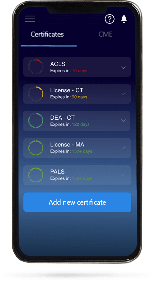
All Your Certificates in One Place
When you have your licenses, certificates and CMEs in one place, it's easier to track your career growth. You can easily share these with hospitals as well, using your medtigo app.