RyR1 Structural Alterations Explain Statin-Associated Muscle Dysfunction
December 16, 2025


Background
Osteoid osteoma is a benign bone tumor that primarily affects young individuals, typically between the ages of 5 and 25 years, with a peak incidence in the second and third decades of life. It is more common in males than females, with a ratio of approximately 2:1. It accounts for about 10% of all the benign bone tumors.
Osteoid osteomas are primarily found in the long bones, especially the femur and tibia. However, they can occur in any bone. The tumors are often located in the cortex (outer layer) of the bone. They are generally small, typically less than 1.5 cm in diameter.
The hallmark symptom of osteoid osteoma is localized pain, which is often more intense at night and may be relieved by nonsteroidal anti-inflammatory drugs (NSAIDs) such as aspirin or ibuprofen.
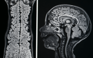
On imaging studies such as CT scans or X-rays, osteoid osteomas typically exhibit a characteristic radiolucent nidus surrounded by sclerotic (dense) bone. The nidus is the central region of the tumor where the active osteoid-forming tissue is concentrated.
Epidemiology
Anatomy
Pathophysiology
Etiology
Genetics
Prognostic Factors
Clinical History
Age Group:
Children and Adolescents:
Adults:
Physical Examination
Pain Assessment:
Range of Motion:
Palpation:
Neurovascular Examination:
Functional Assessment:
Observation of Gait:
Muscle Strength Testing:
Systemic Symptoms:
Age group
Associated comorbidity
No Clear Association with Comorbidities:
Activity Level:
Associated activity
Acuity of presentation
Chronic, Intermittent Pain:
Gradual Onset:
No Systemic Symptoms:
Differential Diagnoses
Laboratory Studies
Imaging Studies
Procedures
Histologic Findings
Staging
Treatment Paradigm
Minimally Invasive Procedures:
Surgical Excision: Surgical removal of the osteoid osteoma is another treatment option, especially if the tumor is in a location that is difficult to access with minimally invasive procedures. This may involve open surgery with direct visualization of the tumor and removal of the nidus.
by Stage
by Modality
Chemotherapy
Radiation Therapy
Surgical Interventions
Hormone Therapy
Immunotherapy
Hyperthermia
Photodynamic Therapy
Stem Cell Transplant
Targeted Therapy
Palliative Care
use-of-a-non-pharmacological-approach-for-treating-osteoid-osteoma
Role of NSAID’s in the treatment of Osteoid Osteoma
Nonsteroidal anti-inflammatory drugs (NSAIDs) play a role in the management of osteoid osteoma, a benign bone tumor. Osteoid osteoma is characterized by a small, painful nidus (localized area of tissue) within the bone, and NSAIDs can be effective in alleviating the associated pain and inflammation.
The primary symptom of osteoid osteoma is pain, often worse at night and typically responsive to NSAIDs. Osteoid osteomas are associated with local inflammation around the tumor nidus. NSAIDs reduce inflammation by preventing the cyclooxygenase (COX) enzymes from doing their job, which is necessary for the production of prostaglandins.
This anti-inflammatory action helps to reduce pain and swelling associated with the tumor. While NSAIDs can provide symptomatic relief, they are often used in conjunction with other treatment modalities.
Surgical removal of the tumor nidus is a common and curative treatment for osteoid osteoma. Minimally invasive methods like laser treatment or radiofrequency ablation may be used in specific situations.
Naproxen
Commonly used to treat pain and decrease inflammation is naproxen, a nonsteroidal anti-inflammatory medication.
Osteoid osteoma is mostly characterized by pain. Nonsteroidal anti-inflammatory drugs (NSAIDs), such as naproxen, function by preventing the synthesis of cytokines, known as compounds that are involved in pain and inflammation.
Naproxen lowers prostaglandin levels, which lessens discomfort and enhances comfort for those who have osteoid osteoma. Osteoid osteomas are associated with local inflammation around the tumor nidus.
Because it is an NSAID, naproxen contains anti-inflammatory qualities that may help reduce inflammation and swelling in the affected region.
use-of-intervention-with-a-procedure-in-treating-osteoid-osteoma
Radionuclide-Guided Excision:
CT-Guided Percutaneous Excision:
Percutaneous Laser Photocoagulation:
Percutaneous Radiofrequency Coagulation:
Cryoablation:
Computer-Assisted Surgery:
Magnetic Resonance–Guided Focused Ultrasound (MRgFUS):
Arthroscopy:
use-of-phases-in-managing-osteoid-osteoma
Diagnosis:
Medical Management:
Minimally Invasive Procedures:
Surgical Intervention:
Post-Treatment Follow-up:
Complications Management:
Medication
Future Trends
References
Osteoid osteoma is a benign bone tumor that primarily affects young individuals, typically between the ages of 5 and 25 years, with a peak incidence in the second and third decades of life. It is more common in males than females, with a ratio of approximately 2:1. It accounts for about 10% of all the benign bone tumors.
Osteoid osteomas are primarily found in the long bones, especially the femur and tibia. However, they can occur in any bone. The tumors are often located in the cortex (outer layer) of the bone. They are generally small, typically less than 1.5 cm in diameter.
The hallmark symptom of osteoid osteoma is localized pain, which is often more intense at night and may be relieved by nonsteroidal anti-inflammatory drugs (NSAIDs) such as aspirin or ibuprofen.
On imaging studies such as CT scans or X-rays, osteoid osteomas typically exhibit a characteristic radiolucent nidus surrounded by sclerotic (dense) bone. The nidus is the central region of the tumor where the active osteoid-forming tissue is concentrated.
Age Group:
Children and Adolescents:
Adults:
Pain Assessment:
Range of Motion:
Palpation:
Neurovascular Examination:
Functional Assessment:
Observation of Gait:
Muscle Strength Testing:
Systemic Symptoms:
No Clear Association with Comorbidities:
Activity Level:
Chronic, Intermittent Pain:
Gradual Onset:
No Systemic Symptoms:
Minimally Invasive Procedures:
Surgical Excision: Surgical removal of the osteoid osteoma is another treatment option, especially if the tumor is in a location that is difficult to access with minimally invasive procedures. This may involve open surgery with direct visualization of the tumor and removal of the nidus.
Nonsteroidal anti-inflammatory drugs (NSAIDs) play a role in the management of osteoid osteoma, a benign bone tumor. Osteoid osteoma is characterized by a small, painful nidus (localized area of tissue) within the bone, and NSAIDs can be effective in alleviating the associated pain and inflammation.
The primary symptom of osteoid osteoma is pain, often worse at night and typically responsive to NSAIDs. Osteoid osteomas are associated with local inflammation around the tumor nidus. NSAIDs reduce inflammation by preventing the cyclooxygenase (COX) enzymes from doing their job, which is necessary for the production of prostaglandins.
This anti-inflammatory action helps to reduce pain and swelling associated with the tumor. While NSAIDs can provide symptomatic relief, they are often used in conjunction with other treatment modalities.
Surgical removal of the tumor nidus is a common and curative treatment for osteoid osteoma. Minimally invasive methods like laser treatment or radiofrequency ablation may be used in specific situations.
Naproxen
Commonly used to treat pain and decrease inflammation is naproxen, a nonsteroidal anti-inflammatory medication.
Osteoid osteoma is mostly characterized by pain. Nonsteroidal anti-inflammatory drugs (NSAIDs), such as naproxen, function by preventing the synthesis of cytokines, known as compounds that are involved in pain and inflammation.
Naproxen lowers prostaglandin levels, which lessens discomfort and enhances comfort for those who have osteoid osteoma. Osteoid osteomas are associated with local inflammation around the tumor nidus.
Because it is an NSAID, naproxen contains anti-inflammatory qualities that may help reduce inflammation and swelling in the affected region.
Radionuclide-Guided Excision:
CT-Guided Percutaneous Excision:
Percutaneous Laser Photocoagulation:
Percutaneous Radiofrequency Coagulation:
Cryoablation:
Computer-Assisted Surgery:
Magnetic Resonance–Guided Focused Ultrasound (MRgFUS):
Arthroscopy:
Diagnosis:
Medical Management:
Minimally Invasive Procedures:
Surgical Intervention:
Post-Treatment Follow-up:
Complications Management:

Both our subscription plans include Free CME/CPD AMA PRA Category 1 credits.

On course completion, you will receive a full-sized presentation quality digital certificate.
A dynamic medical simulation platform designed to train healthcare professionals and students to effectively run code situations through an immersive hands-on experience in a live, interactive 3D environment.

When you have your licenses, certificates and CMEs in one place, it's easier to track your career growth. You can easily share these with hospitals as well, using your medtigo app.