Peripheral Anterior Synechia also known PAS, is a condition where the peripheral iris attaches itself to the anterior surface of the eye’s structures, such as the cornea or anterior lens capsule. This condition is often linked to specific types of glaucoma, eye inflammation, or trauma.
Salzmann first documented PAS in 1914, and its presence can cause various visual disturbances and complications. The severity and location of the adhesions determine whether it obstructs the regular flow of fluid within the eye, which can result in heightened intraocular pressure.
Epidemiology
Prevalence: The occurrence of PAS is usually greater in persons with specific forms of glaucoma, notably angle-closure glaucoma. In communities with primary angle-closure glaucoma, PAS can be detected in roughly 20-30% of instances.
Age and Sex: PAS may arise at any stage of life, but it is more frequently identified in elderly people. The vulnerability to PAS development surges with age, mainly in individuals who are above 40 years old.
Ethnicity: There is some evidence to suggest that the prevalence of angle-closure glaucoma and associated PAS may vary among different ethnic groups.
Anatomy
Pathophysiology
The pathophysiology of peripheral anterior synechia (PAS) involves the formation of adhesions between the peripheral iris and the anterior structures of the eye, typically the cornea or the anterior lens capsule.
Inflammatory Response: The development of PAS is closely linked to the body’s inflammatory response. When conditions like uveitis or inflammation within the eye occur, it can trigger an immune response.
Formation of Fibrous Tissue and Scars: Whenever the eye experiences inflammation or injury, it starts a healing process that involves the buildup of fibrous tissue and the formation of scars. The adhesions that appear in PAS are mostly made up of collagen and fibrous tissue.
Etiology
Inflammation of the uvea, also known as uveitis, can contribute to the formation of PAS. Inflammatory processes related to uveitis can cause adhesions to form between the iris and surrounding structures, including the lens or cornea.
Individuals with certain types of glaucoma, particularly chronic angle-closure glaucoma or angle-closure glaucoma, frequently exhibit PAS. The development of PAS can result from iris ischemia, which occurs when the iris receives insufficient blood supply. Ischemic eye conditions, such as carotid artery disease or ocular ischemic syndrome, can contribute to this.
Genetics
Prognostic Factors
Underlying Cause: The root cause of PAS holds significant importance as a prognostic factor. PAS linked with specific forms of glaucoma, such as angle-closure glaucoma, may have a more cautious prognosis when compared to PAS arising from other reasons such as uveitis or trauma.
Response to Therapy: The response to therapy is a crucial prognostic aspect. If PAS responds well to medical treatments such as glaucoma medications or anti-inflammatory therapies, and the adhesions can be effectively managed or lessened, the prognosis may be enhanced.
Complications: The presence of complications associated with PAS, such as corneal endothelial cell loss, irregular pupil shape, or visual disturbances, can impact the prognosis. The severity and persistence of these complications can affect the visual outcome and quality of life for individuals with PAS.
Clinical History
CLINICAL HISTORY
Age Group: Peripheral Anterior Synechia (PAS) can occur in individuals of various age groups, but it is more commonly observed in older individuals. The risk of developing PAS increases with age, particularly in individuals over the age of 40.
Physical Examination
PHYSICAL EXAMINATION
Assessment of Vision acuity: The sharpness of vision is usually assessed using a Snellen chart or other standardized charts for evaluating visual acuity. This examination aids in identifying any visual impairment and determining the clarity of vision associated with PAS.
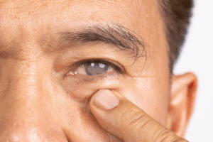
Examination of the Pupil: The assessment involves examining the size, shape, and responsiveness of the pupil. PAS may cause irregularities in the shape of the pupil or partial blockage of the pupil.
Gonioscopy: It is a specialized technique for examination that enables the visualization of the angle between the cornea and iris. It can aid in identifying the type and severity of angle-closure glaucoma associated with PAS.
Age group
Associated comorbidity
Associated Comorbidity or Activity:
Inflammatory Conditions: PAS can occur in the context of various inflammatory conditions affecting the eye, such as iridocyclitis, iritis, or other forms of ocular inflammation.
Glaucoma: PAS is often associated with different types of glaucoma, especially angle-closure glaucoma or persistent angle-closure glaucoma. Therefore, people with PAS may also have a multiplied threat of glaucoma or may also have already got a prognosis of glaucoma.
Trauma: PAS may be an outcome of eye trauma, which include direct harm or ocular surgical operation. Individuals with a record of ocular trauma or current eye surgical operation can be at better threat of growing PAS because of the recuperation technique and scar formation.
Ocular Ischemia: PAS can be observed in individuals with conditions causing reduced blood supply to the iris, such as ocular ischemic syndrome or carotid artery disease.
Associated activity
Acuity of presentation
Acuity of Presentation:
Asymptomatic: In some instances, PAS can be without symptoms, indicating that the person does not encounter any visible disruptions or modifications in visual sharpness.
Sudden Loss of Sight: In specific cases, mainly in acute angle-closure glaucoma with swift formation of PAS, people may encounter an abrupt loss of sight.
Unstable Vision: Unstable visual sharpness can be detected in people with PAS, particularly if the adhesions are in motion or if the degree of pupillary blockage fluctuates over time.
Differential Diagnoses
DIFFERENTIAL DIAGNOSIS
Pseudoexfoliation Syndrome: Pseudoexfoliation syndrome is a condition characterized by the deposition of white, flaky material on various intraocular structures, including the anterior segment.
Posterior synechia: it pertains to the binding of the iris with the lens or the back parts of the eye. It may transpire in instances like posterior uveitis or former eye surgery.
Traumatic Iris Abnormalities: Abnormalities of the iris that occur as a result of prior trauma or surgical procedures may sometimes resemble peripheral anterior synechiae (PAS). It is important to conduct a thorough review of the patient’s medical history, examine previous eye records, and evaluate the degree and position of the adhesions in order to distinguish them from PAS.
Iris tumors: In rare instances, tumors affecting the iris, such as iris melanoma or iris metastases, may lead to adhesions between the iris and adjacent structures.
Iridocorneal Endothelial (ICE) Syndrome: ICE syndrome is a group of eye disorders marked by anomalous growth of corneal endothelial cells, causing alterations in the iris and angle structures.
Laboratory Studies
Imaging Studies
Procedures
Histologic Findings
Staging
Treatment Paradigm
TREATMENT PARADIGM
Modification of Environment:
Lighting Conditions: Adjusting the lighting conditions in the environment can help improve visual comfort for individuals with PAS. Bright or intense lighting may cause discomfort or glare for those with irregular pupils or compromised iris function.
Minimizing Glare: Employing anti-reflective coatings on spectacles, donning shaded lenses or sunglasses, and installing window treatments or blinds to limit direct sunlight can effectively decrease any discomfort caused by glare.
Ideal Seating and Positioning: ensuring appropriate seating and placement can heighten visual ease and optimize visual capabilities for people with PAS. Positioning oneself in a manner that minimizes any need to tilt the head or strain the eyes can advance visual efficacy.
Administration of Pharmaceutical Agents with Drugs:
Antiglaucoma Medications: In cases of PAS associated with glaucoma, antiglaucoma medications may be prescribed to manage intraocular pressure. Medications such as beta-blockers, prostaglandin analogs, carbonic anhydrase inhibitors can be used to reduce intraocular pressure.
Osmotic agents: The agents like mannitol or glycerin are indicated to swiftly reduce intraocular pressure in acute instances of angle-closure glaucoma that involve significant peripheral anterior synechia.
Intervention with a Procedure:
Laser Peripheral Iridotomy (LPI): LPI is a technique frequently employed in instances of PAS linked with angle-closure glaucoma. It consists of generating a minor aperture in the outermost iris by utilizing a laser.
Cyclophotocoagulation: It is a procedure that uses laser energy or cryotherapy to selectively destroy parts of the ciliary body, which produces aqueous humor.
Surgical Iridectomy: It is a medical practice that necessitates the elimination of a segment of the outermost iris. This produces a continuous opening that facilitates better fluid movement and inhibits or decreases pupillary obstruction.
Phase of Management:
Underlying Cause Treatment: The management of PAS often focuses on addressing the underlying cause. For example, if the PAS is associated with uveitis, appropriate treatment with anti-inflammatory medications, such as topical steroids or immunosuppressive agents.
Rehabilitation and Visual Optimization: This stage could include the utilization of visual devices, low vision recuperation, visual training, or vocational therapy to aid people with PAS in optimizing their residual eyesight and accommodating to any visual impairments.
Ongoing Monitoring and Follow-up: Consistent evaluation and aftercare sessions are vital to evaluate the reaction to therapy, oversee any variations in the state, and modify as required.
Peripheral Anterior Synechia also known PAS, is a condition where the peripheral iris attaches itself to the anterior surface of the eye’s structures, such as the cornea or anterior lens capsule. This condition is often linked to specific types of glaucoma, eye inflammation, or trauma.
Salzmann first documented PAS in 1914, and its presence can cause various visual disturbances and complications. The severity and location of the adhesions determine whether it obstructs the regular flow of fluid within the eye, which can result in heightened intraocular pressure.
Prevalence: The occurrence of PAS is usually greater in persons with specific forms of glaucoma, notably angle-closure glaucoma. In communities with primary angle-closure glaucoma, PAS can be detected in roughly 20-30% of instances.
Age and Sex: PAS may arise at any stage of life, but it is more frequently identified in elderly people. The vulnerability to PAS development surges with age, mainly in individuals who are above 40 years old.
Ethnicity: There is some evidence to suggest that the prevalence of angle-closure glaucoma and associated PAS may vary among different ethnic groups.
The pathophysiology of peripheral anterior synechia (PAS) involves the formation of adhesions between the peripheral iris and the anterior structures of the eye, typically the cornea or the anterior lens capsule.
Inflammatory Response: The development of PAS is closely linked to the body’s inflammatory response. When conditions like uveitis or inflammation within the eye occur, it can trigger an immune response.
Formation of Fibrous Tissue and Scars: Whenever the eye experiences inflammation or injury, it starts a healing process that involves the buildup of fibrous tissue and the formation of scars. The adhesions that appear in PAS are mostly made up of collagen and fibrous tissue.
Inflammation of the uvea, also known as uveitis, can contribute to the formation of PAS. Inflammatory processes related to uveitis can cause adhesions to form between the iris and surrounding structures, including the lens or cornea.
Individuals with certain types of glaucoma, particularly chronic angle-closure glaucoma or angle-closure glaucoma, frequently exhibit PAS. The development of PAS can result from iris ischemia, which occurs when the iris receives insufficient blood supply. Ischemic eye conditions, such as carotid artery disease or ocular ischemic syndrome, can contribute to this.
Underlying Cause: The root cause of PAS holds significant importance as a prognostic factor. PAS linked with specific forms of glaucoma, such as angle-closure glaucoma, may have a more cautious prognosis when compared to PAS arising from other reasons such as uveitis or trauma.
Response to Therapy: The response to therapy is a crucial prognostic aspect. If PAS responds well to medical treatments such as glaucoma medications or anti-inflammatory therapies, and the adhesions can be effectively managed or lessened, the prognosis may be enhanced.
Complications: The presence of complications associated with PAS, such as corneal endothelial cell loss, irregular pupil shape, or visual disturbances, can impact the prognosis. The severity and persistence of these complications can affect the visual outcome and quality of life for individuals with PAS.
CLINICAL HISTORY
Age Group: Peripheral Anterior Synechia (PAS) can occur in individuals of various age groups, but it is more commonly observed in older individuals. The risk of developing PAS increases with age, particularly in individuals over the age of 40.
PHYSICAL EXAMINATION
Assessment of Vision acuity: The sharpness of vision is usually assessed using a Snellen chart or other standardized charts for evaluating visual acuity. This examination aids in identifying any visual impairment and determining the clarity of vision associated with PAS.
Examination of the Pupil: The assessment involves examining the size, shape, and responsiveness of the pupil. PAS may cause irregularities in the shape of the pupil or partial blockage of the pupil.
Gonioscopy: It is a specialized technique for examination that enables the visualization of the angle between the cornea and iris. It can aid in identifying the type and severity of angle-closure glaucoma associated with PAS.
Associated Comorbidity or Activity:
Inflammatory Conditions: PAS can occur in the context of various inflammatory conditions affecting the eye, such as iridocyclitis, iritis, or other forms of ocular inflammation.
Glaucoma: PAS is often associated with different types of glaucoma, especially angle-closure glaucoma or persistent angle-closure glaucoma. Therefore, people with PAS may also have a multiplied threat of glaucoma or may also have already got a prognosis of glaucoma.
Trauma: PAS may be an outcome of eye trauma, which include direct harm or ocular surgical operation. Individuals with a record of ocular trauma or current eye surgical operation can be at better threat of growing PAS because of the recuperation technique and scar formation.
Ocular Ischemia: PAS can be observed in individuals with conditions causing reduced blood supply to the iris, such as ocular ischemic syndrome or carotid artery disease.
Acuity of Presentation:
Asymptomatic: In some instances, PAS can be without symptoms, indicating that the person does not encounter any visible disruptions or modifications in visual sharpness.
Sudden Loss of Sight: In specific cases, mainly in acute angle-closure glaucoma with swift formation of PAS, people may encounter an abrupt loss of sight.
Unstable Vision: Unstable visual sharpness can be detected in people with PAS, particularly if the adhesions are in motion or if the degree of pupillary blockage fluctuates over time.
DIFFERENTIAL DIAGNOSIS
Pseudoexfoliation Syndrome: Pseudoexfoliation syndrome is a condition characterized by the deposition of white, flaky material on various intraocular structures, including the anterior segment.
Posterior synechia: it pertains to the binding of the iris with the lens or the back parts of the eye. It may transpire in instances like posterior uveitis or former eye surgery.
Traumatic Iris Abnormalities: Abnormalities of the iris that occur as a result of prior trauma or surgical procedures may sometimes resemble peripheral anterior synechiae (PAS). It is important to conduct a thorough review of the patient’s medical history, examine previous eye records, and evaluate the degree and position of the adhesions in order to distinguish them from PAS.
Iris tumors: In rare instances, tumors affecting the iris, such as iris melanoma or iris metastases, may lead to adhesions between the iris and adjacent structures.
Iridocorneal Endothelial (ICE) Syndrome: ICE syndrome is a group of eye disorders marked by anomalous growth of corneal endothelial cells, causing alterations in the iris and angle structures.
TREATMENT PARADIGM
Modification of Environment:
Lighting Conditions: Adjusting the lighting conditions in the environment can help improve visual comfort for individuals with PAS. Bright or intense lighting may cause discomfort or glare for those with irregular pupils or compromised iris function.
Minimizing Glare: Employing anti-reflective coatings on spectacles, donning shaded lenses or sunglasses, and installing window treatments or blinds to limit direct sunlight can effectively decrease any discomfort caused by glare.
Ideal Seating and Positioning: ensuring appropriate seating and placement can heighten visual ease and optimize visual capabilities for people with PAS. Positioning oneself in a manner that minimizes any need to tilt the head or strain the eyes can advance visual efficacy.
Administration of Pharmaceutical Agents with Drugs:
Antiglaucoma Medications: In cases of PAS associated with glaucoma, antiglaucoma medications may be prescribed to manage intraocular pressure. Medications such as beta-blockers, prostaglandin analogs, carbonic anhydrase inhibitors can be used to reduce intraocular pressure.
Osmotic agents: The agents like mannitol or glycerin are indicated to swiftly reduce intraocular pressure in acute instances of angle-closure glaucoma that involve significant peripheral anterior synechia.
Intervention with a Procedure:
Laser Peripheral Iridotomy (LPI): LPI is a technique frequently employed in instances of PAS linked with angle-closure glaucoma. It consists of generating a minor aperture in the outermost iris by utilizing a laser.
Cyclophotocoagulation: It is a procedure that uses laser energy or cryotherapy to selectively destroy parts of the ciliary body, which produces aqueous humor.
Surgical Iridectomy: It is a medical practice that necessitates the elimination of a segment of the outermost iris. This produces a continuous opening that facilitates better fluid movement and inhibits or decreases pupillary obstruction.
Phase of Management:
Underlying Cause Treatment: The management of PAS often focuses on addressing the underlying cause. For example, if the PAS is associated with uveitis, appropriate treatment with anti-inflammatory medications, such as topical steroids or immunosuppressive agents.
Rehabilitation and Visual Optimization: This stage could include the utilization of visual devices, low vision recuperation, visual training, or vocational therapy to aid people with PAS in optimizing their residual eyesight and accommodating to any visual impairments.
Ongoing Monitoring and Follow-up: Consistent evaluation and aftercare sessions are vital to evaluate the reaction to therapy, oversee any variations in the state, and modify as required.
Both our subscription plans include Free CME/CPD AMA PRA Category 1 credits.
Digital Certificate PDF
On course completion, you will receive a full-sized presentation quality digital certificate.
medtigo Simulation
A dynamic medical simulation platform designed to train healthcare professionals and students to effectively run code situations through an immersive hands-on experience in a live, interactive 3D environment.
medtigo Points
medtigo points is our unique point redemption system created to award users for interacting on our site. These points can be redeemed for special discounts on the medtigo marketplace as well as towards the membership cost itself.
Community Forum post/reply = 5 points
*Redemption of points can occur only through the medtigo marketplace, courses, or simulation system. Money will not be credited to your bank account. 10 points = $1.
All Your Certificates in One Place
When you have your licenses, certificates and CMEs in one place, it's easier to track your career growth. You can easily share these with hospitals as well, using your medtigo app.