Rising Adoption of Continuous Glucose Monitors in Medicare Advantage: Trends from 2021–2023
April 13, 2026


Background
Also called enterobiasis, pinworm infection is caused by Enterobius vermicualris, a small, very slender nematode with a sharply pointed tail. In humans, they are found in the appendix, ascending colon, and cecum. Females measure between 8-13mm, while males range between 2-5mm in length.
Pinworm is predominantly a disease of children, and parents often become infected through their children, as a result of reinfection or incomplete treatment. This infection is common throughout the world particularly in the temperate areas. In the United States, it us the most common helminthic infection.
The usual modes of transmission are by direct contact with contaminated bedclothes, furniture, bedding, towels, doorknobs, toilets, or other inanimate objects. The parasites occasionally are transmitted sexually. Generally, there are no symptoms due to pinworm infection and asymptomatic carriers are many in number. The rate of cure with treatment is approximately 90-95% with common chances of re-infection if all contracted patients are not simultaneously treated.
Epidemiology
Pinworm is a common helminthic infestation in the United States, with general prevalence of 0.2 to 20% in children. Living in crowds and institutionalized individuals show higher prevalence rates, with 50-100% occurrence in institutionalized people. Europe equally reports a similar prevalence. It is highly recorded in cosmopolitan areas in the cool and temperate regions. The rate of carrying eggs may vary between countries and with the most at risk category being school-aged children. In adults, the peak incidence of pin worm infection is among 30-39 years, primarily as a result of the children between ages of 5-9 transmitting it to them, Males are infected in 2:1 ratio over females, except in the ages of 5-14.
Anatomy
Pathophysiology
A prickling sensation or pruritus or in the perianal area that normally occurs at night is actually the most common symptom of pinworm. It is caused by a gravid female pinworm that has migrated towards the anal area to deposit eggs with her tail pin in the mucosa. Enterobius vermicularis is considered to reside mainly in the small intestine, specifically in the ileocecal area.
Intense local itching is due to the movement of the female pinworm and her eggs. The ova can survive for up to three weeks prior to hatching, and the hatched larvae can migrate back into the lower intestine and anus and reach the site of fertilization. Embryonated eggs can be released onto fomites (e.g., clothing, paper money, bedding, toys), air or onto hands, which then can be placed directly into the mouth leading to autoinfection and helping the worm to enter the small intestines.
Typically, pinworms present in the cecum and adjoining structures are asymptomatic, but it has been postulated that acute infection might cause diarrhea by inducing inflammation of the wall of the bowel. Although pinworms were found in the appendix during histologic study of acute appendicitis, this relationship is probably coincidental.
Probable risk factors for pinworm infection include eating without washing hands, poor personal and group hygiene, and living with an egg-positive person.
Etiology
Infection by the Enterobius vermicularis nematode has been known to result in this condition. The major transmission pathway involves the ingestion of the egg stage of the pinworm, mainly via contaminated hands, food, drink, or fomite. Autoinfection occurs when infective eggs on the hands are transferred to the mouth from scratching the perianal area, and retroinfection results from the migration of hatched larvae into the intestines.
The life cycle consists of infective eggs hatching in the small intestine, larvae developing to adults, and adults laying many eggs, which cause intense itching and are subsequently spread from scratching. The risk factors of pinworm infection are a close contact, poor hygienic actions, environmental contamination, and self-infection. The understanding of pinworm infection helps to effect control and prevention through good personal hygiene, regular handwashing, and a clean-living environment.
Genetics
Prognostic Factors
Pinworm infection causes serious morbidity rarely unless ectopic infection takes place, which can happen in patients with inflammatory bowel disease. Parasites can penetrate the bowel wall and can be located in extracolonic sites such as vagina, fallopian tubes, inguinal area, liver, male genital tract, omentum, genital area, pelvic peritoneum, salivary glands, lungs, and male genital tract.
Ectopic enterobiasis has been linked with acute appendicitis, eosinophilic gastroenteritis, and eosinophilic colitis. Infestation by pinworm is rarely fatal and the primary cause of death includes effect of secondary infections. It is very hard to eliminate pinworm entirely in the institutionalized individual, and follow-up examination must be persistent. The success of treatment is higher if treatment of the child is done together with his family and classmates.
Clinical History
History includes prickling or pain in the anal region particularly in the night, severe anal itching, difficulty sleeping or restless sleep, rarely loss of appetite or abdominal discomfort. Other signs may include irritability and vaginal itching in females. However, most patients are asymptomatic.
Physical Examination
One hospital-based study of children aged 2–12 years found that itching in the perianal area was found to be not notably more common in children who are infected than in children who are uninfected, although persons who are egg–positive for Enetrobius typically consult a doctor due to perianal itching.
The female pinworm (10 mm) can be easily visualized in the perianal region with naked eye. Oftentimes the worm is mistaken for a cotton thread residue. Unless viewed under the microscope, eggs (30 μm X 50-60 μm) are not visualized. Perianal excoriations from the itching are commonly found.
Age group
Associated comorbidity
Associated activity
Acuity of presentation
Differential Diagnoses
Proctitis
Anusitis
Laboratory Studies
Imaging Studies
Procedures
Histologic Findings
Staging
Treatment Paradigm
by Stage
by Modality
Chemotherapy
Radiation Therapy
Surgical Interventions
Hormone Therapy
Immunotherapy
Hyperthermia
Photodynamic Therapy
Stem Cell Transplant
Targeted Therapy
Palliative Care
Use of anthelmintics
use-of-phases-of-management-in-treating-pinworm-infections
Management of pinworm infections includes several phases like diagnosis, treatment, prevention, and follow-up. Diagnosis is based on clinical evaluation, visual examination, laboratory tests, examination of faeces, and anthelmintics such as albendazole, ebendazole, Pyrantel Pamoate. Symptomatic relief can be provided using antihistamines. Prevention includes personal hygiene maintenance, cleaning fingernails, environmental cleaning by laundering, cleaning, and dusting, among others.
It enables a reduction in the rate of reinfection through mass treatment and educative programs.
The follow-up should then include re-evaluation, repetition of treatment dose, and monitoring of signs and symptoms. All legs are crucial in effectiveness management, reduction of morbidity, and prevention of reinfection.
Medication
Take a dose of 1.8 gm orally every four hours for a total of three doses daily
This treatment may be repeated in two weeks
A single dose is administered based on body weight, and it is repeated in two to three weeks
Take one tablet orally two times in a day
Administer as a single dose of 12.5 mg/kg orally of total body weight
Take a dose of 100 mg orally as a single dose
for 2 years old:
Take a dose of 600 mg orally in every four hours for a total of three doses daily
for 2 to 8 years old:
Take a dose of 1.2 gm orally in every six hours for a total of two doses daily
for 8 to 14 years old:
Take a dose of 1.2 gm orally in every four hours for a total of three doses daily
A single dose is administered based on body weight, and it is repeated in two to three weeks
For <2 years old: Safety and efficacy not established
For ≥2 years old:
Take a dose of 100 mg orally as a single dose
Future Trends
Also called enterobiasis, pinworm infection is caused by Enterobius vermicualris, a small, very slender nematode with a sharply pointed tail. In humans, they are found in the appendix, ascending colon, and cecum. Females measure between 8-13mm, while males range between 2-5mm in length.
Pinworm is predominantly a disease of children, and parents often become infected through their children, as a result of reinfection or incomplete treatment. This infection is common throughout the world particularly in the temperate areas. In the United States, it us the most common helminthic infection.
The usual modes of transmission are by direct contact with contaminated bedclothes, furniture, bedding, towels, doorknobs, toilets, or other inanimate objects. The parasites occasionally are transmitted sexually. Generally, there are no symptoms due to pinworm infection and asymptomatic carriers are many in number. The rate of cure with treatment is approximately 90-95% with common chances of re-infection if all contracted patients are not simultaneously treated.
Pinworm is a common helminthic infestation in the United States, with general prevalence of 0.2 to 20% in children. Living in crowds and institutionalized individuals show higher prevalence rates, with 50-100% occurrence in institutionalized people. Europe equally reports a similar prevalence. It is highly recorded in cosmopolitan areas in the cool and temperate regions. The rate of carrying eggs may vary between countries and with the most at risk category being school-aged children. In adults, the peak incidence of pin worm infection is among 30-39 years, primarily as a result of the children between ages of 5-9 transmitting it to them, Males are infected in 2:1 ratio over females, except in the ages of 5-14.
A prickling sensation or pruritus or in the perianal area that normally occurs at night is actually the most common symptom of pinworm. It is caused by a gravid female pinworm that has migrated towards the anal area to deposit eggs with her tail pin in the mucosa. Enterobius vermicularis is considered to reside mainly in the small intestine, specifically in the ileocecal area.
Intense local itching is due to the movement of the female pinworm and her eggs. The ova can survive for up to three weeks prior to hatching, and the hatched larvae can migrate back into the lower intestine and anus and reach the site of fertilization. Embryonated eggs can be released onto fomites (e.g., clothing, paper money, bedding, toys), air or onto hands, which then can be placed directly into the mouth leading to autoinfection and helping the worm to enter the small intestines.
Typically, pinworms present in the cecum and adjoining structures are asymptomatic, but it has been postulated that acute infection might cause diarrhea by inducing inflammation of the wall of the bowel. Although pinworms were found in the appendix during histologic study of acute appendicitis, this relationship is probably coincidental.
Probable risk factors for pinworm infection include eating without washing hands, poor personal and group hygiene, and living with an egg-positive person.
Infection by the Enterobius vermicularis nematode has been known to result in this condition. The major transmission pathway involves the ingestion of the egg stage of the pinworm, mainly via contaminated hands, food, drink, or fomite. Autoinfection occurs when infective eggs on the hands are transferred to the mouth from scratching the perianal area, and retroinfection results from the migration of hatched larvae into the intestines.
The life cycle consists of infective eggs hatching in the small intestine, larvae developing to adults, and adults laying many eggs, which cause intense itching and are subsequently spread from scratching. The risk factors of pinworm infection are a close contact, poor hygienic actions, environmental contamination, and self-infection. The understanding of pinworm infection helps to effect control and prevention through good personal hygiene, regular handwashing, and a clean-living environment.
Pinworm infection causes serious morbidity rarely unless ectopic infection takes place, which can happen in patients with inflammatory bowel disease. Parasites can penetrate the bowel wall and can be located in extracolonic sites such as vagina, fallopian tubes, inguinal area, liver, male genital tract, omentum, genital area, pelvic peritoneum, salivary glands, lungs, and male genital tract.
Ectopic enterobiasis has been linked with acute appendicitis, eosinophilic gastroenteritis, and eosinophilic colitis. Infestation by pinworm is rarely fatal and the primary cause of death includes effect of secondary infections. It is very hard to eliminate pinworm entirely in the institutionalized individual, and follow-up examination must be persistent. The success of treatment is higher if treatment of the child is done together with his family and classmates.
History includes prickling or pain in the anal region particularly in the night, severe anal itching, difficulty sleeping or restless sleep, rarely loss of appetite or abdominal discomfort. Other signs may include irritability and vaginal itching in females. However, most patients are asymptomatic.
One hospital-based study of children aged 2–12 years found that itching in the perianal area was found to be not notably more common in children who are infected than in children who are uninfected, although persons who are egg–positive for Enetrobius typically consult a doctor due to perianal itching.
The female pinworm (10 mm) can be easily visualized in the perianal region with naked eye. Oftentimes the worm is mistaken for a cotton thread residue. Unless viewed under the microscope, eggs (30 μm X 50-60 μm) are not visualized. Perianal excoriations from the itching are commonly found.
Proctitis
Anusitis
Infectious Disease
Infectious Disease
Management of pinworm infections includes several phases like diagnosis, treatment, prevention, and follow-up. Diagnosis is based on clinical evaluation, visual examination, laboratory tests, examination of faeces, and anthelmintics such as albendazole, ebendazole, Pyrantel Pamoate. Symptomatic relief can be provided using antihistamines. Prevention includes personal hygiene maintenance, cleaning fingernails, environmental cleaning by laundering, cleaning, and dusting, among others.
It enables a reduction in the rate of reinfection through mass treatment and educative programs.
The follow-up should then include re-evaluation, repetition of treatment dose, and monitoring of signs and symptoms. All legs are crucial in effectiveness management, reduction of morbidity, and prevention of reinfection.

Also called enterobiasis, pinworm infection is caused by Enterobius vermicualris, a small, very slender nematode with a sharply pointed tail. In humans, they are found in the appendix, ascending colon, and cecum. Females measure between 8-13mm, while males range between 2-5mm in length.
Pinworm is predominantly a disease of children, and parents often become infected through their children, as a result of reinfection or incomplete treatment. This infection is common throughout the world particularly in the temperate areas. In the United States, it us the most common helminthic infection.
The usual modes of transmission are by direct contact with contaminated bedclothes, furniture, bedding, towels, doorknobs, toilets, or other inanimate objects. The parasites occasionally are transmitted sexually. Generally, there are no symptoms due to pinworm infection and asymptomatic carriers are many in number. The rate of cure with treatment is approximately 90-95% with common chances of re-infection if all contracted patients are not simultaneously treated.
Pinworm is a common helminthic infestation in the United States, with general prevalence of 0.2 to 20% in children. Living in crowds and institutionalized individuals show higher prevalence rates, with 50-100% occurrence in institutionalized people. Europe equally reports a similar prevalence. It is highly recorded in cosmopolitan areas in the cool and temperate regions. The rate of carrying eggs may vary between countries and with the most at risk category being school-aged children. In adults, the peak incidence of pin worm infection is among 30-39 years, primarily as a result of the children between ages of 5-9 transmitting it to them, Males are infected in 2:1 ratio over females, except in the ages of 5-14.
A prickling sensation or pruritus or in the perianal area that normally occurs at night is actually the most common symptom of pinworm. It is caused by a gravid female pinworm that has migrated towards the anal area to deposit eggs with her tail pin in the mucosa. Enterobius vermicularis is considered to reside mainly in the small intestine, specifically in the ileocecal area.
Intense local itching is due to the movement of the female pinworm and her eggs. The ova can survive for up to three weeks prior to hatching, and the hatched larvae can migrate back into the lower intestine and anus and reach the site of fertilization. Embryonated eggs can be released onto fomites (e.g., clothing, paper money, bedding, toys), air or onto hands, which then can be placed directly into the mouth leading to autoinfection and helping the worm to enter the small intestines.
Typically, pinworms present in the cecum and adjoining structures are asymptomatic, but it has been postulated that acute infection might cause diarrhea by inducing inflammation of the wall of the bowel. Although pinworms were found in the appendix during histologic study of acute appendicitis, this relationship is probably coincidental.
Probable risk factors for pinworm infection include eating without washing hands, poor personal and group hygiene, and living with an egg-positive person.
Infection by the Enterobius vermicularis nematode has been known to result in this condition. The major transmission pathway involves the ingestion of the egg stage of the pinworm, mainly via contaminated hands, food, drink, or fomite. Autoinfection occurs when infective eggs on the hands are transferred to the mouth from scratching the perianal area, and retroinfection results from the migration of hatched larvae into the intestines.
The life cycle consists of infective eggs hatching in the small intestine, larvae developing to adults, and adults laying many eggs, which cause intense itching and are subsequently spread from scratching. The risk factors of pinworm infection are a close contact, poor hygienic actions, environmental contamination, and self-infection. The understanding of pinworm infection helps to effect control and prevention through good personal hygiene, regular handwashing, and a clean-living environment.
Pinworm infection causes serious morbidity rarely unless ectopic infection takes place, which can happen in patients with inflammatory bowel disease. Parasites can penetrate the bowel wall and can be located in extracolonic sites such as vagina, fallopian tubes, inguinal area, liver, male genital tract, omentum, genital area, pelvic peritoneum, salivary glands, lungs, and male genital tract.
Ectopic enterobiasis has been linked with acute appendicitis, eosinophilic gastroenteritis, and eosinophilic colitis. Infestation by pinworm is rarely fatal and the primary cause of death includes effect of secondary infections. It is very hard to eliminate pinworm entirely in the institutionalized individual, and follow-up examination must be persistent. The success of treatment is higher if treatment of the child is done together with his family and classmates.
History includes prickling or pain in the anal region particularly in the night, severe anal itching, difficulty sleeping or restless sleep, rarely loss of appetite or abdominal discomfort. Other signs may include irritability and vaginal itching in females. However, most patients are asymptomatic.
One hospital-based study of children aged 2–12 years found that itching in the perianal area was found to be not notably more common in children who are infected than in children who are uninfected, although persons who are egg–positive for Enetrobius typically consult a doctor due to perianal itching.
The female pinworm (10 mm) can be easily visualized in the perianal region with naked eye. Oftentimes the worm is mistaken for a cotton thread residue. Unless viewed under the microscope, eggs (30 μm X 50-60 μm) are not visualized. Perianal excoriations from the itching are commonly found.
Proctitis
Anusitis
Infectious Disease
Infectious Disease
Infectious Disease
Management of pinworm infections includes several phases like diagnosis, treatment, prevention, and follow-up. Diagnosis is based on clinical evaluation, visual examination, laboratory tests, examination of faeces, and anthelmintics such as albendazole, ebendazole, Pyrantel Pamoate. Symptomatic relief can be provided using antihistamines. Prevention includes personal hygiene maintenance, cleaning fingernails, environmental cleaning by laundering, cleaning, and dusting, among others.
It enables a reduction in the rate of reinfection through mass treatment and educative programs.
The follow-up should then include re-evaluation, repetition of treatment dose, and monitoring of signs and symptoms. All legs are crucial in effectiveness management, reduction of morbidity, and prevention of reinfection.

Both our subscription plans include Free CME/CPD AMA PRA Category 1 credits.
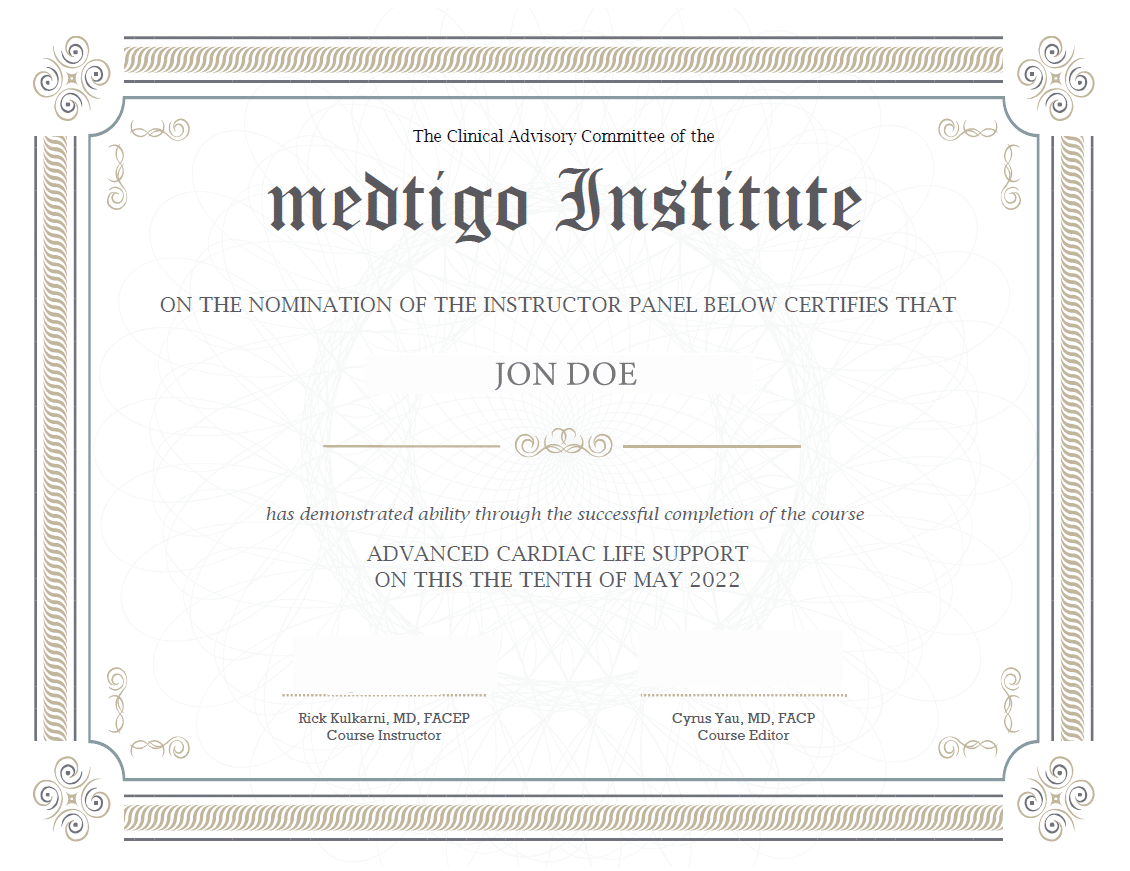
On course completion, you will receive a full-sized presentation quality digital certificate.
A dynamic medical simulation platform designed to train healthcare professionals and students to effectively run code situations through an immersive hands-on experience in a live, interactive 3D environment.

When you have your licenses, certificates and CMEs in one place, it's easier to track your career growth. You can easily share these with hospitals as well, using your medtigo app.