Vascular imaging is a field of medical imaging that shows the body’s vessels and passages containing blood. It is used to diagnose and treat vascular diseases and disorders.
Indications
Peripheral Artery Disease (PAD): To determine the areas and degree of the obstruction of arteries in limbs.
Aneurysms: Aneurysm scan is performed to assess the size of the aorta and other large arteries for aneurysms and to monitor their growth.
Carotid Artery Disease: To evaluate the buildup of plaque in the carotids, which results in stroke.
Deep Vein Thrombosis (DVT): To diagnose deep vein thrombosis, which is a blood clot in the deep veins of the body, commonly in the legs.
Pulmonary Embolism: For checking the ability to diagnose blood clots that have reached the lungs.
Renal Artery Stenosis: To assess stenosis of the arteries that feed kidneys.
Varicose Veins and Venous Insufficiency: To evaluate the physiological status of the veins as well as to determine veins that have defective valves that no longer work.
Tumor Evaluation: This is useful in identifying the blood vessels supply of tumors with a view of planning on how to treat the tumors.
Contraindications
Allergic Reactions to Contrast Media: Some of the risks of any type of imaging are the allergic reactions such as severe anaphylaxis episodes in patients with a known allergy to iodine or contrast agents used in procedure.
Renal Insufficiency: Contrast agents worsen the state of the kidney in patients who are already suffering from renal insufficiency or chronic kidney diseases.
Metal Implants or Devices: MRA is contraindicated in patients with specific types of implanted metals, including pacemakers, cochlear implants, or clips on aneurysms.
Claustrophobia: MRI machines are closed, which can become problematic when treating patients with claustrophobia.
Outcomes
Equipment
Ultrasound (Doppler Ultrasound)
Magnetic Resonance Angiography (MRA)
Computed Tomography Angiography (CTA)
X-ray Angiography (Fluoroscopy)
Positron Emission Tomography (PET)
Patient Preparation
Medical History and Consent:
Data that should be taken include allergy history to the contrast agents, current medications, and other medical conditions.
The procedure must be explained to the patient and informed consent is obtained.
Medications:
Before this procedure some of the medicines might require a change or complete withdrawal such as those that impact blood clotting.
Fasting:
Based on the imaging type that a patient should be subjected to; the patient may have to abstain from eating any food for several hours prior to the imaging.
Technique
Ultrasound Imaging (Doppler Ultrasound)
It creates images of body structures which involves using sound waves in the high-frequency range. Doppler ultrasound is mainly used to analyze blood movement by changes in the frequency of sound waves.
Step 1-Preparation: If the imaging is to be done on the abdominal vessels, then in this case, the patient might require fasting.
Step 2-Procedure: The patient’s skin is first coated with a gel. An interface (probe) is rolled over the skin surface, producing sound waves and receiving sound wave reflections.
Step 3-Imaging: It creates pictures of the blood vessels and the blood over the vessels. Doppler ultrasound can demonstrate the blood flow of vessels and its direction and speed.
Applications:
It is measuring the flow of blood in arteries as well as the veins.
Diagnosing conditions such as deep vein thrombosis and arterial occlusions.
Diagnosis of the conditions associated with heart valves and blood circulation.
Computed Tomography Angiography (CTA)
Computed Tomography Angiography, or CTA, is a type of CT scan done with the help of contrast agents but focuses on blood vessels. The test uses X-ray technology and computer processing to produce images of the vascular system.
Step 1-Preparation: The patient might need to fast during the procedure.
Step 2-Procedure: The patient should lay down on a table. Contrast material is introduced into the blood vessels and X-ray images are made from the way in which it goes through the vascular system.
Step 3-Imaging: Several cross-sectional pictures of the blood arteries are taken by the CT scanner and converted into three-dimensional images.
Applications:
Intervention for diagnosis and assessment of aneurysms, stenosis, and other vascular diseases.
In patients with complex diseases, prepare for surgical or interventional procedures.
Assessing trauma or bleeding.
Magnetic Resonance Angiography (MRA)
MRA uses magnetic fields and radio waves to generate or capture pictures of blood vessels. It can be carried out with or without contrast media.
Step 1-Preparation: Depending on the type of scan that is to be done, the patient may be required to fast to reduce any distortions that may be caused by food and fluids. If the contrast is necessary, it is given intravenously.
Step 2-Procedure: MRI is relatively painless, and the patient remains passive throughout the procedure in an MRI scanner. In some forms of MRA, contrast material is given, and images are taken while the material flows through the blood vessels.
Step 3-Imaging: The MRI scanner provides clear, detailed pictures of the vascular system, usually in 3D.
Applications:
Diagnosing aneurysms, blood clot, and other vascular malformation.
Assessing the blood vessels of the brain, the heart and such other organs.
Follow-up of the vascular monitoring over time.
Complications
Ultrasound:
Discomfort: It might cause skin sensitization on gel application.
Limitations: It may be restricted in depicting more underlying structures and in obese individuals.
CT Angiography (CTA):
Radiation Exposure: Imposes the risk of exposure to ionizing radiation and can be a hazardous if the patient has to undergo several scans.
Contrast Allergies: It is hazardous for patients with allergies and can also cause kidney problems in some people.
Contrast-Induced Nephropathy: Severe but rare, involving acute kidney injury due to the contrast agent.
Magnetic Resonance Angiography (MRA):
Contrast Reactions: Gadolinium-based contrast agents can cause allergic reactions or, in rare cases, nephrogenic systemic fibrosis (NSF) in patients with severe kidney impairment.
Metallic Implants: Patients known to have metallic implants are likely to encounter specific challenges or distortions in the resulting images.
Vascular imaging is a field of medical imaging that shows the body’s vessels and passages containing blood. It is used to diagnose and treat vascular diseases and disorders.
Peripheral Artery Disease (PAD): To determine the areas and degree of the obstruction of arteries in limbs.
Aneurysms: Aneurysm scan is performed to assess the size of the aorta and other large arteries for aneurysms and to monitor their growth.
Carotid Artery Disease: To evaluate the buildup of plaque in the carotids, which results in stroke.
Deep Vein Thrombosis (DVT): To diagnose deep vein thrombosis, which is a blood clot in the deep veins of the body, commonly in the legs.
Pulmonary Embolism: For checking the ability to diagnose blood clots that have reached the lungs.
Renal Artery Stenosis: To assess stenosis of the arteries that feed kidneys.
Varicose Veins and Venous Insufficiency: To evaluate the physiological status of the veins as well as to determine veins that have defective valves that no longer work.
Tumor Evaluation: This is useful in identifying the blood vessels supply of tumors with a view of planning on how to treat the tumors.
Allergic Reactions to Contrast Media: Some of the risks of any type of imaging are the allergic reactions such as severe anaphylaxis episodes in patients with a known allergy to iodine or contrast agents used in procedure.
Renal Insufficiency: Contrast agents worsen the state of the kidney in patients who are already suffering from renal insufficiency or chronic kidney diseases.
Metal Implants or Devices: MRA is contraindicated in patients with specific types of implanted metals, including pacemakers, cochlear implants, or clips on aneurysms.
Claustrophobia: MRI machines are closed, which can become problematic when treating patients with claustrophobia.
Ultrasound (Doppler Ultrasound)
Magnetic Resonance Angiography (MRA)
Computed Tomography Angiography (CTA)
X-ray Angiography (Fluoroscopy)
Positron Emission Tomography (PET)
Medical History and Consent:
Data that should be taken include allergy history to the contrast agents, current medications, and other medical conditions.
The procedure must be explained to the patient and informed consent is obtained.
Medications:
Before this procedure some of the medicines might require a change or complete withdrawal such as those that impact blood clotting.
Fasting:
Based on the imaging type that a patient should be subjected to; the patient may have to abstain from eating any food for several hours prior to the imaging.
Ultrasound Imaging (Doppler Ultrasound)
It creates images of body structures which involves using sound waves in the high-frequency range. Doppler ultrasound is mainly used to analyze blood movement by changes in the frequency of sound waves.
Step 1-Preparation: If the imaging is to be done on the abdominal vessels, then in this case, the patient might require fasting.
Step 2-Procedure: The patient’s skin is first coated with a gel. An interface (probe) is rolled over the skin surface, producing sound waves and receiving sound wave reflections.
Step 3-Imaging: It creates pictures of the blood vessels and the blood over the vessels. Doppler ultrasound can demonstrate the blood flow of vessels and its direction and speed.
Applications:
It is measuring the flow of blood in arteries as well as the veins.
Diagnosing conditions such as deep vein thrombosis and arterial occlusions.
Diagnosis of the conditions associated with heart valves and blood circulation.
Computed Tomography Angiography (CTA)
Computed Tomography Angiography, or CTA, is a type of CT scan done with the help of contrast agents but focuses on blood vessels. The test uses X-ray technology and computer processing to produce images of the vascular system.
Step 1-Preparation: The patient might need to fast during the procedure.
Step 2-Procedure: The patient should lay down on a table. Contrast material is introduced into the blood vessels and X-ray images are made from the way in which it goes through the vascular system.
Step 3-Imaging: Several cross-sectional pictures of the blood arteries are taken by the CT scanner and converted into three-dimensional images.
Applications:
Intervention for diagnosis and assessment of aneurysms, stenosis, and other vascular diseases.
In patients with complex diseases, prepare for surgical or interventional procedures.
Assessing trauma or bleeding.
Magnetic Resonance Angiography (MRA)
MRA uses magnetic fields and radio waves to generate or capture pictures of blood vessels. It can be carried out with or without contrast media.
Step 1-Preparation: Depending on the type of scan that is to be done, the patient may be required to fast to reduce any distortions that may be caused by food and fluids. If the contrast is necessary, it is given intravenously.
Step 2-Procedure: MRI is relatively painless, and the patient remains passive throughout the procedure in an MRI scanner. In some forms of MRA, contrast material is given, and images are taken while the material flows through the blood vessels.
Step 3-Imaging: The MRI scanner provides clear, detailed pictures of the vascular system, usually in 3D.
Applications:
Diagnosing aneurysms, blood clot, and other vascular malformation.
Assessing the blood vessels of the brain, the heart and such other organs.
Follow-up of the vascular monitoring over time.
Ultrasound:
Discomfort: It might cause skin sensitization on gel application.
Limitations: It may be restricted in depicting more underlying structures and in obese individuals.
CT Angiography (CTA):
Radiation Exposure: Imposes the risk of exposure to ionizing radiation and can be a hazardous if the patient has to undergo several scans.
Contrast Allergies: It is hazardous for patients with allergies and can also cause kidney problems in some people.
Contrast-Induced Nephropathy: Severe but rare, involving acute kidney injury due to the contrast agent.
Magnetic Resonance Angiography (MRA):
Contrast Reactions: Gadolinium-based contrast agents can cause allergic reactions or, in rare cases, nephrogenic systemic fibrosis (NSF) in patients with severe kidney impairment.
Metallic Implants: Patients known to have metallic implants are likely to encounter specific challenges or distortions in the resulting images.
Both our subscription plans include Free CME/CPD AMA PRA Category 1 credits.
Digital Certificate PDF
On course completion, you will receive a full-sized presentation quality digital certificate.
medtigo Simulation
A dynamic medical simulation platform designed to train healthcare professionals and students to effectively run code situations through an immersive hands-on experience in a live, interactive 3D environment.
medtigo Points
medtigo points is our unique point redemption system created to award users for interacting on our site. These points can be redeemed for special discounts on the medtigo marketplace as well as towards the membership cost itself.
Community Forum post/reply = 5 points
*Redemption of points can occur only through the medtigo marketplace, courses, or simulation system. Money will not be credited to your bank account. 10 points = $1.
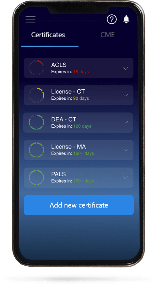
All Your Certificates in One Place
When you have your licenses, certificates and CMEs in one place, it's easier to track your career growth. You can easily share these with hospitals as well, using your medtigo app.