Rising Adoption of Continuous Glucose Monitors in Medicare Advantage: Trends from 2021–2023
April 13, 2026


Background
Pneumonia is a severe acute lower respiratory tract infection that impacts the pulmonary parenchyma in one or both lungs. Pneumonia is the sixth leading cause of mortality in the United States and the only infectious condition in the top ten. It is a prevalent and possibly fatal infectious disease with significant morbidity and fatality.
Viral pneumonia is an infection of the lungs caused by a virus etiology. It is characterized by inflammation of the alveoli, the tiny air sacs in the lungs responsible for facilitating oxygen and carbon dioxide exchange. It commonly affects individuals in vulnerable populations such as infants, the elderly, and those with existing respiratory conditions or compromised immune systems.
Epidemiology
The most common causes of viral pneumonia among adults are influenza A and B, together responsible for more than half of all such hospitalizations annually in the United States. Other important pathogens include RSV (respiratory syncytial virus), adenovirus, parainfluenza virus, hMPV (human metapneumovirus), and coronaviruses. Influenza is particularly severe among the elderly who represent 63% of hospital admissions and 85% deaths despite making up only 10% of the population.
RSV causes most cases of child viral pneumonitis worldwide but is increasingly seen in older people where it leads to 60 000–160 000 hospitalisations annually amongst those and individuals aged 65 years with 6000-10 000 deaths in the US alone each year. Residents in nursing homes get infected with RSV at a rate of 10% per year while adenovirus accounts for 10% childhood pneumonia.
Anatomy
Pathophysiology
Three primary causes can lead to viral pneumonia which include direct introduction of viral particles into the lung, contiguous spread from a nearby infection in the upper respiratory tract, and hematogenous dissemination from a distant site. The microscopic pathogenesis of the disease is characterized by the invasion of viral particles that target pneumocytes causing alveolar damage, inflammation, and secondary submucosal edema together with microhemorrhage as well as a cellular immune response.
Mononuclear lymphocytes are attracted and then followed by polymorphonuclear leukocytes while fibrin release also plays a role in the immune reaction;[4] at the same time, CD4 and CD8 cells become activated resulting into increased vascular permeability which leads to more severe edema. If left untreated this can progress towards intra-alveolar organization with clinical symptoms similar to those seen in obliterans disease states.
Etiology
Viral pneumonia stems from a multitude of DNA and RNA viruses, some of which are known as lung pathogens and have established themselves by causing common clinical and radiographic manifestations. These include families like Coronaviridae (e.g. SARS, MERS, and 2019-nCoV), Adenoviridae, and Orthomyxoviridae (such as the influenza virus). Others such as Paramyxoviridae, Picornaviridae, and Herpesviridae may also be implicated since they cause anything between mild and severe infections.
When it comes to community-acquired viral pneumonia, the most frequently associated pathogens are influenza virus, respiratory syncytial virus, adenovirus, parainfluenza virus, coronavirus, rhinovirus and human metapneumovirus. Depending on the immune status and age of the host, these viruses differ in their means of transmission as well as clinical manifestations severity. Therefore, it is important to understand how different pathogenic mechanisms work if we are to effectively manage this condition through diagnosis, treatment or prevention strategies.
Genetics
Prognostic Factors
Several factors affect the outlook for viral pneumonia, which include the virulence of viral species, immune competence of the patient, underlying pathology such as diabetes, COPD, diabetes mellitus (DM), dyscrasias, and haematological disorder and whether there is a coexisting bacterial infection or not and if recognized, the progress of infection and specific therapy started where available. Patients who are immunocompromised due to having been infected with HIV have an even worse prognosis than those without any other underlying medical conditions.
Clinical History
Pathognomonic history cues are not available for diagnosing viral pneumonia compared to bacterial pneumonia. However, some cues hint towards viral pneumonia during differential diagnosis as follows:
Physical Examination
These findings resemble to those of pyogenic pneumonia and are not specific. In physical examination increased fremitus, bronchial breath, crackles and wheezing may be noticed. Some patients may show mild fever, respiratory failure or multiple organ failure. Other observations may include:
Chills
Fever
Dyspnea and/or tachypnea
Rhonchi
Wheezing
Dullness to percussion
Pleurisy
Cough with presence or absence of sputum
Cyanosis
Rash
Pleural friction rub
Acute respiratory distress
Age group
Associated comorbidity
Associated activity
Acuity of presentation
Differential Diagnoses
Laboratory Studies
Imaging Studies
Procedures
Histologic Findings
Staging
Treatment Paradigm
Approach considerations: All the patients suffering from viral pneumonia should be considered for supportive care with oxygen, analgesics, nutrition, antipyretics and rest.
Supportive care: The patients need oxygen if they have hypoxemia or are unable to breathe adequately, which must be given by the EMTs. Prehospital providers can deliver drugs that help breathing through the airways. Intubation and a breathing machine are required for someone who cannot breathe on their own because of their lungs not working. If a person is in shock without heart failure, then they should be given fluids intravenously such as normal saline solution. In acute care settings this might involve giving medications like acyclovir (Zovirax), putting them on antibiotics if there is an infection and isolating them from others who may become infected while also using mechanical ventilation when necessary due to respiratory failure.
by Stage
by Modality
Chemotherapy
Radiation Therapy
Surgical Interventions
Hormone Therapy
Immunotherapy
Hyperthermia
Photodynamic Therapy
Stem Cell Transplant
Targeted Therapy
Palliative Care
Use of antiviral agents
Use of monoclonal antibodies
premature babies who are at an elevated risk for severe RSV-associated conditions.
Nirsevimab: It is given as an IM injection each year with an indication to prevent lower respiratory syncytial virus (RSV) disease in neonates and infants born during or entering their first RSV season as well as in those children up to 24 months old still at high risk for serious RSV infection during their second RSV season.
Use of immune globulins
Immune globulin IV: Anti-idiotypic antibodies neutralize the circulating myelin antibodies in immune globulin IV. They reduce INF-gamma and proinflammatory cytokines; suppress inducer T cells and B cells; block Fc receptors on macrophages; and increase suppressor T cells. This medication also promotes remyelination, obstructs the complement cascade, and may cause an elevation in CSF IgG levels (10%).
Use of beta-agonists
Albuterol: It is a beta-agonist indicated in the treatment of bronchospasm. It acts on β2 receptors by relaxing bronchial smooth muscles.
Use of vaccines
use-of-phases-of-management-in-treating-viral-pneumonia
Oxygen is crucial for patients experiencing hypoxemia or shortness of breath, and emergency medical personnel should administer it to dyspnoeic patients. Prehospital providers can deliver beta-agonist treatments to improve breathing. Respiratory failure patients require endotracheal intubation and ventilator support. Isotonic sodium chloride solution is used for shock without heart failure. Acute care may include oxygen, beta-agonists, dehydration fluids, Acyclovir, respiratory isolation, antibiotics, and mechanical ventilation.
Medication
(Off-label) :
75
mg
Orally
every 12 hrs
5
days
Note: oseltamivir is an antiviral medication commonly used to treat influenza virus infections. It is not typically used to treat viral pneumonia, as most cases are caused by viruses other than influenza
However, oseltamivir may be prescribed if a patient with viral pneumonia is diagnosed with an influenza virus infection
(Off label):
900
mg
orally
every 12 hrs
21
days
Dose Adjustments
Renal impairment:
The dosing may need to be adjusted based on the patient's creatinine clearance.
The dose may be reduced to 450 mg orally twice daily for patients with a creatinine clearance of 50 mL/min or less.
(Off label):
For children two weeks to 12 months old: The recommended dose is 3 mg/kg/dose twice daily for five days
For children 1 to 12 years old: The recommended dose is 2 mg/kg/dose twice daily for five days
Do not exceed 150 mg per day
For pediatric patients with normal renal function and BSA ≥ 1.5 m²: The recommended dose is 900 mg/m² orally twice daily for 21 days.
For pediatric patients with normal renal function and BSA < 1.5 m²: The recommended dose is 16 mg/kg orally twice daily for 21 days, up to a maximum of 900 mg per dose.
Dose Adjustments
For pediatric patients with renal impairment:
The dosing may need to be adjusted based on the patient's creatinine clearance.
The dose may be reduced to 7.5 mg/kg orally twice daily for patients with a creatinine clearance of 10 mL/min or less.
Future Trends
Pneumonia is a severe acute lower respiratory tract infection that impacts the pulmonary parenchyma in one or both lungs. Pneumonia is the sixth leading cause of mortality in the United States and the only infectious condition in the top ten. It is a prevalent and possibly fatal infectious disease with significant morbidity and fatality.
Viral pneumonia is an infection of the lungs caused by a virus etiology. It is characterized by inflammation of the alveoli, the tiny air sacs in the lungs responsible for facilitating oxygen and carbon dioxide exchange. It commonly affects individuals in vulnerable populations such as infants, the elderly, and those with existing respiratory conditions or compromised immune systems.
The most common causes of viral pneumonia among adults are influenza A and B, together responsible for more than half of all such hospitalizations annually in the United States. Other important pathogens include RSV (respiratory syncytial virus), adenovirus, parainfluenza virus, hMPV (human metapneumovirus), and coronaviruses. Influenza is particularly severe among the elderly who represent 63% of hospital admissions and 85% deaths despite making up only 10% of the population.
RSV causes most cases of child viral pneumonitis worldwide but is increasingly seen in older people where it leads to 60 000–160 000 hospitalisations annually amongst those and individuals aged 65 years with 6000-10 000 deaths in the US alone each year. Residents in nursing homes get infected with RSV at a rate of 10% per year while adenovirus accounts for 10% childhood pneumonia.
Three primary causes can lead to viral pneumonia which include direct introduction of viral particles into the lung, contiguous spread from a nearby infection in the upper respiratory tract, and hematogenous dissemination from a distant site. The microscopic pathogenesis of the disease is characterized by the invasion of viral particles that target pneumocytes causing alveolar damage, inflammation, and secondary submucosal edema together with microhemorrhage as well as a cellular immune response.
Mononuclear lymphocytes are attracted and then followed by polymorphonuclear leukocytes while fibrin release also plays a role in the immune reaction;[4] at the same time, CD4 and CD8 cells become activated resulting into increased vascular permeability which leads to more severe edema. If left untreated this can progress towards intra-alveolar organization with clinical symptoms similar to those seen in obliterans disease states.
Viral pneumonia stems from a multitude of DNA and RNA viruses, some of which are known as lung pathogens and have established themselves by causing common clinical and radiographic manifestations. These include families like Coronaviridae (e.g. SARS, MERS, and 2019-nCoV), Adenoviridae, and Orthomyxoviridae (such as the influenza virus). Others such as Paramyxoviridae, Picornaviridae, and Herpesviridae may also be implicated since they cause anything between mild and severe infections.
When it comes to community-acquired viral pneumonia, the most frequently associated pathogens are influenza virus, respiratory syncytial virus, adenovirus, parainfluenza virus, coronavirus, rhinovirus and human metapneumovirus. Depending on the immune status and age of the host, these viruses differ in their means of transmission as well as clinical manifestations severity. Therefore, it is important to understand how different pathogenic mechanisms work if we are to effectively manage this condition through diagnosis, treatment or prevention strategies.
Several factors affect the outlook for viral pneumonia, which include the virulence of viral species, immune competence of the patient, underlying pathology such as diabetes, COPD, diabetes mellitus (DM), dyscrasias, and haematological disorder and whether there is a coexisting bacterial infection or not and if recognized, the progress of infection and specific therapy started where available. Patients who are immunocompromised due to having been infected with HIV have an even worse prognosis than those without any other underlying medical conditions.
Pathognomonic history cues are not available for diagnosing viral pneumonia compared to bacterial pneumonia. However, some cues hint towards viral pneumonia during differential diagnosis as follows:
These findings resemble to those of pyogenic pneumonia and are not specific. In physical examination increased fremitus, bronchial breath, crackles and wheezing may be noticed. Some patients may show mild fever, respiratory failure or multiple organ failure. Other observations may include:
Chills
Fever
Dyspnea and/or tachypnea
Rhonchi
Wheezing
Dullness to percussion
Pleurisy
Cough with presence or absence of sputum
Cyanosis
Rash
Pleural friction rub
Acute respiratory distress
Approach considerations: All the patients suffering from viral pneumonia should be considered for supportive care with oxygen, analgesics, nutrition, antipyretics and rest.
Supportive care: The patients need oxygen if they have hypoxemia or are unable to breathe adequately, which must be given by the EMTs. Prehospital providers can deliver drugs that help breathing through the airways. Intubation and a breathing machine are required for someone who cannot breathe on their own because of their lungs not working. If a person is in shock without heart failure, then they should be given fluids intravenously such as normal saline solution. In acute care settings this might involve giving medications like acyclovir (Zovirax), putting them on antibiotics if there is an infection and isolating them from others who may become infected while also using mechanical ventilation when necessary due to respiratory failure.
Pulmonary Medicine
Pulmonary Medicine
premature babies who are at an elevated risk for severe RSV-associated conditions.
Nirsevimab: It is given as an IM injection each year with an indication to prevent lower respiratory syncytial virus (RSV) disease in neonates and infants born during or entering their first RSV season as well as in those children up to 24 months old still at high risk for serious RSV infection during their second RSV season.
Pulmonary Medicine
Immune globulin IV: Anti-idiotypic antibodies neutralize the circulating myelin antibodies in immune globulin IV. They reduce INF-gamma and proinflammatory cytokines; suppress inducer T cells and B cells; block Fc receptors on macrophages; and increase suppressor T cells. This medication also promotes remyelination, obstructs the complement cascade, and may cause an elevation in CSF IgG levels (10%).
Albuterol: It is a beta-agonist indicated in the treatment of bronchospasm. It acts on β2 receptors by relaxing bronchial smooth muscles.
Pulmonary Medicine
Oxygen is crucial for patients experiencing hypoxemia or shortness of breath, and emergency medical personnel should administer it to dyspnoeic patients. Prehospital providers can deliver beta-agonist treatments to improve breathing. Respiratory failure patients require endotracheal intubation and ventilator support. Isotonic sodium chloride solution is used for shock without heart failure. Acute care may include oxygen, beta-agonists, dehydration fluids, Acyclovir, respiratory isolation, antibiotics, and mechanical ventilation.
Both our subscription plans include Free CME/CPD AMA PRA Category 1 credits.
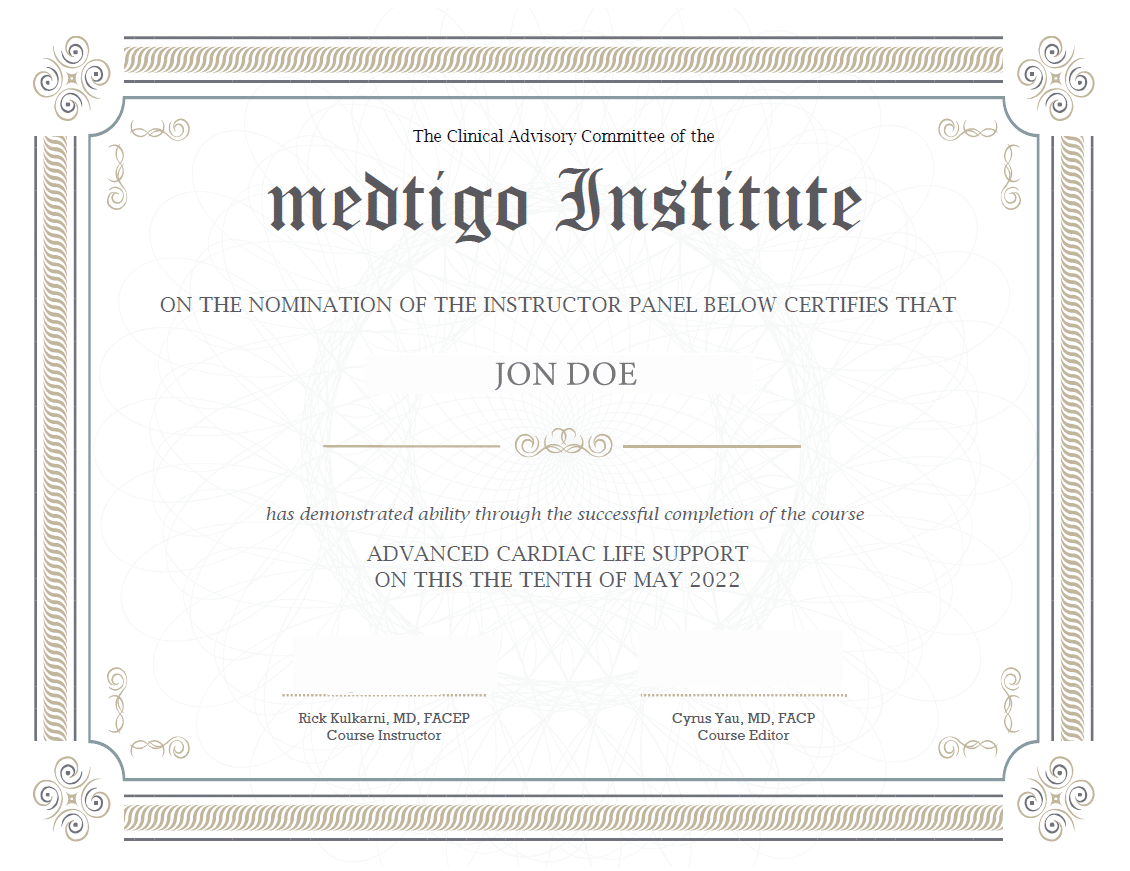
On course completion, you will receive a full-sized presentation quality digital certificate.
A dynamic medical simulation platform designed to train healthcare professionals and students to effectively run code situations through an immersive hands-on experience in a live, interactive 3D environment.
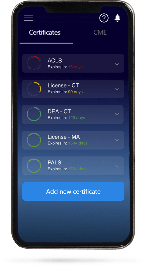
When you have your licenses, certificates and CMEs in one place, it's easier to track your career growth. You can easily share these with hospitals as well, using your medtigo app.