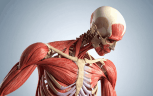
Wyburn-Mason Syndrome is a sporadic, non-hereditary congenital disease distinguished by arteriovenous malformations in the brain and face. AVMs are aneurysmal malformations that are the networks of blood vessels that supply artery to vein without passing through the capillary bed. In the context of Wyburn-Mason Syndrome, the referred AVMs are in the blood vessels that are the retina of the eye and the brain.
The exact cause of this syndrome is not well understood. Hence, it is thought to be random research without a pattern of transmittal. Considering the syndrome’s symptoms, they may be as mild as some vision changes and as severe as bleeding in the brain or vision loss.
Epidemiology
Wyburn-Mason Syndrome, other refer as racemose angioma or Bonnet-Dechaume-Blanc syndrome is a non-hereditary neurocutaneous syndrome associated with arteriovenous anomaly in the brain and retina.
The incidence of the syndrome is rare globally and it affects less than 100 people in the world as per the literature. The frequency of the disorder is low, and its occurrence is global and non-affected by gender. Clinical manifestations are based on vascular dysgenesis and affect the retina and the brain.
Anatomy
Pathophysiology
Wyburn-Mason Syndrome also known as Bonnet-Dechaume-Blanc Syndrome has a pathophysiology of arteriovenous malformation in the brain and face. AVMs are unusually formed shunts, which are direct anastomoses between arteries and veins directly through the formation of capillaries. These AVMs in the case of Wyburn-Mason Syndrome are principally confining to the blood vessels in the retina of the eye and the brain.
The AVMs located in the WM syndrome might result in the disturbance of blood flow and oxygen supply near the tissues. These abnormal blood vessels in retina can cause changes in vision and in some instances, even vision threat. AVMs in the brain can disrupt the average circulation of the cerebrospinal fluid and is quite prone to bleed thereby causing some form of neurological impairment.
Etiology
Genetic Factors: Although specific research is lacking, there are perspectives that help to reveal predisposing factors of AVMs in Wyburn-Mason Syndrome, inheritance is one of the characteristics that could slightly increase an individual’s probability of developing an AVM, although they are not hereditary. The following are some possible genotypic characteristics that could cause AVMs in this syndrome, a mutation in the genes involved in blood vessel formation.
Embryonic Developmental Abnormalities: Abnormal vascular procedures resulting in the embryogenesis process and blood vessels may be connected to generating AVMs in Wyburn-Mason Syndrome. Defects in vascular differentiation and organization can lead to the formation of aberrant artery-to-vein connections.
Vascular Growth Factors: AVMs in Wyburn-Mason Syndrome may be caused by abnormal signaling in blood vessel development, such as problems with vascular endothelial growth factor.
Genetics
Prognostic Factors
Regarding this syndrome with involvement of AVMs in both the brain and eye, the outcome is generally determined by the lesion site, size, involvement, and other related findings.
Clinical History
It is crucial to emphasize that the clinical presentation of Wyburn-Mason Syndrome will depend on the age of onset, the presence or absence of comorbidities or activities that provoke or aggravate the manifestation, and the overall understanding and perception of the presentation’s acuity.
Age Group: Individuals with Wyburn-Mason Syndrome can be diagnosed at any age depending on the severity of the illness; however, the age defining its onset determines its characteristics of manifestation. Possible symptoms may appear in infancy, and some may appear as adults, depending on the cause.
Physical Examination
Eye Examination
Ophthalmoscopy
Visual Acuity Testing
Visual Field Testing
Neurological Examination
Cranial Nerve Assessment
Motor and Sensory Testing
Coordination and Balance Testing
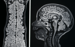
Imaging Studies
Skin Examination
Age group
Associated comorbidity
Comorbidities: People, who have other vascular diseases or systemic diseases, could have a higher chance of getting Wyburn-Mason Syndrome, or worsening of severity of the disease.
Pregnancy: Hormonal changes in pregnancy also influence the course of the syndrome, specifically, impulsive blood flow changes may be due to voluminous blood circulation to AVMs, if they are in the brain or eye.
Associated activity
Acuity of presentation
Acute Presentation: In some cases, some people may experience sudden and aggressive manifestations indicating a rupture of an AVM, specifically in the brain which manifests neurological abnormalities like weakness, numbness or even loss of consciousness resulting from bleeding. In the eye, symptoms such as vision change or if there was any bleeding may at times develop suddenly.
Subacute or Chronic Presentation: Some may develop symptoms slowly and more subtly; this is why there are more people who get diagnosed with chronic diseases such as high blood pressure or diabetes. For instance, vision changes may occur as to AVM’s influence on the eye’s blood vessels and become progressively worse as time passes.
Differential Diagnoses
Sturge-Weber Syndrome
Hereditary Hemorrhagic Telangiectasia
Retinal Arterial Macroaneurysms
Capillary Telangiectasia
Cavernous Hemangioma
Retinoblastoma
Laboratory Studies
Imaging Studies
Procedures
Histologic Findings
Staging
Treatment Paradigm
Multidisciplinary Care: The patient with Wyburn-Mason syndrome needs the attention of an ophthalmologist, neurologist, neurosurgeon, and an interventional radiologist since the condition affects the retina and the brain.
Symptomatic Management: There is no known cure for Wyburn-Mason syndrome; therefore, patients are advised to live with medications, eyesight issues, seizures, and headaches.
Visual Symptoms: AVM involvement is the common site in the eye retina; therefore, ophthalmologists oversee the patients’ condition and may direct interventions such as laser treatment or anti-VEGF.
Neurological Symptoms: This category deals with patients who have symptoms related to the brain such as epilepsy and headaches with the help of drugs and occasionally operations.
Interventional Radiology: Those may be done for significant symptoms or risks, depending on characteristics of AVMs, so procedures like embolization can be done.
Individualized Approach: The treatment plans should therefore be assessed individually for treatment depending on the person’s situation, where it needs to be conducted by a specialized medical team.
Lighting: Sufficient lighting ensures that vision is enhanced besides reducing strain on the eyes, that is, by use of task lighting, such as for reading.
Contrast: Contrast between objects and their backgrounds can be improved, which will help in visibility, for instance in dark plates on a light table.
Magnification: Reading strain is eliminated by using magnifying devices or larger fonts.
Safety Precautions:
Fall Prevention: Several changes should be made; get rid of any tripping hazards and install handrails particularly for individuals with balance disorders or neurological disorders.
Seizure Precautions: Ensure a safe environment since a seizure might cause injuries; avoid knives or sharp objects.
Stress Reduction:
Clutter Reduction: Cook healthy meals while avoiding accumulating items in the living zones for tranquillity.
Noise Reduction: Reduce noise pollution as a way of helping the body and the mind to focus and to rest.
Use of Radiosurgery for Treating Wyburn-Mason Syndrome
Radiosurgery can be used in managing patients with AVMs in WMS, including in this patient.
Radiosurgery Planning: The specialists in the treatment team apply a cutting-edge technology known as image-guided therapy to help them locate the AVM accurately while sparing the normal brain cells from exposure to radiation. Some techniques for instance gamma knife or CyberKnife, are used to radiate the AVM.
Radiosurgery Session: A customised head frame or mask is worn by the patient to ensure precise alignment. The aberrant blood vessels are damaged and eventually shut down by the regulated radiation dosage that the radiation machine administers to the AVM.
Embolization: A catheter is guided through the blood vessels to reach the AVM area to minimize or stop blood within the area using agents. It is effective in case of selectively accessible AVMs with easily identifiable feeding arteries to reduce the possibilities of bleeding episodes and manifestations. There may be need to have several sessions for the activity.
Laser Therapy: The high-energy lasers are used to focus on the affected blood vessels within the eye to shrink them and seal them off. This can enhance central vision and can help avoid dangers originating from retinal AVMs.
Surgical Resection: Through this process, surgically accessible AVMs that are causing severe symptoms, particularly in the brain are removed. It entails cutting off the aberrant arteries without damaging the surrounding healthy tissue. Complex and dangerous, especially if the AVM is in a crucial portion of the brain.
use-of-phases-in-managing-wyburn-mason-syndrome
Diagnosis and Evaluation:
Those diagnosed with WMS have clinical examination based on the medical history and physical examination in addition to tests such as the angiography and MRI.
Ophthalmologists, neurologists, radiologists diagnose AVMs in the brain and eye and evaluate their effects.
Treatment Planning:
A treatment plan is developed by a team of healthcare professionals based on the characteristics of the AVM and the patient: the position and size of the lesion, the manifestation of the disease, and potential danger.
Depending on the case, the treatment will involve the obstruction of blood flow by using a procedure known as embolization, the use of laser light in treating tumor, surgery or radiation will be recommended.
Post-Procedure Monitoring and Recovery:
Outcome measures of treatment include assessment of treatment uptake and response in the patients.
Rehabilitation could be physical or occupational, or even any other type of supporting activity.
Ongoing Follow-Up and Long-Term Management:
Further management includes follow up including clinical examination, neurological assessment and imaging studies to look for presence of new AVMs, recurrent haemorrhage or new symptoms.
The members of the patient’s team adapt the required treatments as necessary.
Wyburn-Mason Syndrome is a sporadic, non-hereditary congenital disease distinguished by arteriovenous malformations in the brain and face. AVMs are aneurysmal malformations that are the networks of blood vessels that supply artery to vein without passing through the capillary bed. In the context of Wyburn-Mason Syndrome, the referred AVMs are in the blood vessels that are the retina of the eye and the brain.
The exact cause of this syndrome is not well understood. Hence, it is thought to be random research without a pattern of transmittal. Considering the syndrome’s symptoms, they may be as mild as some vision changes and as severe as bleeding in the brain or vision loss.
Wyburn-Mason Syndrome, other refer as racemose angioma or Bonnet-Dechaume-Blanc syndrome is a non-hereditary neurocutaneous syndrome associated with arteriovenous anomaly in the brain and retina.
The incidence of the syndrome is rare globally and it affects less than 100 people in the world as per the literature. The frequency of the disorder is low, and its occurrence is global and non-affected by gender. Clinical manifestations are based on vascular dysgenesis and affect the retina and the brain.
Wyburn-Mason Syndrome also known as Bonnet-Dechaume-Blanc Syndrome has a pathophysiology of arteriovenous malformation in the brain and face. AVMs are unusually formed shunts, which are direct anastomoses between arteries and veins directly through the formation of capillaries. These AVMs in the case of Wyburn-Mason Syndrome are principally confining to the blood vessels in the retina of the eye and the brain.
The AVMs located in the WM syndrome might result in the disturbance of blood flow and oxygen supply near the tissues. These abnormal blood vessels in retina can cause changes in vision and in some instances, even vision threat. AVMs in the brain can disrupt the average circulation of the cerebrospinal fluid and is quite prone to bleed thereby causing some form of neurological impairment.
Genetic Factors: Although specific research is lacking, there are perspectives that help to reveal predisposing factors of AVMs in Wyburn-Mason Syndrome, inheritance is one of the characteristics that could slightly increase an individual’s probability of developing an AVM, although they are not hereditary. The following are some possible genotypic characteristics that could cause AVMs in this syndrome, a mutation in the genes involved in blood vessel formation.
Embryonic Developmental Abnormalities: Abnormal vascular procedures resulting in the embryogenesis process and blood vessels may be connected to generating AVMs in Wyburn-Mason Syndrome. Defects in vascular differentiation and organization can lead to the formation of aberrant artery-to-vein connections.
Vascular Growth Factors: AVMs in Wyburn-Mason Syndrome may be caused by abnormal signaling in blood vessel development, such as problems with vascular endothelial growth factor.
Regarding this syndrome with involvement of AVMs in both the brain and eye, the outcome is generally determined by the lesion site, size, involvement, and other related findings.
It is crucial to emphasize that the clinical presentation of Wyburn-Mason Syndrome will depend on the age of onset, the presence or absence of comorbidities or activities that provoke or aggravate the manifestation, and the overall understanding and perception of the presentation’s acuity.
Age Group: Individuals with Wyburn-Mason Syndrome can be diagnosed at any age depending on the severity of the illness; however, the age defining its onset determines its characteristics of manifestation. Possible symptoms may appear in infancy, and some may appear as adults, depending on the cause.
Eye Examination
Ophthalmoscopy
Visual Acuity Testing
Visual Field Testing
Neurological Examination
Cranial Nerve Assessment
Motor and Sensory Testing
Coordination and Balance Testing
Imaging Studies
Skin Examination
Comorbidities: People, who have other vascular diseases or systemic diseases, could have a higher chance of getting Wyburn-Mason Syndrome, or worsening of severity of the disease.
Pregnancy: Hormonal changes in pregnancy also influence the course of the syndrome, specifically, impulsive blood flow changes may be due to voluminous blood circulation to AVMs, if they are in the brain or eye.
Acute Presentation: In some cases, some people may experience sudden and aggressive manifestations indicating a rupture of an AVM, specifically in the brain which manifests neurological abnormalities like weakness, numbness or even loss of consciousness resulting from bleeding. In the eye, symptoms such as vision change or if there was any bleeding may at times develop suddenly.
Subacute or Chronic Presentation: Some may develop symptoms slowly and more subtly; this is why there are more people who get diagnosed with chronic diseases such as high blood pressure or diabetes. For instance, vision changes may occur as to AVM’s influence on the eye’s blood vessels and become progressively worse as time passes.
Sturge-Weber Syndrome
Hereditary Hemorrhagic Telangiectasia
Retinal Arterial Macroaneurysms
Capillary Telangiectasia
Cavernous Hemangioma
Retinoblastoma
Multidisciplinary Care: The patient with Wyburn-Mason syndrome needs the attention of an ophthalmologist, neurologist, neurosurgeon, and an interventional radiologist since the condition affects the retina and the brain.
Symptomatic Management: There is no known cure for Wyburn-Mason syndrome; therefore, patients are advised to live with medications, eyesight issues, seizures, and headaches.
Visual Symptoms: AVM involvement is the common site in the eye retina; therefore, ophthalmologists oversee the patients’ condition and may direct interventions such as laser treatment or anti-VEGF.
Neurological Symptoms: This category deals with patients who have symptoms related to the brain such as epilepsy and headaches with the help of drugs and occasionally operations.
Interventional Radiology: Those may be done for significant symptoms or risks, depending on characteristics of AVMs, so procedures like embolization can be done.
Individualized Approach: The treatment plans should therefore be assessed individually for treatment depending on the person’s situation, where it needs to be conducted by a specialized medical team.
Neurology
Ophthalmology
Vascular Medicine
Vision Support:
Lighting: Sufficient lighting ensures that vision is enhanced besides reducing strain on the eyes, that is, by use of task lighting, such as for reading.
Contrast: Contrast between objects and their backgrounds can be improved, which will help in visibility, for instance in dark plates on a light table.
Magnification: Reading strain is eliminated by using magnifying devices or larger fonts.
Safety Precautions:
Fall Prevention: Several changes should be made; get rid of any tripping hazards and install handrails particularly for individuals with balance disorders or neurological disorders.
Seizure Precautions: Ensure a safe environment since a seizure might cause injuries; avoid knives or sharp objects.
Stress Reduction:
Clutter Reduction: Cook healthy meals while avoiding accumulating items in the living zones for tranquillity.
Noise Reduction: Reduce noise pollution as a way of helping the body and the mind to focus and to rest.
Neurology
Ophthalmology
Vascular Medicine
Radiosurgery can be used in managing patients with AVMs in WMS, including in this patient.
Radiosurgery Planning: The specialists in the treatment team apply a cutting-edge technology known as image-guided therapy to help them locate the AVM accurately while sparing the normal brain cells from exposure to radiation. Some techniques for instance gamma knife or CyberKnife, are used to radiate the AVM.
Radiosurgery Session: A customised head frame or mask is worn by the patient to ensure precise alignment. The aberrant blood vessels are damaged and eventually shut down by the regulated radiation dosage that the radiation machine administers to the AVM.
Neurology
Ophthalmology
Vascular Medicine
Embolization: A catheter is guided through the blood vessels to reach the AVM area to minimize or stop blood within the area using agents. It is effective in case of selectively accessible AVMs with easily identifiable feeding arteries to reduce the possibilities of bleeding episodes and manifestations. There may be need to have several sessions for the activity.
Laser Therapy: The high-energy lasers are used to focus on the affected blood vessels within the eye to shrink them and seal them off. This can enhance central vision and can help avoid dangers originating from retinal AVMs.
Surgical Resection: Through this process, surgically accessible AVMs that are causing severe symptoms, particularly in the brain are removed. It entails cutting off the aberrant arteries without damaging the surrounding healthy tissue. Complex and dangerous, especially if the AVM is in a crucial portion of the brain.
Vascular Medicine
Diagnosis and Evaluation:
Those diagnosed with WMS have clinical examination based on the medical history and physical examination in addition to tests such as the angiography and MRI.
Ophthalmologists, neurologists, radiologists diagnose AVMs in the brain and eye and evaluate their effects.
Treatment Planning:
A treatment plan is developed by a team of healthcare professionals based on the characteristics of the AVM and the patient: the position and size of the lesion, the manifestation of the disease, and potential danger.
Depending on the case, the treatment will involve the obstruction of blood flow by using a procedure known as embolization, the use of laser light in treating tumor, surgery or radiation will be recommended.
Post-Procedure Monitoring and Recovery:
Outcome measures of treatment include assessment of treatment uptake and response in the patients.
Rehabilitation could be physical or occupational, or even any other type of supporting activity.
Ongoing Follow-Up and Long-Term Management:
Further management includes follow up including clinical examination, neurological assessment and imaging studies to look for presence of new AVMs, recurrent haemorrhage or new symptoms.
The members of the patient’s team adapt the required treatments as necessary.
Both our subscription plans include Free CME/CPD AMA PRA Category 1 credits.
Digital Certificate PDF
On course completion, you will receive a full-sized presentation quality digital certificate.
medtigo Simulation
A dynamic medical simulation platform designed to train healthcare professionals and students to effectively run code situations through an immersive hands-on experience in a live, interactive 3D environment.
medtigo Points
medtigo points is our unique point redemption system created to award users for interacting on our site. These points can be redeemed for special discounts on the medtigo marketplace as well as towards the membership cost itself.
Community Forum post/reply = 5 points
*Redemption of points can occur only through the medtigo marketplace, courses, or simulation system. Money will not be credited to your bank account. 10 points = $1.
All Your Certificates in One Place
When you have your licenses, certificates and CMEs in one place, it's easier to track your career growth. You can easily share these with hospitals as well, using your medtigo app.