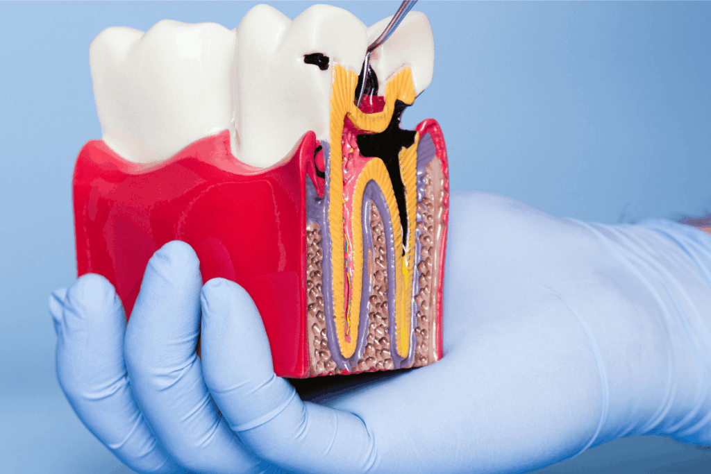
Dental enamel is the hardest and most mineralized tissue in vertebrates. It is formed through the organized 3D assembly of amelogenin. It guides hierarchical nanocrystal growth, providing strength and resistance to thermal, physical, and chemical insults. This protective layer safeguards the teeth throughout life. However, enamel loss occurs due to trauma, acid erosion, or mechanical wear. This leads to oral problems that affect half of the world’s population, at an estimated annual cost of approximately $544 billion. Remineralization methods capable of restoring complex architecture and full functional properties in patients. But clinic-friendly remains an unsolved challenge.
A recent study published in Nature Communications introduces a clinically friendly supramolecular protein matrix based on elastin-like recombinamers (ELRs). This matrix mimics amelogenin’s β-rich fibrillar organization, enabling epitaxial growth of enamel-like layers ex vivo. Human molar teeth were extracted and ethically approved by the University of Nottingham Research Ethics Committee with informed consent.
ELRs were fabricated using either a dimethyl sulfoxide (DMSO)/dimethyl formamide (DMF) or water/ethanol system with calcium (Ca2+) ions and crosslinkers, then analyzed through different techniques like isothermal titration calorimetry (ITC), dynamic light scattering (DLS), transmission electron microscopy (TEM), small- and wide-angle X-ray scattering (SAXS/WAXS), attenuated total reflectance-Fourier transform infrared spectroscopy (ATR-FTIR), X-ray photoelectron spectroscopy (XPS), and scanning electron microscopy (SEM).
Mineralization and remineralization experiments were performed on acid-etched enamel and dentine under natural or artificial saliva. Biological, mechanical, and chemical resistance were evaluated using water immersion tests and nanoindentation. Cytotoxicity assays were also conducted to examine the durability, restorative capacity, and biocompatibility properties of ELR coating.
WAXS analysis revealed that ELR fibrils exhibited β-sheet structures with a 4.7 Å strand spacing and 10 Å inter-sheet distance. In contrast, small-angle analysis identified 48.7 Å-wide filament-forming smooth fibrillar networks. Simulations confirmed Ca2+ dependent ELR self-assembly into six parallel filaments with hydrophobic cores and charged outer regions. This organization replicated the structural hierarchy of natural enamel. Mineralization and remineralization processes confirmed that the ELR matrix was a versatile, clinically adaptable platform for full enamel regeneration across varying levels of erosion.
The ELR matrix formed an aprismatic enamel-like layer with mechanical properties like Young’s modulus (E = 58.3 ± 16.7 GPa vs 50 – 90 GPa), hardness (H = 1.4 ± 0.3 GPa vs 2.5 – 4 GPa), wear strength (WS = 119.1 ± 24.3 GPa vs 153.9 ± 3.2 GPa), and coefficient of friction (CoF = 0.29 ± 0.07 vs 0.1–0.4), comparable to native enamel. After 60 minutes of brushing teeth coated with remineralized enamel, it was shown that there was no structural damage and retained E H of 2.76 ± 0.8 GPa and E of 68.6 ± 16.4 GPa compared to native enamel (H = 3.0 ± 0.9 GPa and E = 70.2 ± 15.2 GPa), confirming mechanical stability.
Physical resistance testing demonstrated that remineralized teeth exhibited a significant increase in fracture toughness from 0.7 ± 0.1 MPa.m0.5 to 1.1 ± 0.1 MPa.m0.5 which matched healthy enamel values of 1.2 ± 0.2 MPa.m0.5.
To simulate long-term grinding and chewing, samples of enamel and dentine were subjected to 75 N frictional forces for two weeks (approximately 3.5 years). The remineralized enamel showed wear in terms of height loss (HL = 30.5 ± 8.9 µm) and volume loss (VL = 3.5 ± 1.4 mm3), similar to native enamel (HL = 29.1 ± 9.3 µm, VL = 2.9 ± 1.0 mm3). Whereas mineralized dentine exhibited less wear (HL = 16.8 ± 6.7 µm and VL = 1.6 ± 0.6 mm3) compared to native dentine (HL = 28.0 ± 10.3 µm and VL = 2.5 ± 0.9 mm3). Exposure to 0.1 M acetic acid for 2 days or 15 mins confirmed that the remineralized layers exhibit greater mechanical stability and structural integrity over short- and long-term periods.
In conclusion, this study developed an ELR matrix that regenerated enamel tissue with restored function and structure. The findings highlight its superior mechanical stability and clinical potential for effective, bioinspired enamel remineralization compared with existing alternatives.
References: Hasan A, Chuvilin A, Van Teijlingen A, et al. Biomimetic supramolecular protein matrix restores structure and properties of human dental enamel. Nat Commun. 2025;16:9434. doi:10.1038/s41467-025-64982-y


