Modoc virus is a rodent-associated virus with no known vector, and it can infect humans accidentally with infected rodents or their excreta. The deer mouse (Peromyscus maniculatus) serves as the primary reservoir and host for the Modoc virus, and it is widely distributed across North America. Other rodent species, such as the white-footed mouse (Peromyscus leucopus) and the cotton rat (Sigmodon hispidus), may also be susceptible to Modoc virus infection.
First isolated in 1958 from the mammary gland tissue of a deer mouse in Modoc County, California, the Modoc virus has since been found in other regions, including Oregon, Colorado, and Montana. However, due to the lack of systematic surveillance and serological studies, the geographic distribution and prevalence of the Modoc virus in rodents still need to be discovered.
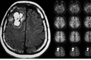
Depending on the severity and location of the infection, the Modoc virus can cause various clinical symptoms in humans. Aseptic meningitis is the most frequent and mild kind, characterized by inflammation of the brain & spinal cord membranes. Symptoms typically appear within 1-2 weeks after exposure and last 3-10 days, presenting as headache, fever, stiff neck, nausea, vomiting, photophobia, and altered mental status.
However, the true incidence and prevalence of Modoc virus infection in humans are still being determined, as reliable diagnostic tests and reporting systems need to be made available. Consequently, many cases may go undiagnosed or be misidentified as other viral or bacterial infections.Despite limited data, sporadic cases and outbreaks of Modoc virus disease have been reported in the literature, mainly in North America.
For instance, an outbreak of aseptic meningitis occurred among children at a summer camp in Oregon in 1969, with eight cases showing serological evidence of recent Modoc virus infection. In other instances, cases of encephalitis, meningoencephalomyelitis, and myelitis were reported in individuals with contact with deer mice, and they recovered after treatment with corticosteroids.
Classification and Structure:
Kingdom: Virus
Phylum: Kitrinoviricota
Class: Flasuviricetes
Order: Amarillovirales
Family: Flaviviridae
Genus: Flavivirus
Species: Modoc virus
Modoc virus has a spherical shape and a particle size of approximately 45 nm, within the range of other flaviviruses that vary from 40 to 60 nm in diameter.
A lipid bilayer membrane formed from the host cell encloses the viral DNA as well as numerous copies of three structural proteins, including C (capsid), M (membrane), & E (envelope).
The capsid protein, coded by the viral genome, encapsulates the viral RNA to form the nucleocapsid. The membrane protein anchors the nucleocapsid to the host-derived lipid membrane.
The Modoc virus genome is a single-stranded positive-sense RNA molecule, approximately 11,000 nucleotides in length. It has only one open reading frame (ORF) with 3374 amino acids encoded.
The Modoc virus‘s viral genome contains a single big polypeptide processed in the endoplasmic reticulum (ER) membrane via either host or viral proteases. This process produces three structural proteins (capsid, prM, & E) and a collection of non-structural proteins (NS1-NS5). The capsid protein joins forces with the viral genomic RNA to create a nucleocapsid complex, allowing the genome to be packaged into mature virus particles.
The prM & E proteins are required for the viral particle to form spherical particles. The roles of the NS proteins are unknown, but they are thought to play a role in forming virus particle replicating organelles. For example, the vast ectodomain of the NS1 protein is assumed to be involved throughout the deformation of an ER membrane from the luminal side.
Modoc virus is antigenically related to, but distinct from, other rodent-associated flaviviruses like Cowbone Ridge, Jutiapa, San Perlita, and Sal Vieja viruses. The NS4B protein can inhibit interferon signaling, and the NS5 protein is an RNA-dependent RNA polymerase and methyltransferase.
The E protein contains epitopes that determine the antigenic type of the virus, and the C protein interacts with host factors like nucleolin and heat shock proteins, potentially aiding in viral replication or evasion of host defense.
Modoc, a flavivirus associated with rodents, can enter host cells via endocytosis. Once inside the cell, the virus uses membrane fusion to release its positive-sense single-stranded RNA genomes into the cytoplasm. The viral genome encodes a single open reading frame (ORF), which cellular and viral proteases process to provide three structural proteins (C, M, & E) and seven non-structural proteins. The ORF’s untranslated regions (UTRs) fold into complicated stem-loop structures essential for replication. The viral genome is transcribed using the positive (+) RNA strand structure.
The Modoc virus is believed to assemble and replicate its genome in the host cell’s rough endoplasmic reticulum. After replication, newly synthesized RNA interacts with the capsid and buds into the ER lumen along with immature prM and E proteins. Maturation of prM and E proteins occurs in the Golgi and endosomes, with prM cleaved by cellular proteases to generate mature virions. The mature virions are then released from the cell via exocytosis.
Modoc virus spreads to humans by contact with sick rats or their excrement. Deer mice (Peromyscus maniculatus) are the principal reservoir and host. However, other rodent species, such as the white-footed mouse and cotton rat, may be vulnerable to infection. The precise route of transmission has yet to be discovered. However, it is thought to occur by direct or indirect contact with rodents, such as salivary secretions, fomites, aerosols, or urine. Transmission can also be promoted in mouse nests throughout the winter, which provides ideal circumstances for viral spread.
According to the present literature, specific information about the human host resistance against the Modoc virus is scarce. Modoc virus, a relatively rare and poorly studied virus, needs more comprehensive research on its interactions with the human immune system. While the human immune response to similar flaviviruses can be inferred, comprehensive data regardingModoc virus-specific human defenses are yet to be elucidated.
Modoc virus, a rodent-associated flavivirus, can cause human disease like other family members. One of the observed clinical manifestations in humans is aseptic meningitis, characterized by inflammation of the membranes covering the brain and spinal cord. This condition typically presents with symptoms such as headache, fever, stiff neck, nausea, vomiting, sensitivity to light, and altered mental status. Aseptic meningitis is the most common and usually mild Modoc virus disease, lasting 3-10 days.
Modoc virus can lead to encephalitis, an inflammation of brain tissue. Encephalitis is a rare but severe manifestation of the infection and may occur as a complication of aseptic meningitis or as a primary infection without meningitis. Symptoms of encephalitis include headache, fever, confusion, seizures, coma, and neurological deficits. Encephalitis can have prolonged effects, lasting several weeks, & cause permanent brain damage or even death.
Another severe manifestation is myelitis, an inflammation of the spinal cord. Myelitis can occur as a complication of aseptic meningitis or as a primary infection without meningitis. Symptoms may include back pain, weakness, numbness, tingling, bladder or bowel dysfunction, and paralysis. Like encephalitis, myelitis can also cause lasting effects, resulting in spinal cord damage or death.
Hemagglutination-Inhibition (HI) Test: The antibody responses of hamsters to Modoc virus infection were measured using the HI test. Antigens for the HI test were prepared from the brains of Modoc virus-infected newborn mice. Hamster serum samples were tested at serial two-fold dilutions, and the results were expressed as the inverse of the last dilution of sera that resulted in hemagglutination inhibition. This test helps detect specific antibodies against the Modoc virus in the serum.
IgM-Capture Enzyme-Linked Immunosorbent Assay (ELISA): The IgM ELISA is used to screen serum samples at a 1:40 dilution. The results are recorded based on the optical density at 405 nm (OD405). Absorbance values ≥ 0.20 -OD405 are considered positive for IgM antibodies, indicating recent or acute infection with the Modoc virus.
Co-cultivation for Virus Detection: Tissue samples of kidneys & lungs from experimentally infected hamsters were co-cultivated for the Modoc virus. The tissue fragments were dissociated using a trypsin-EDTA solution before being injected into culture flasks containing Vero cells. Co-cultures were evaluated for cytopathic effect (CPE) evidence. If CPE was seen, the presence of Modoc virus in the culture media was confirmed by sub-culturing the medium over an additional monolayer of Vero cells and using an immunofluorescence assay (IFA) to look for the presence of Modoc viral antigen.
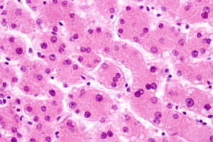
Histologic and Immunohistochemical Examinations: Tissues will be taken from infected individuals for histopathologic & immunohistochemical examination. Tissue samples from several organs are fixed in 10% buffered formalin & processed for histologic slides, including the lung, liver, spleen, kidney, spinal cord, & brain. The primary antibody used in immunohistochemical staining for Modoc virus antigen was mouse hyperimmune ascitic fluid, and the bound primary antibody was identified using a commercially available kit. This test detects the presence of Modoc virus antigen in diverse organs, hence confirming infection.
Implementing effective rodent control measures is essential to reduce the reservoir and transmission of the Modoc virus. It may include trapping, baiting, and eliminating rodent nesting sites in and around human dwellings, agricultural areas, and other locations where rodents are prevalent.
Individuals at a higher risk of Modoc virus exposure, including field workers, veterinarians, and laboratory professionals, should take necessary personal protective measures.
Modoc virus is a rodent-associated virus with no known vector, and it can infect humans accidentally with infected rodents or their excreta. The deer mouse (Peromyscus maniculatus) serves as the primary reservoir and host for the Modoc virus, and it is widely distributed across North America. Other rodent species, such as the white-footed mouse (Peromyscus leucopus) and the cotton rat (Sigmodon hispidus), may also be susceptible to Modoc virus infection.
First isolated in 1958 from the mammary gland tissue of a deer mouse in Modoc County, California, the Modoc virus has since been found in other regions, including Oregon, Colorado, and Montana. However, due to the lack of systematic surveillance and serological studies, the geographic distribution and prevalence of the Modoc virus in rodents still need to be discovered.
Depending on the severity and location of the infection, the Modoc virus can cause various clinical symptoms in humans. Aseptic meningitis is the most frequent and mild kind, characterized by inflammation of the brain & spinal cord membranes. Symptoms typically appear within 1-2 weeks after exposure and last 3-10 days, presenting as headache, fever, stiff neck, nausea, vomiting, photophobia, and altered mental status.
However, the true incidence and prevalence of Modoc virus infection in humans are still being determined, as reliable diagnostic tests and reporting systems need to be made available. Consequently, many cases may go undiagnosed or be misidentified as other viral or bacterial infections.Despite limited data, sporadic cases and outbreaks of Modoc virus disease have been reported in the literature, mainly in North America.
For instance, an outbreak of aseptic meningitis occurred among children at a summer camp in Oregon in 1969, with eight cases showing serological evidence of recent Modoc virus infection. In other instances, cases of encephalitis, meningoencephalomyelitis, and myelitis were reported in individuals with contact with deer mice, and they recovered after treatment with corticosteroids.
Classification and Structure:
Kingdom: Virus
Phylum: Kitrinoviricota
Class: Flasuviricetes
Order: Amarillovirales
Family: Flaviviridae
Genus: Flavivirus
Species: Modoc virus
Modoc virus has a spherical shape and a particle size of approximately 45 nm, within the range of other flaviviruses that vary from 40 to 60 nm in diameter.
A lipid bilayer membrane formed from the host cell encloses the viral DNA as well as numerous copies of three structural proteins, including C (capsid), M (membrane), & E (envelope).
The capsid protein, coded by the viral genome, encapsulates the viral RNA to form the nucleocapsid. The membrane protein anchors the nucleocapsid to the host-derived lipid membrane.
The Modoc virus genome is a single-stranded positive-sense RNA molecule, approximately 11,000 nucleotides in length. It has only one open reading frame (ORF) with 3374 amino acids encoded.
The Modoc virus‘s viral genome contains a single big polypeptide processed in the endoplasmic reticulum (ER) membrane via either host or viral proteases. This process produces three structural proteins (capsid, prM, & E) and a collection of non-structural proteins (NS1-NS5). The capsid protein joins forces with the viral genomic RNA to create a nucleocapsid complex, allowing the genome to be packaged into mature virus particles.
The prM & E proteins are required for the viral particle to form spherical particles. The roles of the NS proteins are unknown, but they are thought to play a role in forming virus particle replicating organelles. For example, the vast ectodomain of the NS1 protein is assumed to be involved throughout the deformation of an ER membrane from the luminal side.
Modoc virus is antigenically related to, but distinct from, other rodent-associated flaviviruses like Cowbone Ridge, Jutiapa, San Perlita, and Sal Vieja viruses. The NS4B protein can inhibit interferon signaling, and the NS5 protein is an RNA-dependent RNA polymerase and methyltransferase.
The E protein contains epitopes that determine the antigenic type of the virus, and the C protein interacts with host factors like nucleolin and heat shock proteins, potentially aiding in viral replication or evasion of host defense.
Modoc, a flavivirus associated with rodents, can enter host cells via endocytosis. Once inside the cell, the virus uses membrane fusion to release its positive-sense single-stranded RNA genomes into the cytoplasm. The viral genome encodes a single open reading frame (ORF), which cellular and viral proteases process to provide three structural proteins (C, M, & E) and seven non-structural proteins. The ORF’s untranslated regions (UTRs) fold into complicated stem-loop structures essential for replication. The viral genome is transcribed using the positive (+) RNA strand structure.
The Modoc virus is believed to assemble and replicate its genome in the host cell’s rough endoplasmic reticulum. After replication, newly synthesized RNA interacts with the capsid and buds into the ER lumen along with immature prM and E proteins. Maturation of prM and E proteins occurs in the Golgi and endosomes, with prM cleaved by cellular proteases to generate mature virions. The mature virions are then released from the cell via exocytosis.
Modoc virus spreads to humans by contact with sick rats or their excrement. Deer mice (Peromyscus maniculatus) are the principal reservoir and host. However, other rodent species, such as the white-footed mouse and cotton rat, may be vulnerable to infection. The precise route of transmission has yet to be discovered. However, it is thought to occur by direct or indirect contact with rodents, such as salivary secretions, fomites, aerosols, or urine. Transmission can also be promoted in mouse nests throughout the winter, which provides ideal circumstances for viral spread.
According to the present literature, specific information about the human host resistance against the Modoc virus is scarce. Modoc virus, a relatively rare and poorly studied virus, needs more comprehensive research on its interactions with the human immune system. While the human immune response to similar flaviviruses can be inferred, comprehensive data regardingModoc virus-specific human defenses are yet to be elucidated.
Modoc virus, a rodent-associated flavivirus, can cause human disease like other family members. One of the observed clinical manifestations in humans is aseptic meningitis, characterized by inflammation of the membranes covering the brain and spinal cord. This condition typically presents with symptoms such as headache, fever, stiff neck, nausea, vomiting, sensitivity to light, and altered mental status. Aseptic meningitis is the most common and usually mild Modoc virus disease, lasting 3-10 days.
Modoc virus can lead to encephalitis, an inflammation of brain tissue. Encephalitis is a rare but severe manifestation of the infection and may occur as a complication of aseptic meningitis or as a primary infection without meningitis. Symptoms of encephalitis include headache, fever, confusion, seizures, coma, and neurological deficits. Encephalitis can have prolonged effects, lasting several weeks, & cause permanent brain damage or even death.
Another severe manifestation is myelitis, an inflammation of the spinal cord. Myelitis can occur as a complication of aseptic meningitis or as a primary infection without meningitis. Symptoms may include back pain, weakness, numbness, tingling, bladder or bowel dysfunction, and paralysis. Like encephalitis, myelitis can also cause lasting effects, resulting in spinal cord damage or death.
Hemagglutination-Inhibition (HI) Test: The antibody responses of hamsters to Modoc virus infection were measured using the HI test. Antigens for the HI test were prepared from the brains of Modoc virus-infected newborn mice. Hamster serum samples were tested at serial two-fold dilutions, and the results were expressed as the inverse of the last dilution of sera that resulted in hemagglutination inhibition. This test helps detect specific antibodies against the Modoc virus in the serum.
IgM-Capture Enzyme-Linked Immunosorbent Assay (ELISA): The IgM ELISA is used to screen serum samples at a 1:40 dilution. The results are recorded based on the optical density at 405 nm (OD405). Absorbance values ≥ 0.20 -OD405 are considered positive for IgM antibodies, indicating recent or acute infection with the Modoc virus.
Co-cultivation for Virus Detection: Tissue samples of kidneys & lungs from experimentally infected hamsters were co-cultivated for the Modoc virus. The tissue fragments were dissociated using a trypsin-EDTA solution before being injected into culture flasks containing Vero cells. Co-cultures were evaluated for cytopathic effect (CPE) evidence. If CPE was seen, the presence of Modoc virus in the culture media was confirmed by sub-culturing the medium over an additional monolayer of Vero cells and using an immunofluorescence assay (IFA) to look for the presence of Modoc viral antigen.
Histologic and Immunohistochemical Examinations: Tissues will be taken from infected individuals for histopathologic & immunohistochemical examination. Tissue samples from several organs are fixed in 10% buffered formalin & processed for histologic slides, including the lung, liver, spleen, kidney, spinal cord, & brain. The primary antibody used in immunohistochemical staining for Modoc virus antigen was mouse hyperimmune ascitic fluid, and the bound primary antibody was identified using a commercially available kit. This test detects the presence of Modoc virus antigen in diverse organs, hence confirming infection.
Implementing effective rodent control measures is essential to reduce the reservoir and transmission of the Modoc virus. It may include trapping, baiting, and eliminating rodent nesting sites in and around human dwellings, agricultural areas, and other locations where rodents are prevalent.
Individuals at a higher risk of Modoc virus exposure, including field workers, veterinarians, and laboratory professionals, should take necessary personal protective measures.
Both our subscription plans include Free CME/CPD AMA PRA Category 1 credits.
Digital Certificate PDF
On course completion, you will receive a full-sized presentation quality digital certificate.
medtigo Simulation
A dynamic medical simulation platform designed to train healthcare professionals and students to effectively run code situations through an immersive hands-on experience in a live, interactive 3D environment.
medtigo Points
medtigo points is our unique point redemption system created to award users for interacting on our site. These points can be redeemed for special discounts on the medtigo marketplace as well as towards the membership cost itself.
Community Forum post/reply = 5 points
*Redemption of points can occur only through the medtigo marketplace, courses, or simulation system. Money will not be credited to your bank account. 10 points = $1.
All Your Certificates in One Place
When you have your licenses, certificates and CMEs in one place, it's easier to track your career growth. You can easily share these with hospitals as well, using your medtigo app.