The ankle-brachial index (ABI) is a noninvasive measurement test which compares upper and lower limbs blood pressure level.
ABI detects resistance to blood flow in lower extremities due to arterial narrowing from atherosclerosis.
ABI measurement simplicity and low cost make it an effective test for early peripheral arterial disease (PAD) or arterial injury detection.
ABI < 0.90 indicates significant PAD with 79-95% sensitivity and >95% specificity in patients.
Diabetic patients with neuropathy or ulcers show variable ABI accuracy for PAD detection, with sensitivity 29-100% and specificity 58-97%.
ABI helps in identifying and assessing risk for lower-extremity arterial injuries in trauma.
Potential arterial injury in stable patients following lower extremity blunt or penetrating trauma.
In diabetic patients the normal ABI results require cautious interpretation and potential further testing for guideline-directed medical therapy.
Indications
Diagnosis of peripheral arterial disease
Assessment of non-compressible arteries
Evaluation of vascular complication
Detection of arterial injury
Pre-surgical risk assessment
Contraindications
Acute limb ischemia
Severe pain/open wound
Non-Compressible Arteries
Recent surgery
Severe edema
Infection/cellulitis
Patients unable to stay supine during examination or those at risk of worsening extremity injuries with cuffs.
Avoid extremity compression in patients with DVT to prevent thrombus dislodging and embolization risks.
Outcomes
Low ABI suggests higher risk of heart attack, stroke, and cardiovascular mortality while high ABI indicates stiffness.
Associated with higher mortality and atherosclerosis marker. The ABI is a ratio of highest ankle to highest brachial systolic blood pressure.
ABI values reflect arterial disease progression or successful treatment outcomes.
Equipment required:
Blood Pressure Cuffs
Doppler Ultrasound Device
Doppler Gel
Measurement Tool
Sanitization Supplies
Patient Preparation:
Patient should wear loose clothing for easy arm and ankle access.
No anesthesia recommended as sedatives may affect blood pressure and ABI accuracy.
Position the patient supine, legs at heart level and ensure stillness for accurate measurements without movement interference.
Patient Positioning:
The patient must lie supine on an examination table, arms and legs level with the heart, resting for at least 10 minutes before measurement.
Standard Technique for ABI Measurement:
Choose a blood pressure cuff that is at least 40% of the limb’s circumference.
The ankle cuff should be positioned between the malleolus and calf, that allows space beneath for ultrasound gel to enable proper detection of brachial, dorsalis pedis, and posterior tibial pulses.
Measure brachial SBP in both arms, select the higher value, and palpate the pulse on the medial antecubital fossa.
Measure anterior and posterior tibial systolic pressures, then choose the higher value as the ankle pressure.
The DP pulse is felt on the dorsum between the first and second metatarsals, while the PT pulse is found near the medial malleolus.
Select the higher ankle SBP value, then divide it by brachial SBP to calculate the ABI.
ABI measurement for upper limb
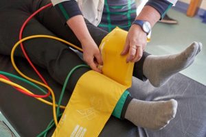
ABI measurement for lower limb
Automated Technique for ABI Measurement:
Patient should rest adequately before procedure.
Attach four cuffs to limbs, then press “start” for the automated multicuff device to cycle. Results will appear on screen in 1 minute.
»
Home » Procedure » Ankle-Brachial Index (ABI) Measurement
Ankle-Brachial Index (ABI) Measurement
Updated :
December 19, 2025
The ankle-brachial index (ABI) is a noninvasive measurement test which compares upper and lower limbs blood pressure level.
ABI detects resistance to blood flow in lower extremities due to arterial narrowing from atherosclerosis.
ABI measurement simplicity and low cost make it an effective test for early peripheral arterial disease (PAD) or arterial injury detection.
ABI < 0.90 indicates significant PAD with 79-95% sensitivity and >95% specificity in patients.
Diabetic patients with neuropathy or ulcers show variable ABI accuracy for PAD detection, with sensitivity 29-100% and specificity 58-97%.
ABI helps in identifying and assessing risk for lower-extremity arterial injuries in trauma.
Potential arterial injury in stable patients following lower extremity blunt or penetrating trauma.
In diabetic patients the normal ABI results require cautious interpretation and potential further testing for guideline-directed medical therapy.
Diagnosis of peripheral arterial disease
Assessment of non-compressible arteries
Evaluation of vascular complication
Detection of arterial injury
Pre-surgical risk assessment
Acute limb ischemia
Severe pain/open wound
Non-Compressible Arteries
Recent surgery
Severe edema
Infection/cellulitis
Patients unable to stay supine during examination or those at risk of worsening extremity injuries with cuffs.
Avoid extremity compression in patients with DVT to prevent thrombus dislodging and embolization risks.
Low ABI suggests higher risk of heart attack, stroke, and cardiovascular mortality while high ABI indicates stiffness.
Associated with higher mortality and atherosclerosis marker. The ABI is a ratio of highest ankle to highest brachial systolic blood pressure.
ABI values reflect arterial disease progression or successful treatment outcomes.
Blood Pressure Cuffs
Doppler Ultrasound Device
Doppler Gel
Measurement Tool
Sanitization Supplies
Patient Preparation:
Patient should wear loose clothing for easy arm and ankle access.
No anesthesia recommended as sedatives may affect blood pressure and ABI accuracy.
Position the patient supine, legs at heart level and ensure stillness for accurate measurements without movement interference.
Patient Positioning:
The patient must lie supine on an examination table, arms and legs level with the heart, resting for at least 10 minutes before measurement.
Choose a blood pressure cuff that is at least 40% of the limb’s circumference.
The ankle cuff should be positioned between the malleolus and calf, that allows space beneath for ultrasound gel to enable proper detection of brachial, dorsalis pedis, and posterior tibial pulses.
Measure brachial SBP in both arms, select the higher value, and palpate the pulse on the medial antecubital fossa.
Measure anterior and posterior tibial systolic pressures, then choose the higher value as the ankle pressure.
The DP pulse is felt on the dorsum between the first and second metatarsals, while the PT pulse is found near the medial malleolus.
Select the higher ankle SBP value, then divide it by brachial SBP to calculate the ABI.
ABI measurement for upper limb
ABI measurement for lower limb
Automated Technique for ABI Measurement:
Patient should rest adequately before procedure.
Attach four cuffs to limbs, then press “start” for the automated multicuff device to cycle. Results will appear on screen in 1 minute.
Both our subscription plans include Free CME/CPD AMA PRA Category 1 credits.
Digital Certificate PDF
On course completion, you will receive a full-sized presentation quality digital certificate.
medtigo Simulation
A dynamic medical simulation platform designed to train healthcare professionals and students to effectively run code situations through an immersive hands-on experience in a live, interactive 3D environment.
medtigo Points
medtigo points is our unique point redemption system created to award users for interacting on our site. These points can be redeemed for special discounts on the medtigo marketplace as well as towards the membership cost itself.
Community Forum post/reply = 5 points
*Redemption of points can occur only through the medtigo marketplace, courses, or simulation system. Money will not be credited to your bank account. 10 points = $1.
All Your Certificates in One Place
When you have your licenses, certificates and CMEs in one place, it's easier to track your career growth. You can easily share these with hospitals as well, using your medtigo app.