Aortic bifemoral bypass is a surgery for patients with atherosclerosis in the aorta and iliacs.
Patients typically show claudication, impotence, or poorly healing ulcers with weak femoral pulses.
Endovascular revascularization is a primary treatment for aortoiliac occlusive disease, but aortic bypass grafting effectively repairs vessel disease.
Aortobifemoral bypass now preferred over unilateral iliac vessel reconstruction.
Bilateral femoral bypass prevents future revisions due to contralateral progression.
The aortic bifemoral bypass is the most durable peripheral bypass.
Ideal procedure for aortoiliac disease, aortic aneurysms, or aortic atherosclerosis patients.
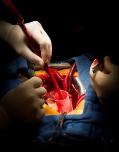
Surgery restores lower limb blood flow using synthetic graft bypass.
Aortic bifemoral bypass advances from vascular surgery and graft technology.
Dr. Arthur Voorhees pioneered synthetic grafts for arterial replacement mid-century.
Indications
Nonhealing ulcers in the extremities
Acute abdominal aortic occlusion
Atherosclerosis of the abdominal aorta or iliac arteries
Presence of severe claudication symptoms
Impotence
Critical limb-threatening ischemia
Failed endovascular interventions
Severe symptomatic aortoiliac occlusive disease with claudication, rest pain, or tissue loss
Aortic and iliac artery aneurysms when endovascular repair is not feasible
Contraindications
Severe Cardiac Dysfunction
Severe Pulmonary Disease
End-stage renal disease without dialysis access
Active Systemic Infection or Sepsis
Intra-abdominal Sepsis or Bowel Ischemia
Severe Frailty or Poor Functional Status
Severe Peripheral Artery Disease Beyond Femoral Arteries
Morbid Obesity
Severe Liver Disease
Outcomes
Experienced surgeons have a 2-5% mortality rate for aortic bifemoral bypass with less experienced hospitals face higher rates.
Surgery in infrequently performing hospitals has higher mortality due to myocardial infarction and stroke.
Aortic bifemoral bypass grafts have 91% patency at 5 years and 80% at 10 years.
Patency rates decrease in patients with ischemic rest pain, ulcerated toes, or distal disease.
Equipment required
Vascular Surgical Instruments
Synthetic Graft Materials
Bifurcated Dacron or PTFE
Vascular sutures
Anesthesia & Hemodynamic Monitoring Equipment
Patient Preparation:
Smoking patients should stop tobacco use 4 weeks before surgery.
Patients on nonsteroidal anti-inflammatory drugs should discontinue them 5-7 days before surgery.
Warfarin patients need heparin started 2-3 days prior to surgery.
Discontinue heparin at midnight before the procedure. Patients must stop all herbal products pre-procedure.
Patients with heart disease risk factors need cardiologist clearance thus heavy smokers require pulmonary consultation.
Start IV fluids at midnight and administer a broad-spectrum antibiotic before incision.
Autologous blood donation is possible three weeks before elective surgery.
A central venous line monitors fluid access, while a radial line continuously measures blood pressure.
Epidural catheter placement for days helps minimize pain and reduce narcotic needs significantly.
Informed Consent:
Explain the procedure’s risks and potential complications clearly to the patient.
Patient Positioning:
Patient is supine with arms extended for aortic bifemoral bypass procedure.
The infrarenal aorta is typically accessed through a midline abdominal incision from xiphoid to pubis.
Bypass of aneurysmal portion of aorta
Technique
Step 1: Aortobifemoral Bypass
Aortic bifemoral bypass can use transperitoneal or retroperitoneal methods.
Step 2: Operative exposure
Make two small incisions in the groin to access the common femoral artery. Cut the inguinal ligament if the bifurcation is high.
Incise the abdomen from xiphoid to below the umbilicus, lift transverse colon, and move small bowel right into a bowel bag.
In the retroperitoneal space, the surgeon can either elevate the left kidney and abdominal contents or navigate anteriorly towards the aorta.
Avoid lateral dissection to prevent damage to the IVC and IMA.
Always visualize the left renal vein anteriorly before dissecting below the proximal aorta.
Anaesthesiologist prepares for possible blood pressure surge during clamping.
Step 3: Proximal anastomosis
Avoid dissection behind the aorta after applying the proximal clamp below vessels.
Remove a small segment of anterior aorta for inner visualization and trim the prosthetic graft to leave approximately 3 cm trunk.
For aortic disease near renal arteries, position the proximal clamp above them and retract the left renal vein with a loop for better exposure.
Execute proximal aortic anastomosis as end-to-side or end-to-end; choose end-to-side for aberrant renal arteries or significant mesenteric artery preservation.
End-to-end anastomosis is necessary for aortic aneurysmal disease or complete occlusion by renal arteries.
Realize proximal anastomosis using 3-0 polypropylene suture, beginning posterior to anterior wall.
Clamp the distal graft, then gently release the proximal aortic clamp.
ePTFE grafts are reliable, but all vascular prosthetic grafts need regular monitoring for potential expansion.
Step 4: Distal anastomoses
Insert a long-curved clamp from each groin, bringing the graft into the retroperitoneum also avoid injury to the colon, ureters, and iliac veins.
Distal anastomoses in the groin typically occur at the common femoral artery bifurcation and the proximal deep femoral artery, irrespective of superficial femoral artery status.
In case of backflow doubt, perform embolectomy or select a distal target on the deep femoral artery without ligation.
Flush to clear emboli and air bubbles then alert anesthesiologist before unclamping.
Step 5: Closure
Administer protamine, count instruments, return bowel, and close peritoneal layer carefully.
Close the abdominal wall with 1-0 suture, then reassess groin pulses.
Groin closure in three layers with graft coverage.
»
Home » Procedure » Aortic Bifemoral (Aortobifemoral) Bypass
Aortic Bifemoral (Aortobifemoral) Bypass
Updated :
December 10, 2025
Aortic bifemoral bypass is a surgery for patients with atherosclerosis in the aorta and iliacs.
Patients typically show claudication, impotence, or poorly healing ulcers with weak femoral pulses.
Endovascular revascularization is a primary treatment for aortoiliac occlusive disease, but aortic bypass grafting effectively repairs vessel disease.
Aortobifemoral bypass now preferred over unilateral iliac vessel reconstruction.
Bilateral femoral bypass prevents future revisions due to contralateral progression.
The aortic bifemoral bypass is the most durable peripheral bypass.
Ideal procedure for aortoiliac disease, aortic aneurysms, or aortic atherosclerosis patients.
Surgery restores lower limb blood flow using synthetic graft bypass.
Aortic bifemoral bypass advances from vascular surgery and graft technology.
Dr. Arthur Voorhees pioneered synthetic grafts for arterial replacement mid-century.
Nonhealing ulcers in the extremities
Acute abdominal aortic occlusion
Atherosclerosis of the abdominal aorta or iliac arteries
Presence of severe claudication symptoms
Impotence
Critical limb-threatening ischemia
Failed endovascular interventions
Severe symptomatic aortoiliac occlusive disease with claudication, rest pain, or tissue loss
Aortic and iliac artery aneurysms when endovascular repair is not feasible
Severe Cardiac Dysfunction
Severe Pulmonary Disease
End-stage renal disease without dialysis access
Active Systemic Infection or Sepsis
Intra-abdominal Sepsis or Bowel Ischemia
Severe Frailty or Poor Functional Status
Severe Peripheral Artery Disease Beyond Femoral Arteries
Morbid Obesity
Severe Liver Disease
Experienced surgeons have a 2-5% mortality rate for aortic bifemoral bypass with less experienced hospitals face higher rates.
Surgery in infrequently performing hospitals has higher mortality due to myocardial infarction and stroke.
Aortic bifemoral bypass grafts have 91% patency at 5 years and 80% at 10 years.
Patency rates decrease in patients with ischemic rest pain, ulcerated toes, or distal disease.
Vascular Surgical Instruments
Synthetic Graft Materials
Bifurcated Dacron or PTFE
Vascular sutures
Anesthesia & Hemodynamic Monitoring Equipment
Patient Preparation:
Smoking patients should stop tobacco use 4 weeks before surgery.
Patients on nonsteroidal anti-inflammatory drugs should discontinue them 5-7 days before surgery.
Warfarin patients need heparin started 2-3 days prior to surgery.
Discontinue heparin at midnight before the procedure. Patients must stop all herbal products pre-procedure.
Patients with heart disease risk factors need cardiologist clearance thus heavy smokers require pulmonary consultation.
Start IV fluids at midnight and administer a broad-spectrum antibiotic before incision.
Autologous blood donation is possible three weeks before elective surgery.
A central venous line monitors fluid access, while a radial line continuously measures blood pressure.
Epidural catheter placement for days helps minimize pain and reduce narcotic needs significantly.
Informed Consent:
Explain the procedure’s risks and potential complications clearly to the patient.
Patient Positioning:
Patient is supine with arms extended for aortic bifemoral bypass procedure.
The infrarenal aorta is typically accessed through a midline abdominal incision from xiphoid to pubis.
Bypass of aneurysmal portion of aorta
Step 1: Aortobifemoral Bypass
Aortic bifemoral bypass can use transperitoneal or retroperitoneal methods.
Step 2: Operative exposure
Make two small incisions in the groin to access the common femoral artery. Cut the inguinal ligament if the bifurcation is high.
Incise the abdomen from xiphoid to below the umbilicus, lift transverse colon, and move small bowel right into a bowel bag.
In the retroperitoneal space, the surgeon can either elevate the left kidney and abdominal contents or navigate anteriorly towards the aorta.
Avoid lateral dissection to prevent damage to the IVC and IMA.
Always visualize the left renal vein anteriorly before dissecting below the proximal aorta.
Anaesthesiologist prepares for possible blood pressure surge during clamping.
Step 3: Proximal anastomosis
Avoid dissection behind the aorta after applying the proximal clamp below vessels.
Remove a small segment of anterior aorta for inner visualization and trim the prosthetic graft to leave approximately 3 cm trunk.
For aortic disease near renal arteries, position the proximal clamp above them and retract the left renal vein with a loop for better exposure.
Execute proximal aortic anastomosis as end-to-side or end-to-end; choose end-to-side for aberrant renal arteries or significant mesenteric artery preservation.
End-to-end anastomosis is necessary for aortic aneurysmal disease or complete occlusion by renal arteries.
Realize proximal anastomosis using 3-0 polypropylene suture, beginning posterior to anterior wall.
Clamp the distal graft, then gently release the proximal aortic clamp.
ePTFE grafts are reliable, but all vascular prosthetic grafts need regular monitoring for potential expansion.
Step 4: Distal anastomoses
Insert a long-curved clamp from each groin, bringing the graft into the retroperitoneum also avoid injury to the colon, ureters, and iliac veins.
Distal anastomoses in the groin typically occur at the common femoral artery bifurcation and the proximal deep femoral artery, irrespective of superficial femoral artery status.
In case of backflow doubt, perform embolectomy or select a distal target on the deep femoral artery without ligation.
Flush to clear emboli and air bubbles then alert anesthesiologist before unclamping.
Step 5: Closure
Administer protamine, count instruments, return bowel, and close peritoneal layer carefully.
Close the abdominal wall with 1-0 suture, then reassess groin pulses.
Groin closure in three layers with graft coverage.
Both our subscription plans include Free CME/CPD AMA PRA Category 1 credits.
Digital Certificate PDF
On course completion, you will receive a full-sized presentation quality digital certificate.
medtigo Simulation
A dynamic medical simulation platform designed to train healthcare professionals and students to effectively run code situations through an immersive hands-on experience in a live, interactive 3D environment.
medtigo Points
medtigo points is our unique point redemption system created to award users for interacting on our site. These points can be redeemed for special discounts on the medtigo marketplace as well as towards the membership cost itself.
Community Forum post/reply = 5 points
*Redemption of points can occur only through the medtigo marketplace, courses, or simulation system. Money will not be credited to your bank account. 10 points = $1.
All Your Certificates in One Place
When you have your licenses, certificates and CMEs in one place, it's easier to track your career growth. You can easily share these with hospitals as well, using your medtigo app.