An auricular hematoma is also termed as “cauliflower ear” is an injury that is marked by enlarged ears and stagnant accumulation of blood beneath the cartilage and skin of the ear. This frequently follows injuries, like blunt force, or friction which can further tear blood vessels of the ear tissues. Without being treated auricular hematoma may lead to deformation of the ear and other complications like infection and dying.
Indications
Tender anterior auricular swelling after trauma: This will induce a remarkable swelling and deformation of the normal anatomy of the pinna (the outer ear).
Pain and Tenderness: The patient may feel some pain or local ache when touching or manipulating the affected ear.
Contraindications
Hematoma that are older than 7 days: Once the absence of blood continues for a longer period, blood starts to clot, and extraction becomes even more complicated.
Unrepaired laceration, particularly with exposed cartilage: If the cartilage remains visible under the broken skin, the doctor should perform remedial work on the laceration first.
Outcomes
Equipment
Sutures
Compression dressings
Sterile gauze pads
Antiseptic solution
Suction device
Aspiration syringe
Anesthesia
Patient preparation
The doctor shall explain to the patient and the patient will sign the consent form which the surgeon will handover to.
In order to establish the size and site of the hematoma, usually, the physician carry out the physical ear examination.
Physical ear examination
The surgeon can either choose needle aspiration or surgical drainage based on the size of the hematoma.
Patient position
The patient position is lateral decubitus.
Technical considerations
Step 1: Patient Positioning and anesthesia: A patient will be lying down with a head that is elevated above and his or her affected ear upwards facing.
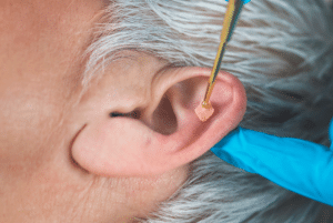
Step 2: Drainage Procedure: Incision: Through a small hole between the natural folds of the cartilage, that leaving behind a scaring is targeted.
Hematoma Removal: To access the hematoma, the surgeon will cautiously detach the skin and underlying tissue from the cartilage.
Irrigation and Cleaning: The area surrounding the hematoma is washed out with sterile saline solution to remove any specific blood or debris. Step 3: Closure and Dressing:
Dressing for auricular hematoma with bandage
Antibiotic Ointment: The incision will be covered with a thin layer of antibiotic ointment.
Compression Dressing: A pressure bandage will be applied to the ear to prevent blood reaccumulation and aid healing, possibly consisting of gauze pads and dental rolls.
Laboratory tests
Auricular hematoma drainage does not necessitate any laboratory testing.
Complications
Infection: This is the most common complication of any procedure with a skin puncture.
Recurrence of hematoma: There is a possibility of a recurrence of the clot symptoms after the blood clot is drained. This is more likely to become true if the person does not follow the surgeon’s instructions about caring for the treatment after the patient is discharged from the hospital, including wearing a pressure dressing.
Cauliflower ear: It is an abnormality in the cartilage of the ear that results if the cartilage formed is not firmly fixed on the blood clot after draining. Several patients who have sustained multiple hemorrhages around their ears are at a higher risk.
An auricular hematoma is also termed as “cauliflower ear” is an injury that is marked by enlarged ears and stagnant accumulation of blood beneath the cartilage and skin of the ear. This frequently follows injuries, like blunt force, or friction which can further tear blood vessels of the ear tissues. Without being treated auricular hematoma may lead to deformation of the ear and other complications like infection and dying.
Tender anterior auricular swelling after trauma: This will induce a remarkable swelling and deformation of the normal anatomy of the pinna (the outer ear).
Pain and Tenderness: The patient may feel some pain or local ache when touching or manipulating the affected ear.
Hematoma that are older than 7 days: Once the absence of blood continues for a longer period, blood starts to clot, and extraction becomes even more complicated.
Unrepaired laceration, particularly with exposed cartilage: If the cartilage remains visible under the broken skin, the doctor should perform remedial work on the laceration first.
Sutures
Compression dressings
Sterile gauze pads
Antiseptic solution
Suction device
Aspiration syringe
Anesthesia
The doctor shall explain to the patient and the patient will sign the consent form which the surgeon will handover to.
In order to establish the size and site of the hematoma, usually, the physician carry out the physical ear examination.
Physical ear examination
The surgeon can either choose needle aspiration or surgical drainage based on the size of the hematoma.
Patient position
The patient position is lateral decubitus.
Step 1: Patient Positioning and anesthesia: A patient will be lying down with a head that is elevated above and his or her affected ear upwards facing.
Step 2: Drainage Procedure: Incision: Through a small hole between the natural folds of the cartilage, that leaving behind a scaring is targeted.
Hematoma Removal: To access the hematoma, the surgeon will cautiously detach the skin and underlying tissue from the cartilage.
Irrigation and Cleaning: The area surrounding the hematoma is washed out with sterile saline solution to remove any specific blood or debris. Step 3: Closure and Dressing:
Dressing for auricular hematoma with bandage
Antibiotic Ointment: The incision will be covered with a thin layer of antibiotic ointment.
Compression Dressing: A pressure bandage will be applied to the ear to prevent blood reaccumulation and aid healing, possibly consisting of gauze pads and dental rolls.
Auricular hematoma drainage does not necessitate any laboratory testing.
Infection: This is the most common complication of any procedure with a skin puncture.
Recurrence of hematoma: There is a possibility of a recurrence of the clot symptoms after the blood clot is drained. This is more likely to become true if the person does not follow the surgeon’s instructions about caring for the treatment after the patient is discharged from the hospital, including wearing a pressure dressing.
Cauliflower ear: It is an abnormality in the cartilage of the ear that results if the cartilage formed is not firmly fixed on the blood clot after draining. Several patients who have sustained multiple hemorrhages around their ears are at a higher risk.
Both our subscription plans include Free CME/CPD AMA PRA Category 1 credits.
Digital Certificate PDF
On course completion, you will receive a full-sized presentation quality digital certificate.
medtigo Simulation
A dynamic medical simulation platform designed to train healthcare professionals and students to effectively run code situations through an immersive hands-on experience in a live, interactive 3D environment.
medtigo Points
medtigo points is our unique point redemption system created to award users for interacting on our site. These points can be redeemed for special discounts on the medtigo marketplace as well as towards the membership cost itself.
Community Forum post/reply = 5 points
*Redemption of points can occur only through the medtigo marketplace, courses, or simulation system. Money will not be credited to your bank account. 10 points = $1.
All Your Certificates in One Place
When you have your licenses, certificates and CMEs in one place, it's easier to track your career growth. You can easily share these with hospitals as well, using your medtigo app.