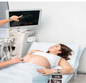
Basic obstetric ultrasound is a crucial part of prenatal care to visualize the fetus and monitor the mother’s wellbeing. Obstetricians started using ultrasound in the 1960s and 1970s for the purpose of making diagnosis of early intrauterine pregnancies. In the 1990s, emergency physicians started using ultrasonic images for point-of-care analysis. It is a noninvasive, quick method for confirming intrauterine pregnancies at the patient’s bedside and can be used as a means for decreasing the average length of stay for pregnant patients in the emergency department. First-trimester diagnosis is most accurate offering point-of-care pelvic ultrasound.
Indications
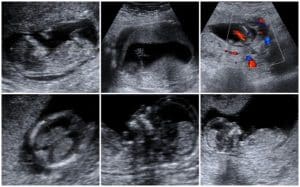
First Trimester Ultrasound
Vagina Bleeding: The first-trimester ultrasound is performed mainly for the assessment of a viable intrauterine pregnancy when the gestational age of the fetus is less than 13 weeks and 6 days. It can be done trans-abdominally or trans-vaginally and is mandatory in patients with pelvic pain or vaginal spotting.
Ectopic Pregnancy: It can diagnose whether a pregnancy is ectopic or if a woman is pregnant with something other than a normal fetus such as a molar pregnancy or pregnancy that has already miscarried.
A diagnosis is considered definitive when the gestational sac along with yolk sac is visualized. If only the gestational sac is visualized, then further assessment is required. When pregnancy tests are positive and no findings are seen, follow-up of symptoms and levels of serum beta human chorionic Gonadotropin (bHCG) may have to be carried out.
For viable pregnancies, it can be used for dating the pregnancy using the mean sac diameter or crown-rump length (CRL) in case an embryo is visible. This is important to calculate the due dates especially in cases where there is a variation on the menstrual method of dating.
Fetal sonography
Indications with evidence
Vaginal Bleeding: Vaginal bleeding is a common occurrence in early pregnancy, a study shows that approximately 25 percent of pregnant women experience this condition. In trauma patients the FAST exam is done to check for intra-abdominal hemorrhage and if fluid is present, then surgeon intervention is needed. In non-trauma cases, absence or presence of free fluid may indicate ectopic pregnancy.
Indications with evidence
The review involved 11 studies, encompassing 96,633 fetuses. Of these, 52 false positive diagnoses occurred and none of the fatal anomalies were detected. Some were linked to potential survival and either long-term or short-term complications with a few being treatable in the womb. Anomaly Detection:
First trimester (less than 15 weeks): All analyses were within normal range and no specific abnormalities were noted.
Second trimester (less than 24 weeks): The overall sensitivity was 41.3%, while higher detection rates were recorded after 18 weeks. Specificity was 99.94%.
Detection rates varied by system:
Central nervous system (CNS): 76.4%
Urinary tract: 67.3%
Pulmonary: 50%
Gastrointestinal: 41.9%
Skeletal: 23. 8%
Cardiac: 17. 4%
Chromosomal: 18.8%
Third trimester (greater than 24 weeks):
Sensitivity was 18. 6 % and specificity is at 100 %.
Detection rates:
CNS: 71%
Urinary tract: 69.7%
Gastrointestinal: 50%
Pulmonary: 44.4%
Skeletal and Cardiac: 26. 1%
Chromosomal: 10.4%
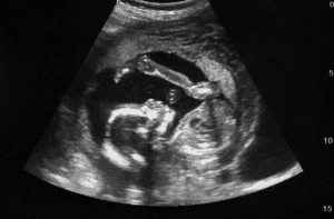
Second and third trimester ultrasound
During the second and third trimester, ultrasonography is used to measure fetal biometry to determine growth and detect anatomic abnormalities; biparietal diameter, abdominal circumference, fetal presentation, and amniotic fluid volume.
Fetal anomalies are best detected by a detailed anatomic scan which should be done between 18 and 20 weeks of gestation. Targeted scans can be done at any point, however standard fetal anomaly scans occur at 11 to 13 weeks, and fetal growth scans are usually performed at 20 to 24 weeks and later at approximately 3 to 4 weeks intervals.
Second trimester sonogram
Types of Ultrasound Scans
Early Pregnancy or Dating Scan (8-11 weeks): Pregnancy is confirmed using this scan, and the estimated due date is established. It usually identifies the fetal heartbeat, giving some comfort to the parents. This information is also used in the non-invasive prenatal test (NIPT) that helps in determining the likelihood of a baby being born with disorders such as Down syndrome. It is normally done externally but may require an internal examination for better pictures.
Nuchal Translucency Scan (11-14 weeks): This is mainly done abdominally but assesses some features:
Provides the assurance of fetal heartbeat.
Yields a better assessment of gestational age particularly among pregnancies that are not induced by in-vitro fertilization (IVF).
Estimates the size of the fetus and helps detect if the woman is pregnant with multiple fetuses.
Examines the shape of the fetus especially amniotic fluid in the spine at the back to check on chromosomal issues including down syndrome.
Prenatal diagnostic tests that involve blood tests for screening Down syndrome may also be given.
Early Anomaly Scan (14-18 weeks): Like the fetal anomaly scan, this preliminary examination allows for detecting possible anomalies. However, some structures may be too small to be imagined so they may require a repeat scan at 19-22 weeks. This scan is usually advised when there are certain risk factors that indicate conditions such as spina bifida.
Fetal Anomaly Scan (18-24 weeks): Often performed between the 19 and 22 weeks of pregnancy, this scan ensures that all organs and body parts of the fetus look normal. The position of the placenta and blood flow of the fetus are also assessed for the management of the pregnant woman during pregnancy.
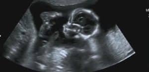
Fourth month fetal sonogram
Growth Scan or Fetal Well-Being Scan (24-42 weeks): These scans assess the size and health of the fetus. Head, abdominal, and thigh measurements aid in determining the weight of the fetus and the volume of amniotic fluid. Umbilical cord blood flow is assessed and fetal positioning towards the delivery is observed for better birth preparation. Subsequent scans may be done at 4-week intervals if results are not suggestive of earlier evaluations.
Growth Scan
Contraindications
The Federal Drug Administration (FDA) first raised concern in 1994 about the abuse of diagnostic ultrasound exams and the equipment in non-medical applications. They particularly pointed out that marketing or offering for sale or rental of ultrasound equipment to capture “baby incredibly moving, laughing or kicking or for ‘keepsake’ fetal videos without the prescription from a doctor may be in violation of some laws and regulations.
Outcomes
Equipment
The main component in the construction of the ultrasound machine is the transducer that is placed directly onto the patient’s skin. This device consists of piezoelectric crystals through which sound pulses are created when the crystals are touched with an electric current. These sound waves are introduced at a certain frequency, propagate through the human tissues, and have an impact on tissues depending on their physical properties. When the sound waves interact with the various tissues, some ultrasonic energy is absorbed by the tissues while others are reflected to the transducer. The piezoelectric crystals then turn these returning signals into electrical impulses, and the electrical impulses are processed and transformed to the images that are displayed on the screen.
Patient Preparation
Anesthesia
Most of the diagnostic ultrasound examinations and the procedure that require ultrasound assistance such as amniocentesis do not require the administration of anesthesia.
Positioning
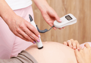
During abdominal ultrasound studies the patient could either stand or sit depending on which form is preferred, after which the abdomen is exposed to enable contact with the transducer.
In transvaginal USG, the patient must undress from the lower part of her body and lie down on her back or on her side with legs in stirrups. Stirrups can be standardized or else the patient may be positioned in the frog leg position. To ensure that the patient does not feel uncomfortable or embarrassed a blanket is placed, which wraps the pelvic area. This is done in such a way that the transvaginal probe must undergo coupling gel which is then followed by a vinyl or a latex cover. To increase smooth insertion and to increase acoustic impedance, another layer of gel is competitively placed.
Transabdominal Pelvic Ultrasound
Probe Selection: It is also recommended to use a low-frequency convex probe. A phased array probe can be used if it is not available at the time of scanning. Switch the ultrasound machine setting to “obstetrics” or “pregnancy.”
Initial Positioning: Position the convex probe slightly above the pubic symphysis with the marker facing upwards. Always maintain good contact throughout the procedure adhesion with coupling gel.
Sagittal View of the Uterus: To scan its interior, position the probe slightly angled in the inferior direction. Superficial structures are depicted at the top of the monitor in the anterior direction while deep structures are depicted at the bottom in the posterior direction. The top of the screen corresponds to the cephalad and the bottom corresponds to the caudal position.
Identify Landmarks: A full bladder can be observed in the anterior part while the vaginal stripe is behind the bladder. The cervix and uterus are no exceptions to this line.
Uterus Assessment: To ensure that the entire uterus had been illuminated and visualized, fan the probe left and right. If the gestational sac is visualized, try to identify a yolk sac or to determine fetal cardiac activity. Evaluate the endomyometrial mantle around the gestational sac to assess the presence of intrauterine pregnancy (IUP).
Fetal Heartbeat: If present, use M-mode to measure the fetal heartbeat. Make the M-mode line parallel to the flicker of the fetal heart, then ‘lock’ the image and count the number of beats per minute using the caliper.
Transabdominal Ultrasound
Transvaginal Examination
Prepare the Patient: An initial step involves performing a transabdominal ultrasound to clarify the location of the uterus. Before transvaginal imaging, ensure that the patient has an empty bladder after imaging.
Position the Patient: Put an inverted bedpan under the patient’s lumbar region and insert the probe in such a manner that the indicator faces upwards. It is preferable to use a small amount of coupling gel in application and to have a sterile sheath on the probe.
Bladder and Uterus Visualization: The bladder should disappear from the screen as the probe is directed in the right manner. Start with an orientation of the uterus in the sagittal plane by moving the probe in fanlike motion toward the right and left. The superior area on the screen depicts the neck of the womb, the inferior part of the screen represents the superior aspect of uterus.
Pouch of Douglas: To locate free fluid in the Pouch of Douglas, look at the right lower quadrant of the screen. As part of the initial assessment, look for the presence of IUP with the yolk sac or fetal heart rate.
Rotate Probe for Coronal View: Flip the probe 90 degrees to slide it into the transverse (coronal) plane to get an orientation of the uterus. On the screen left side is patient’s right side and the right side of the screen reflects the patient’s left or vice versa depending on the orientation of the screen.
Ovary Identification: The position of the ovaries is close to the iliac vessels, and they are essentially hypoechoic structures with anechoic follicles. There may be a need to apply some pressure to make out the shape of the ovaries.
Ovarian Dimensions and Cysts: Ovarian dimensions that are normally found are 1. 5 cm x 1. 5 x 3 cm. A simple ovarian cyst can be as small as 2. A nodule is usually bigger than 5 cm or larger while a follicle is much smaller in size. Identify these corpus luteum cysts, their walls are thicker, and they are common in the early pregnancy.
Approach considerations
Approach to First-Trimester Ultrasound
The evaluation of first-trimester ultrasound mainly involves diagnosing IUP, estimating gestational age, and measuring nuchal thickness. During an examination, the uterus, cervix, and the adnexa are inspected to check for the position of the gestational sac and the presence of a yolk sac. Beta-human chorionic gonadotropin (b-hCG) levels set up have been found to be consistent with standard anticipated fetal sonographic appearances and help in assessing the likelihood of pregnancy continuation.
Some more important observations are visibility of yolk sac at 5-6 weeks of pregnancy and embryo at 6+ weeks of pregnancy. The fetal heart activity, the number, location, and chorionicity should be documented, especially if there are twins at pregnancy. Other findings, including a corpus luteum cyst or uterine fibroid(s) must also be documented.
Ultrasound is also useful in the identification of abnormal pregnancies, for instance, embryo demise characterised by CRL of 6mm and no fetal cardiac activity, ectopic pregnancy or even severe fetal anomalies such as anencephaly. Also, the first-trimester ultrasound is employed for the aneuploidy biomarker, including those such as nuchal translucency, absent nasal bone, and other Doppler signs.
Approach consideration to Second and Third trimester
A second-trimester ultrasound, which is done around 18 to 20 weeks of pregnancy, aims at evaluating fetal structures, defining pregnancy duration, the number of fetuses in twin pregnancies, chorionicity. It can also identify conditions like placenta previa, where the placenta lies over the cervix, and abruptio placentae, where the placenta separates from the wall of the uterus. Maternal or fetal positioning, obesity, or uterine fibroids make it challenging to visualize the target structure and may necessitate additional ultrasounds.
The ultrasound assesses maternal and fetal images, focusing on the CNS, calculating its dimensions, such as the cerebellum or ventricles, that may indicate problems like spina bifida, hydrocephalus, or trisomy 18. Ultrasound measurements including biparietal diameter, head circumference and femur length are usually used in tracking fetal growth particularly in cases categorized under high-risk pregnancy.
Further, congenital anomalies such as cardiac, pulmonary, gastrointestinal, renal and bladder malformations and functions can also be evaluated; birth defects like CDH, bowel obstruction, PKD or renal agenesis are detectable.
Complications
Evaluating the pelvis using a pelvic ultrasound, like many other types of ultrasound procedures, involves practically no risk. As for the patients, they might experience certain discomfort when the probe is applied during both transabdominal and transvaginal scans.
Basic obstetric ultrasound is a crucial part of prenatal care to visualize the fetus and monitor the mother’s wellbeing. Obstetricians started using ultrasound in the 1960s and 1970s for the purpose of making diagnosis of early intrauterine pregnancies. In the 1990s, emergency physicians started using ultrasonic images for point-of-care analysis. It is a noninvasive, quick method for confirming intrauterine pregnancies at the patient’s bedside and can be used as a means for decreasing the average length of stay for pregnant patients in the emergency department. First-trimester diagnosis is most accurate offering point-of-care pelvic ultrasound.
First Trimester Ultrasound
Vagina Bleeding: The first-trimester ultrasound is performed mainly for the assessment of a viable intrauterine pregnancy when the gestational age of the fetus is less than 13 weeks and 6 days. It can be done trans-abdominally or trans-vaginally and is mandatory in patients with pelvic pain or vaginal spotting.
Ectopic Pregnancy: It can diagnose whether a pregnancy is ectopic or if a woman is pregnant with something other than a normal fetus such as a molar pregnancy or pregnancy that has already miscarried.
A diagnosis is considered definitive when the gestational sac along with yolk sac is visualized. If only the gestational sac is visualized, then further assessment is required. When pregnancy tests are positive and no findings are seen, follow-up of symptoms and levels of serum beta human chorionic Gonadotropin (bHCG) may have to be carried out.
For viable pregnancies, it can be used for dating the pregnancy using the mean sac diameter or crown-rump length (CRL) in case an embryo is visible. This is important to calculate the due dates especially in cases where there is a variation on the menstrual method of dating.
Fetal sonography
Indications with evidence
Vaginal Bleeding: Vaginal bleeding is a common occurrence in early pregnancy, a study shows that approximately 25 percent of pregnant women experience this condition. In trauma patients the FAST exam is done to check for intra-abdominal hemorrhage and if fluid is present, then surgeon intervention is needed. In non-trauma cases, absence or presence of free fluid may indicate ectopic pregnancy.
Indications with evidence
The review involved 11 studies, encompassing 96,633 fetuses. Of these, 52 false positive diagnoses occurred and none of the fatal anomalies were detected. Some were linked to potential survival and either long-term or short-term complications with a few being treatable in the womb. Anomaly Detection:
First trimester (less than 15 weeks): All analyses were within normal range and no specific abnormalities were noted.
Second trimester (less than 24 weeks): The overall sensitivity was 41.3%, while higher detection rates were recorded after 18 weeks. Specificity was 99.94%.
Detection rates varied by system:
Central nervous system (CNS): 76.4%
Urinary tract: 67.3%
Pulmonary: 50%
Gastrointestinal: 41.9%
Skeletal: 23. 8%
Cardiac: 17. 4%
Chromosomal: 18.8%
Third trimester (greater than 24 weeks):
Sensitivity was 18. 6 % and specificity is at 100 %.
Detection rates:
CNS: 71%
Urinary tract: 69.7%
Gastrointestinal: 50%
Pulmonary: 44.4%
Skeletal and Cardiac: 26. 1%
Chromosomal: 10.4%
Second and third trimester ultrasound
During the second and third trimester, ultrasonography is used to measure fetal biometry to determine growth and detect anatomic abnormalities; biparietal diameter, abdominal circumference, fetal presentation, and amniotic fluid volume.
Fetal anomalies are best detected by a detailed anatomic scan which should be done between 18 and 20 weeks of gestation. Targeted scans can be done at any point, however standard fetal anomaly scans occur at 11 to 13 weeks, and fetal growth scans are usually performed at 20 to 24 weeks and later at approximately 3 to 4 weeks intervals.
Second trimester sonogram
Types of Ultrasound Scans
Early Pregnancy or Dating Scan (8-11 weeks): Pregnancy is confirmed using this scan, and the estimated due date is established. It usually identifies the fetal heartbeat, giving some comfort to the parents. This information is also used in the non-invasive prenatal test (NIPT) that helps in determining the likelihood of a baby being born with disorders such as Down syndrome. It is normally done externally but may require an internal examination for better pictures.
Nuchal Translucency Scan (11-14 weeks): This is mainly done abdominally but assesses some features:
Provides the assurance of fetal heartbeat.
Yields a better assessment of gestational age particularly among pregnancies that are not induced by in-vitro fertilization (IVF).
Estimates the size of the fetus and helps detect if the woman is pregnant with multiple fetuses.
Examines the shape of the fetus especially amniotic fluid in the spine at the back to check on chromosomal issues including down syndrome.
Prenatal diagnostic tests that involve blood tests for screening Down syndrome may also be given.
Early Anomaly Scan (14-18 weeks): Like the fetal anomaly scan, this preliminary examination allows for detecting possible anomalies. However, some structures may be too small to be imagined so they may require a repeat scan at 19-22 weeks. This scan is usually advised when there are certain risk factors that indicate conditions such as spina bifida.
Fetal Anomaly Scan (18-24 weeks): Often performed between the 19 and 22 weeks of pregnancy, this scan ensures that all organs and body parts of the fetus look normal. The position of the placenta and blood flow of the fetus are also assessed for the management of the pregnant woman during pregnancy.
Fourth month fetal sonogram
Growth Scan or Fetal Well-Being Scan (24-42 weeks): These scans assess the size and health of the fetus. Head, abdominal, and thigh measurements aid in determining the weight of the fetus and the volume of amniotic fluid. Umbilical cord blood flow is assessed and fetal positioning towards the delivery is observed for better birth preparation. Subsequent scans may be done at 4-week intervals if results are not suggestive of earlier evaluations.
Growth Scan
The Federal Drug Administration (FDA) first raised concern in 1994 about the abuse of diagnostic ultrasound exams and the equipment in non-medical applications. They particularly pointed out that marketing or offering for sale or rental of ultrasound equipment to capture “baby incredibly moving, laughing or kicking or for ‘keepsake’ fetal videos without the prescription from a doctor may be in violation of some laws and regulations.
The main component in the construction of the ultrasound machine is the transducer that is placed directly onto the patient’s skin. This device consists of piezoelectric crystals through which sound pulses are created when the crystals are touched with an electric current. These sound waves are introduced at a certain frequency, propagate through the human tissues, and have an impact on tissues depending on their physical properties. When the sound waves interact with the various tissues, some ultrasonic energy is absorbed by the tissues while others are reflected to the transducer. The piezoelectric crystals then turn these returning signals into electrical impulses, and the electrical impulses are processed and transformed to the images that are displayed on the screen.
Patient Preparation
Anesthesia
Most of the diagnostic ultrasound examinations and the procedure that require ultrasound assistance such as amniocentesis do not require the administration of anesthesia.
Positioning
During abdominal ultrasound studies the patient could either stand or sit depending on which form is preferred, after which the abdomen is exposed to enable contact with the transducer.
In transvaginal USG, the patient must undress from the lower part of her body and lie down on her back or on her side with legs in stirrups. Stirrups can be standardized or else the patient may be positioned in the frog leg position. To ensure that the patient does not feel uncomfortable or embarrassed a blanket is placed, which wraps the pelvic area. This is done in such a way that the transvaginal probe must undergo coupling gel which is then followed by a vinyl or a latex cover. To increase smooth insertion and to increase acoustic impedance, another layer of gel is competitively placed.
Probe Selection: It is also recommended to use a low-frequency convex probe. A phased array probe can be used if it is not available at the time of scanning. Switch the ultrasound machine setting to “obstetrics” or “pregnancy.”
Initial Positioning: Position the convex probe slightly above the pubic symphysis with the marker facing upwards. Always maintain good contact throughout the procedure adhesion with coupling gel.
Sagittal View of the Uterus: To scan its interior, position the probe slightly angled in the inferior direction. Superficial structures are depicted at the top of the monitor in the anterior direction while deep structures are depicted at the bottom in the posterior direction. The top of the screen corresponds to the cephalad and the bottom corresponds to the caudal position.
Identify Landmarks: A full bladder can be observed in the anterior part while the vaginal stripe is behind the bladder. The cervix and uterus are no exceptions to this line.
Uterus Assessment: To ensure that the entire uterus had been illuminated and visualized, fan the probe left and right. If the gestational sac is visualized, try to identify a yolk sac or to determine fetal cardiac activity. Evaluate the endomyometrial mantle around the gestational sac to assess the presence of intrauterine pregnancy (IUP).
Fetal Heartbeat: If present, use M-mode to measure the fetal heartbeat. Make the M-mode line parallel to the flicker of the fetal heart, then ‘lock’ the image and count the number of beats per minute using the caliper.
Transabdominal Ultrasound
Transvaginal Examination
Prepare the Patient: An initial step involves performing a transabdominal ultrasound to clarify the location of the uterus. Before transvaginal imaging, ensure that the patient has an empty bladder after imaging.
Position the Patient: Put an inverted bedpan under the patient’s lumbar region and insert the probe in such a manner that the indicator faces upwards. It is preferable to use a small amount of coupling gel in application and to have a sterile sheath on the probe.
Bladder and Uterus Visualization: The bladder should disappear from the screen as the probe is directed in the right manner. Start with an orientation of the uterus in the sagittal plane by moving the probe in fanlike motion toward the right and left. The superior area on the screen depicts the neck of the womb, the inferior part of the screen represents the superior aspect of uterus.
Pouch of Douglas: To locate free fluid in the Pouch of Douglas, look at the right lower quadrant of the screen. As part of the initial assessment, look for the presence of IUP with the yolk sac or fetal heart rate.
Rotate Probe for Coronal View: Flip the probe 90 degrees to slide it into the transverse (coronal) plane to get an orientation of the uterus. On the screen left side is patient’s right side and the right side of the screen reflects the patient’s left or vice versa depending on the orientation of the screen.
Ovary Identification: The position of the ovaries is close to the iliac vessels, and they are essentially hypoechoic structures with anechoic follicles. There may be a need to apply some pressure to make out the shape of the ovaries.
Ovarian Dimensions and Cysts: Ovarian dimensions that are normally found are 1. 5 cm x 1. 5 x 3 cm. A simple ovarian cyst can be as small as 2. A nodule is usually bigger than 5 cm or larger while a follicle is much smaller in size. Identify these corpus luteum cysts, their walls are thicker, and they are common in the early pregnancy.
Approach considerations
Approach to First-Trimester Ultrasound
The evaluation of first-trimester ultrasound mainly involves diagnosing IUP, estimating gestational age, and measuring nuchal thickness. During an examination, the uterus, cervix, and the adnexa are inspected to check for the position of the gestational sac and the presence of a yolk sac. Beta-human chorionic gonadotropin (b-hCG) levels set up have been found to be consistent with standard anticipated fetal sonographic appearances and help in assessing the likelihood of pregnancy continuation.
Some more important observations are visibility of yolk sac at 5-6 weeks of pregnancy and embryo at 6+ weeks of pregnancy. The fetal heart activity, the number, location, and chorionicity should be documented, especially if there are twins at pregnancy. Other findings, including a corpus luteum cyst or uterine fibroid(s) must also be documented.
Ultrasound is also useful in the identification of abnormal pregnancies, for instance, embryo demise characterised by CRL of 6mm and no fetal cardiac activity, ectopic pregnancy or even severe fetal anomalies such as anencephaly. Also, the first-trimester ultrasound is employed for the aneuploidy biomarker, including those such as nuchal translucency, absent nasal bone, and other Doppler signs.
Approach consideration to Second and Third trimester
A second-trimester ultrasound, which is done around 18 to 20 weeks of pregnancy, aims at evaluating fetal structures, defining pregnancy duration, the number of fetuses in twin pregnancies, chorionicity. It can also identify conditions like placenta previa, where the placenta lies over the cervix, and abruptio placentae, where the placenta separates from the wall of the uterus. Maternal or fetal positioning, obesity, or uterine fibroids make it challenging to visualize the target structure and may necessitate additional ultrasounds.
The ultrasound assesses maternal and fetal images, focusing on the CNS, calculating its dimensions, such as the cerebellum or ventricles, that may indicate problems like spina bifida, hydrocephalus, or trisomy 18. Ultrasound measurements including biparietal diameter, head circumference and femur length are usually used in tracking fetal growth particularly in cases categorized under high-risk pregnancy.
Further, congenital anomalies such as cardiac, pulmonary, gastrointestinal, renal and bladder malformations and functions can also be evaluated; birth defects like CDH, bowel obstruction, PKD or renal agenesis are detectable.
Complications
Evaluating the pelvis using a pelvic ultrasound, like many other types of ultrasound procedures, involves practically no risk. As for the patients, they might experience certain discomfort when the probe is applied during both transabdominal and transvaginal scans.
Both our subscription plans include Free CME/CPD AMA PRA Category 1 credits.
Digital Certificate PDF
On course completion, you will receive a full-sized presentation quality digital certificate.
medtigo Simulation
A dynamic medical simulation platform designed to train healthcare professionals and students to effectively run code situations through an immersive hands-on experience in a live, interactive 3D environment.
medtigo Points
medtigo points is our unique point redemption system created to award users for interacting on our site. These points can be redeemed for special discounts on the medtigo marketplace as well as towards the membership cost itself.
Community Forum post/reply = 5 points
*Redemption of points can occur only through the medtigo marketplace, courses, or simulation system. Money will not be credited to your bank account. 10 points = $1.
All Your Certificates in One Place
When you have your licenses, certificates and CMEs in one place, it's easier to track your career growth. You can easily share these with hospitals as well, using your medtigo app.