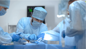
Cardiothoracic surgery is a surgical specialty that involves operations on the heart, lungs and chest area organs including the esophagus. This field includes cardiovascular and thoracic area and focuses on a various diseases associated with the heart and lungs. Cardiothoracic surgeons undergo their training in general surgery and the specialized procedures that will enable them to operate on sensitive structures in the thoracic area.
Indications
Coronary Artery Disease (CAD):
Coronary artery bypass grafting (CABG): It occurs when coronary arteries are heavily narrowed or blocked, for the symptoms like angina or the prevention of more advanced problems like heart attack.
Valvular Heart Disease:
Valve repair or replacement: Prescribed for conditions that may include aortic stenosis, mitral regurgitation or other valvular disorders that lead to heart failure signs or complications, such as infective endocarditis.
Aortic Aneurysms or Dissections:
Aortic surgery: Used for the prevention of rupture in patients with thoracic aortic aneurysms (TAA) or acute aortic dissections.
Congenital Heart Disease:
Repair or palliative surgery: Prescribed for children born with heart disease such as atrial /ventricular septal defect, Tetralogy of Fallot, Transposition of the great arteries.
Arrhythmias:
Surgical ablation (e.g., Maze procedure):
Recommended for those with persistent atrial fibrillation that remains poorly managed by pharmacological therapy, and catheter ablation.
Pacemaker or implantable cardioverter-defibrillator (ICD) placement: Approved for the treatment of life-threatening ventricular arrhythmias or heart failure with left ventricular systolic dysfunction.
Heart Failure:
Heart transplant: Used in the last stage of heart failure.
Left ventricular assist devices (LVADs): Prescribed as a temporary measure before heart transplantation or as chronic treatment for critical heart disease.
Pulmonary Diseases:
In patients with severe emphysema or end-stage.
lung disease: LVRS (Lung volume reduction surgery) or lung transplant.
Esophageal Disorders:
Esophageal Disorders Surgery for treatment of esophageal cancer, severe GERD or Achalasia such as esophagectomy.
Contraindications
Severe Pulmonary Dysfunction: Critically ill COPD patients and patients with severe Pulmonary Hypertension may not be able to tolerate the stress of the surgery since they might lead to respiratory failure.
Advanced Age and Frailty: Age alone is not a definite cause for a contraindication, but reduced functioning and condition accompanied by comorbidities make risks outweigh benefits in elderly patients.
Uncontrolled Infection:
Systemic infections especially sepsis add post operative complications such as mediastinitis or wound infections.
Malignancy:
The presence of active cancer, uncontrolled or newly diagnosed, metastatic cancer, or untreated cancer is a contraindication to elective cardiothoracic surgery except when it is mainly for the purpose of alleviating pain and suffering or substantially enhances the quality of life of the patient.
Outcomes
Periprocedural care
Equipment
Scalpel
Scissors
Forceps
Needle Holders
Sternal Retractors
Rib Spreaders
Vascular Clamps
Suction Devices
Bulldog clamps
Debakey forceps
Patient preparation:
Medical History & Physical Exam: Assessment to have a clear history of patient’s existing medical conditions, past operations and prescribed drugs.
Cardiovascular Assessment: Covers echocardiogram, electrocardiogram, chest radiography, and stress testing to monitor the heart and lung.
Laboratory Tests: Complete blood count, prothrombin time, Bleeding time, Serum electrolytes, Kidney- Liver profile.
Imaging: High-resolution images for surgery planning if the case requires CT scan, MRI, or angiography.
Pulmonary Function Tests: Spirometry or some other test evaluates the lungs for dealing with surgery.
Preoperative Counselling: Explanation about the surgery, complications, benefits, and management of expected outcomes and post-operative complications.
This is usually a family member or a special caregiver who has been assigned that responsibility with the patient.
Pre-anesthesia Consultation: An anesthesiologist who is specialized in giving anesthesia for patients assembles information about the patient’s airway, allergies, and history of previous anesthetic experiences with the view of developing a proper anesthesia plan.
Patient position:
Supine Position:
This position is used for almost all cardiothoracic surgeries, such as CABG, heart valve replacement, and lung surgeries.
The patient lies flat on the back with either arms tucked in at the side or on arm boards, extended. The legs remain straight. Left Lateral Decubitus Position:
These are used for Lung surgeries such as lobectomy or pneumonectomy.
Patient placed on left lateral side with right arm raised and the left arm extended. The legs were bent slightly to enable stability. Trendelenburg Position:
Primarily applied in selected cardiac surgeries such as CABG, where venous return can be enhanced, and access might be easier.
The patient’s head is dropped, and the legs are elevated at an angle providing forward blood flow in the head and upper body.
Aortic Valve Replacement
Cardiothoracic surgery
Step 1-Preoperative Preparation: Assess the patient (history, physical exam, imaging, lab tests).
Obtain informed consent.
Administer general anesthesia. Step 2-Incision:
Perform a median sternotomy through breastbone to reach the heart.
Alternatively, perform minimally invasive approach like a right anterior thoracotomy or TAVR. Step 3-Cardiopulmonary Bypass (CPB):
Initiate CPB by attaching the patient to the machine that oxygenates and circulates blood when the heart is arrested. Step 4-Aortic Cross-Clamping:
Place a clamp on the ascending aorta to stop blood flow and allow for the removal of the diseased valve.
Step 5-Excision of the Aortic Valve:
Aorta is opened, and the old valve is excised.
Determination of the size of the annulus for the prosthetic valve Step 6-Valve Implantation:
Insertion of the prosthetic valve:
Insertion of either a bioprosthetic or a mechanical valve by suturing.
Assure that the new valve is correctly aligned and functional in the annulus. Step 7-De-airing:
Get rid of all the air in the heart chambers by aspiration and gentle filling of the chambers with blood. Step 8-Reinstituting Cardiopulmonary circulation:
Gradually eases off the aortic cross-clamp and restarted the heart either spontaneously or with electrical pacing as necessary.
Wean the patient off the bypass machine. Step 9-Closure:
Closes the aorta and the chest cavity in layers.
Ensure proper hemostasis. Step 10-Postoperative Care:
Monitor in the ICU for recovery.
Manage anticoagulation if a mechanical valve is used.
Mitral valve repair
Mitral valve repair is a significant procedure in the cardiothoracic surgery area due to its potential to restore normal heart function in patients with mitral valve regurgitation or stenosis.
The mitral valve is one of four valves in the heart and is situated between the left atrium and left ventricle of the heart. In some condition, the mitral valve becomes damaged in some ways such as its ability to prolapse, in rheumatic heart disease, or in ischemic conditions, with a result of inefficient functioning of the heart, high pressure in the pulmonary artery, and congestive heart failure.
Aortic dissection
Aortic dissection is a critical emergency in which there is a tear in the aortic wall and blood is pumped through the intimal and medial layers of the vessel. In extreme cases this may result rupture and severe consequences. The management of aortic dissection usually requires a surgical intervention especially if the dissection is of type A; involving the ascending aorta. It mainly seeks to increase the patient’s stability, reduce chances of rupture, as well as improving blood flow.
Cardiopulmonary Bypass
Cardiopulmonary Bypass Surgical procedure is needed where one or both functions of the heart and/or lungs is removed or bypassed temporarily. In this way blood is deliberately shunted through a ‘heart and lung’ machine which adds oxygen and removes carbon dioxide from the blood while the heart is being operated on.
Applied in CABG or valve replacements surgery for patients with various complications. They are especially placed in large veins and arteries then the blood is channelled to the bypass machine. After the surgery is done, the heart is restarted, and the patient is gradually separated from the bypass equipment.
Congenital heart surgery
This form of heart surgery is a subspecialty of cardiothoracic surgery whereby the surgeon operates on structural abnormalities in the heart at birth. These abnormalities can involve the chambers, valves, arteries, or veins in the heart, and usually medical treatment is needed to improve the heart function or prevent heart deficit, or to address the disorder.
This field continues to advance in approaches to surgical treatment and the postoperative period and the subsequent quality of life for those born with congenital heart disease.
Heart transplant
Heart transplant techniques are an essential part of cardiothoracic surgery, and their importance originates to offer lifesaving measures for patients suffering from end-stage heart failure or severe heart diseases that cannot be treated by other forms of medication.
Consequently, heart transplant techniques offering long term solutions to heart failure, increasing the survival rates of a wide variety of chronic heart disease and generally, improving the quality of life among affected patients.
Complications
Bleeding (Hemorrhage)
Postoperative hemorrhage can be intraoperative or occur at any time after surgery and may necessitate surgical re-exploration. Intraoperative and postoperative hemorrhage may also give rise to hematomas.
Infection
Surgical site infections: Such as infection around or within the incision site or even more within the chest cavity; mediastinitis.
Pneumonia: Pneumonia after the operation, particularly such operations that involve intubation or patients with compromised lung capacities.
Endocarditis: For example, endocarditis or other infections within the valves and especially after their surgery.
Arrhythmias
Atrial fibrillation: A common complication following cardiac surgeries, it may lead to stroke or may need to be treated with antiarrhythmic or anticoagulant therapy.
Other arrhythmias: Bradycardia or tachycardia can also happen and might promptly require a temporary or permanent pacemaker.
Pneumothorax:
Pleural collection of air in the chest other than lung, which needs to be managed with chest tube.
Pleural effusion:
Build-up of clear fluids that creates a situation requiring necessary aspirations at times.
Pericardial effusion and tamponade:
Fluid that has congregated around the heart that results in poor performance of the heart.
Sternal Wound Dehiscence:
Injures to the sternum, which can be separating at the surgical site could necessitate reoperation and is also related to infection.
Gastrointestinal Complications:
Ileus, gastrointestinal bleeding, stress ulcers, and poorly controlled diabetes are seen with the emergence of postoperative complications.
Cardiothoracic surgery is a surgical specialty that involves operations on the heart, lungs and chest area organs including the esophagus. This field includes cardiovascular and thoracic area and focuses on a various diseases associated with the heart and lungs. Cardiothoracic surgeons undergo their training in general surgery and the specialized procedures that will enable them to operate on sensitive structures in the thoracic area.
Coronary Artery Disease (CAD):
Coronary artery bypass grafting (CABG): It occurs when coronary arteries are heavily narrowed or blocked, for the symptoms like angina or the prevention of more advanced problems like heart attack.
Valvular Heart Disease:
Valve repair or replacement: Prescribed for conditions that may include aortic stenosis, mitral regurgitation or other valvular disorders that lead to heart failure signs or complications, such as infective endocarditis.
Aortic Aneurysms or Dissections:
Aortic surgery: Used for the prevention of rupture in patients with thoracic aortic aneurysms (TAA) or acute aortic dissections.
Congenital Heart Disease:
Repair or palliative surgery: Prescribed for children born with heart disease such as atrial /ventricular septal defect, Tetralogy of Fallot, Transposition of the great arteries.
Arrhythmias:
Surgical ablation (e.g., Maze procedure):
Recommended for those with persistent atrial fibrillation that remains poorly managed by pharmacological therapy, and catheter ablation.
Pacemaker or implantable cardioverter-defibrillator (ICD) placement: Approved for the treatment of life-threatening ventricular arrhythmias or heart failure with left ventricular systolic dysfunction.
Heart Failure:
Heart transplant: Used in the last stage of heart failure.
Left ventricular assist devices (LVADs): Prescribed as a temporary measure before heart transplantation or as chronic treatment for critical heart disease.
Pulmonary Diseases:
In patients with severe emphysema or end-stage.
lung disease: LVRS (Lung volume reduction surgery) or lung transplant.
Esophageal Disorders:
Esophageal Disorders Surgery for treatment of esophageal cancer, severe GERD or Achalasia such as esophagectomy.
Severe Pulmonary Dysfunction: Critically ill COPD patients and patients with severe Pulmonary Hypertension may not be able to tolerate the stress of the surgery since they might lead to respiratory failure.
Advanced Age and Frailty: Age alone is not a definite cause for a contraindication, but reduced functioning and condition accompanied by comorbidities make risks outweigh benefits in elderly patients.
Uncontrolled Infection:
Systemic infections especially sepsis add post operative complications such as mediastinitis or wound infections.
Malignancy:
The presence of active cancer, uncontrolled or newly diagnosed, metastatic cancer, or untreated cancer is a contraindication to elective cardiothoracic surgery except when it is mainly for the purpose of alleviating pain and suffering or substantially enhances the quality of life of the patient.
Equipment
Scalpel
Scissors
Forceps
Needle Holders
Sternal Retractors
Rib Spreaders
Vascular Clamps
Suction Devices
Bulldog clamps
Debakey forceps
Patient preparation:
Medical History & Physical Exam: Assessment to have a clear history of patient’s existing medical conditions, past operations and prescribed drugs.
Cardiovascular Assessment: Covers echocardiogram, electrocardiogram, chest radiography, and stress testing to monitor the heart and lung.
Laboratory Tests: Complete blood count, prothrombin time, Bleeding time, Serum electrolytes, Kidney- Liver profile.
Imaging: High-resolution images for surgery planning if the case requires CT scan, MRI, or angiography.
Pulmonary Function Tests: Spirometry or some other test evaluates the lungs for dealing with surgery.
Preoperative Counselling: Explanation about the surgery, complications, benefits, and management of expected outcomes and post-operative complications.
This is usually a family member or a special caregiver who has been assigned that responsibility with the patient.
Pre-anesthesia Consultation: An anesthesiologist who is specialized in giving anesthesia for patients assembles information about the patient’s airway, allergies, and history of previous anesthetic experiences with the view of developing a proper anesthesia plan.
Patient position:
Supine Position:
This position is used for almost all cardiothoracic surgeries, such as CABG, heart valve replacement, and lung surgeries.
The patient lies flat on the back with either arms tucked in at the side or on arm boards, extended. The legs remain straight. Left Lateral Decubitus Position:
These are used for Lung surgeries such as lobectomy or pneumonectomy.
Patient placed on left lateral side with right arm raised and the left arm extended. The legs were bent slightly to enable stability. Trendelenburg Position:
Primarily applied in selected cardiac surgeries such as CABG, where venous return can be enhanced, and access might be easier.
The patient’s head is dropped, and the legs are elevated at an angle providing forward blood flow in the head and upper body.
Cardiothoracic surgery
Step 1-Preoperative Preparation: Assess the patient (history, physical exam, imaging, lab tests).
Obtain informed consent.
Administer general anesthesia. Step 2-Incision:
Perform a median sternotomy through breastbone to reach the heart.
Alternatively, perform minimally invasive approach like a right anterior thoracotomy or TAVR. Step 3-Cardiopulmonary Bypass (CPB):
Initiate CPB by attaching the patient to the machine that oxygenates and circulates blood when the heart is arrested. Step 4-Aortic Cross-Clamping:
Place a clamp on the ascending aorta to stop blood flow and allow for the removal of the diseased valve.
Step 5-Excision of the Aortic Valve:
Aorta is opened, and the old valve is excised.
Determination of the size of the annulus for the prosthetic valve Step 6-Valve Implantation:
Insertion of the prosthetic valve:
Insertion of either a bioprosthetic or a mechanical valve by suturing.
Assure that the new valve is correctly aligned and functional in the annulus. Step 7-De-airing:
Get rid of all the air in the heart chambers by aspiration and gentle filling of the chambers with blood. Step 8-Reinstituting Cardiopulmonary circulation:
Gradually eases off the aortic cross-clamp and restarted the heart either spontaneously or with electrical pacing as necessary.
Wean the patient off the bypass machine. Step 9-Closure:
Closes the aorta and the chest cavity in layers.
Ensure proper hemostasis. Step 10-Postoperative Care:
Monitor in the ICU for recovery.
Manage anticoagulation if a mechanical valve is used.
Mitral valve repair is a significant procedure in the cardiothoracic surgery area due to its potential to restore normal heart function in patients with mitral valve regurgitation or stenosis.
The mitral valve is one of four valves in the heart and is situated between the left atrium and left ventricle of the heart. In some condition, the mitral valve becomes damaged in some ways such as its ability to prolapse, in rheumatic heart disease, or in ischemic conditions, with a result of inefficient functioning of the heart, high pressure in the pulmonary artery, and congestive heart failure.
Aortic dissection is a critical emergency in which there is a tear in the aortic wall and blood is pumped through the intimal and medial layers of the vessel. In extreme cases this may result rupture and severe consequences. The management of aortic dissection usually requires a surgical intervention especially if the dissection is of type A; involving the ascending aorta. It mainly seeks to increase the patient’s stability, reduce chances of rupture, as well as improving blood flow.
Cardiopulmonary Bypass Surgical procedure is needed where one or both functions of the heart and/or lungs is removed or bypassed temporarily. In this way blood is deliberately shunted through a ‘heart and lung’ machine which adds oxygen and removes carbon dioxide from the blood while the heart is being operated on.
Applied in CABG or valve replacements surgery for patients with various complications. They are especially placed in large veins and arteries then the blood is channelled to the bypass machine. After the surgery is done, the heart is restarted, and the patient is gradually separated from the bypass equipment.
This form of heart surgery is a subspecialty of cardiothoracic surgery whereby the surgeon operates on structural abnormalities in the heart at birth. These abnormalities can involve the chambers, valves, arteries, or veins in the heart, and usually medical treatment is needed to improve the heart function or prevent heart deficit, or to address the disorder.
This field continues to advance in approaches to surgical treatment and the postoperative period and the subsequent quality of life for those born with congenital heart disease.
Heart transplant techniques are an essential part of cardiothoracic surgery, and their importance originates to offer lifesaving measures for patients suffering from end-stage heart failure or severe heart diseases that cannot be treated by other forms of medication.
Consequently, heart transplant techniques offering long term solutions to heart failure, increasing the survival rates of a wide variety of chronic heart disease and generally, improving the quality of life among affected patients.
Complications
Bleeding (Hemorrhage)
Postoperative hemorrhage can be intraoperative or occur at any time after surgery and may necessitate surgical re-exploration. Intraoperative and postoperative hemorrhage may also give rise to hematomas.
Infection
Surgical site infections: Such as infection around or within the incision site or even more within the chest cavity; mediastinitis.
Pneumonia: Pneumonia after the operation, particularly such operations that involve intubation or patients with compromised lung capacities.
Endocarditis: For example, endocarditis or other infections within the valves and especially after their surgery.
Arrhythmias
Atrial fibrillation: A common complication following cardiac surgeries, it may lead to stroke or may need to be treated with antiarrhythmic or anticoagulant therapy.
Other arrhythmias: Bradycardia or tachycardia can also happen and might promptly require a temporary or permanent pacemaker.
Pneumothorax:
Pleural collection of air in the chest other than lung, which needs to be managed with chest tube.
Pleural effusion:
Build-up of clear fluids that creates a situation requiring necessary aspirations at times.
Pericardial effusion and tamponade:
Fluid that has congregated around the heart that results in poor performance of the heart.
Sternal Wound Dehiscence:
Injures to the sternum, which can be separating at the surgical site could necessitate reoperation and is also related to infection.
Gastrointestinal Complications:
Ileus, gastrointestinal bleeding, stress ulcers, and poorly controlled diabetes are seen with the emergence of postoperative complications.
Both our subscription plans include Free CME/CPD AMA PRA Category 1 credits.
Digital Certificate PDF
On course completion, you will receive a full-sized presentation quality digital certificate.
medtigo Simulation
A dynamic medical simulation platform designed to train healthcare professionals and students to effectively run code situations through an immersive hands-on experience in a live, interactive 3D environment.
medtigo Points
medtigo points is our unique point redemption system created to award users for interacting on our site. These points can be redeemed for special discounts on the medtigo marketplace as well as towards the membership cost itself.
Community Forum post/reply = 5 points
*Redemption of points can occur only through the medtigo marketplace, courses, or simulation system. Money will not be credited to your bank account. 10 points = $1.
All Your Certificates in One Place
When you have your licenses, certificates and CMEs in one place, it's easier to track your career growth. You can easily share these with hospitals as well, using your medtigo app.