Central Venous Access via Tunneled Anterior Approach to Internal Jugular Vein
Updated :
December 22, 2025
Background
Central venous access is crucial for quality care in intensive therapy patients.
In many situations, Semi-permanent tunneled central line is often preferred.
An anterior approach to the internal jugular vein provides the easiest route and low complication risk.
A tunnelled catheter is surgically inserted into a neck or chest vein under skin. Catheter allows administration of medicines, fluids, and blood sampling tasks.
Subcutaneous catheter placement enhances security, lowers infection rates, and allows catheter port mobility.
Tunnelled catheter placement requires experienced practitioners for safety and efficacy.
The internal jugular vein is preferred for ultrasound guidance due to consistent anatomy, ease of access, and lower complication risks.
Central approach involves direct needle insertion over clavicular head, while posterior approach targets behind sternocleidomastoid for IJV cannulation.
Entry point positioned lower in neck or upper chest creates subcutaneous tunnel to vein.
The anterior approach targets the IJV lower near the clavicle, unlike the traditional central method.
Real-time ultrasound imaging optimizes vein identification and minimizes arterial puncture.
A tunnelling device forms a subcutaneous track from the neck or chest to the vein.
Indications
Tunnelled catheters enhance comfort and lower infection risk in chemotherapy patients.
Long-term TPN is essential for patients with intestinal failure or severe gastrointestinal diseases.
Tunnelled dialysis catheters are necessary when other access sites are unsuitable due to stenosis or exhaustion.
Stem Cell Transplantation
Immunocompromised Patients
Poor Peripheral Access
Venous Thrombosis
Chemotherapy
Parenteral Nutrition
Contraindications
Infection at the insertion or tunneling site
Thrombosis or complete occlusion of the internal jugular vein
Anatomic distortion or tumor invasion at the insertion site
Uncorrected pneumothorax on the ipsilateral side
Obesity or difficult neck anatomy
Previous radiation to the neck or chest
Previous central venous catheter placement in the same vein
Active bacteremia/sepsis
Severe respiratory distress
Outcomes
Ultrasound-guided internal jugular vein catheter placement has high success.
The anterior approach with tunnelling presents a challenge, yet success rates stay high in experts.
The anterior tunnelled approach decreases infection risk by placing the exit site lower on the chest.
Tunnelled IJV catheters function for months if maintained. Thrombosis risks in oncology and dialysis patients can be reduced with anticoagulation.
Equipment required
Gauze
Mepore adhesive tape
Scalpel
Vascular forceps
Needle driver
Antiseptic solution with skin swab
Sterile drapes or towels
Central Venous Catheter Tray
Sterile gloves
Polypropylene suture
Patient Preparation:
Imaging neck veins is necessary for long-term venous access patients.
Doppler ultrasonography and magnetic resonance venography help assess venous patency and anatomy for better access planning.
In patients with multiple issues, include CBC, clotting profile, and renal/liver function tests pre-procedure.
2% chlorhexidine in alcohol is superior for skin antisepsis compared to povidone-iodine or 70% alcohol.
Expose the neck from jaw angle superiorly to nipples inferiorly and midaxillary line laterally. Then cover area with sterile drape.
Informed Consent:
Explain the procedure’s risks and potential complications clearly to the patient.
Patient Positioning:
Patients should be positioned in supine positions with mild trendelenburg to engorge vein.
central venous pressure
Puncture internal carotid vein for central venous pressure
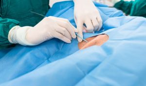
Technique
Step 1: Locate and access vein
To locate the target vein for percutaneous placement of a CVL via the anterior approach, use landmarks or ultrasonographic guidance.
Use landmarks or ultrasound guidance to find target vein for CVL placement.
The IJV is accessed at the apex of the triangle formed by the sternocleidomastoid and clavicle.
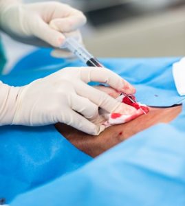
Perform IJV puncture aspiration using a saline-filled syringe and a 20- to 22-gauge needle, with or without ultrasound guidance.
Insert the needle at a 45º angle between the sternocleidomastoid heads towards the nipple.
Blood present but difficult to aspirate indicates the needle may be against the vein wall or not fully in the lumen.
Step 2: Insertion of central venous line with landmark guidance
Insert the guide wire through the needle into the vein, positioning the tip in the upper inferior vena cava without advancing into the retro hepatic vena cava.
Monitor for ectopic beats on ECG to prevent sinoatrial node stimulation then place guide wire tips in IVC for safe manipulation.
Confirm guide wire position, withdraw needle, and use an image intensifier to introduce dilator sheath into the vein correctly.
Remove guide wire and trocar, then aspirate blood from side arm to confirm vein positioning.
Create a 5-mm incision midway between the nipple and humeral head, using an artery clip for 3-4 cm dilatation.
Introduce a tunneller to finalize tunnelling at the neck puncture, then pull the central venous line tip behind the sheath.
Position the line cuff 2-3 cm from the incision entry point. Align with the image intensifier and trim excess from the sheath.
The assistant removes the trocar from the peel-off sheath while the surgeon prepares to insert the line.
Ensure the line at the neck loops smoothly into the vein to prevent future occlusion issues.
Attach line to skin with suture to keep undisturbed for 3 weeks under dressing.
Step 3: Insertion of central venous line under ultrasound guidance
Hold the probe for good skin contact without compressing the veins for imaging.
The line can be inserted low on the neck, with the probe on the clavicle, accessing the widest part of the IJV joining the subclavian vein.
Hold the needle at an acute angle to the ultrasound probe for accurate visualization of its travel direction.
»
Home » Procedure » Central Venous Access via Tunneled Anterior Approach to Internal Jugular Vein
Central Venous Access via Tunneled Anterior Approach to Internal Jugular Vein
Updated :
December 22, 2025
Central venous access is crucial for quality care in intensive therapy patients.
In many situations, Semi-permanent tunneled central line is often preferred.
An anterior approach to the internal jugular vein provides the easiest route and low complication risk.
A tunnelled catheter is surgically inserted into a neck or chest vein under skin. Catheter allows administration of medicines, fluids, and blood sampling tasks.
Subcutaneous catheter placement enhances security, lowers infection rates, and allows catheter port mobility.
Tunnelled catheter placement requires experienced practitioners for safety and efficacy.
The internal jugular vein is preferred for ultrasound guidance due to consistent anatomy, ease of access, and lower complication risks.
Central approach involves direct needle insertion over clavicular head, while posterior approach targets behind sternocleidomastoid for IJV cannulation.
Entry point positioned lower in neck or upper chest creates subcutaneous tunnel to vein.
The anterior approach targets the IJV lower near the clavicle, unlike the traditional central method.
Real-time ultrasound imaging optimizes vein identification and minimizes arterial puncture.
A tunnelling device forms a subcutaneous track from the neck or chest to the vein.
Tunnelled catheters enhance comfort and lower infection risk in chemotherapy patients.
Long-term TPN is essential for patients with intestinal failure or severe gastrointestinal diseases.
Tunnelled dialysis catheters are necessary when other access sites are unsuitable due to stenosis or exhaustion.
Stem Cell Transplantation
Immunocompromised Patients
Poor Peripheral Access
Venous Thrombosis
Chemotherapy
Parenteral Nutrition
Infection at the insertion or tunneling site
Thrombosis or complete occlusion of the internal jugular vein
Anatomic distortion or tumor invasion at the insertion site
Uncorrected pneumothorax on the ipsilateral side
Obesity or difficult neck anatomy
Previous radiation to the neck or chest
Previous central venous catheter placement in the same vein
Active bacteremia/sepsis
Severe respiratory distress
Ultrasound-guided internal jugular vein catheter placement has high success.
The anterior approach with tunnelling presents a challenge, yet success rates stay high in experts.
The anterior tunnelled approach decreases infection risk by placing the exit site lower on the chest.
Tunnelled IJV catheters function for months if maintained. Thrombosis risks in oncology and dialysis patients can be reduced with anticoagulation.
Gauze
Mepore adhesive tape
Scalpel
Vascular forceps
Needle driver
Antiseptic solution with skin swab
Sterile drapes or towels
Central Venous Catheter Tray
Sterile gloves
Polypropylene suture
Patient Preparation:
Imaging neck veins is necessary for long-term venous access patients.
Doppler ultrasonography and magnetic resonance venography help assess venous patency and anatomy for better access planning.
In patients with multiple issues, include CBC, clotting profile, and renal/liver function tests pre-procedure.
2% chlorhexidine in alcohol is superior for skin antisepsis compared to povidone-iodine or 70% alcohol.
Expose the neck from jaw angle superiorly to nipples inferiorly and midaxillary line laterally. Then cover area with sterile drape.
Informed Consent:
Explain the procedure’s risks and potential complications clearly to the patient.
Patient Positioning:
Patients should be positioned in supine positions with mild trendelenburg to engorge vein.
central venous pressure
Puncture internal carotid vein for central venous pressure
Step 1: Locate and access vein
To locate the target vein for percutaneous placement of a CVL via the anterior approach, use landmarks or ultrasonographic guidance.
Use landmarks or ultrasound guidance to find target vein for CVL placement.
The IJV is accessed at the apex of the triangle formed by the sternocleidomastoid and clavicle.
Perform IJV puncture aspiration using a saline-filled syringe and a 20- to 22-gauge needle, with or without ultrasound guidance.
Insert the needle at a 45º angle between the sternocleidomastoid heads towards the nipple.
Blood present but difficult to aspirate indicates the needle may be against the vein wall or not fully in the lumen.
Step 2: Insertion of central venous line with landmark guidance
Insert the guide wire through the needle into the vein, positioning the tip in the upper inferior vena cava without advancing into the retro hepatic vena cava.
Monitor for ectopic beats on ECG to prevent sinoatrial node stimulation then place guide wire tips in IVC for safe manipulation.
Confirm guide wire position, withdraw needle, and use an image intensifier to introduce dilator sheath into the vein correctly.
Remove guide wire and trocar, then aspirate blood from side arm to confirm vein positioning.
Create a 5-mm incision midway between the nipple and humeral head, using an artery clip for 3-4 cm dilatation.
Introduce a tunneller to finalize tunnelling at the neck puncture, then pull the central venous line tip behind the sheath.
Position the line cuff 2-3 cm from the incision entry point. Align with the image intensifier and trim excess from the sheath.
The assistant removes the trocar from the peel-off sheath while the surgeon prepares to insert the line.
Ensure the line at the neck loops smoothly into the vein to prevent future occlusion issues.
Attach line to skin with suture to keep undisturbed for 3 weeks under dressing.
Step 3: Insertion of central venous line under ultrasound guidance
Hold the probe for good skin contact without compressing the veins for imaging.
The line can be inserted low on the neck, with the probe on the clavicle, accessing the widest part of the IJV joining the subclavian vein.
Hold the needle at an acute angle to the ultrasound probe for accurate visualization of its travel direction.
Both our subscription plans include Free CME/CPD AMA PRA Category 1 credits.
Digital Certificate PDF
On course completion, you will receive a full-sized presentation quality digital certificate.
medtigo Simulation
A dynamic medical simulation platform designed to train healthcare professionals and students to effectively run code situations through an immersive hands-on experience in a live, interactive 3D environment.
medtigo Points
medtigo points is our unique point redemption system created to award users for interacting on our site. These points can be redeemed for special discounts on the medtigo marketplace as well as towards the membership cost itself.
Community Forum post/reply = 5 points
*Redemption of points can occur only through the medtigo marketplace, courses, or simulation system. Money will not be credited to your bank account. 10 points = $1.
All Your Certificates in One Place
When you have your licenses, certificates and CMEs in one place, it's easier to track your career growth. You can easily share these with hospitals as well, using your medtigo app.