Background
Congestive Heart Failure (CHF) is a complex clinical syndrome in which the heart fails to efficiently pump blood, resulting in the accumulation of fluid in the lungs and other tissues. Imaging is essential in diagnosing, staging, and monitoring the course of CHF. With imaging studies, clinicians can evaluate heart function, determine underlying causes, quantify the severity of the disease, and monitor treatment response.
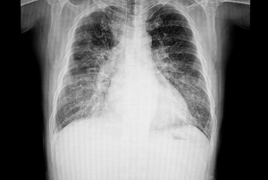
A chest x-ray film of a patient with congestive heart failure
Key imaging modalities for CHF include echocardiography, cardiac MRI, nuclear imaging, and CT scans, each providing unique insights into the heart’s structure and function. Early and accurate imaging is essential for effective management, allowing for tailored therapies and improving patient outcomes. This introduction establishes a general understanding of the value of imaging in the assessment of Congestive Heart Failure with emphasis on the contribution of imaging to enhanced diagnosis and treatment protocol.
Indications
Diagnosis of CHF: Imaging is used to validate the presence of heart failure in the case of clinical signs and symptoms of the condition, to assess structure and function of the heart.
Evaluation of Left and Right Ventricular Function: Echocardiography, cardiac MRI, or CT imaging can measure the ejection fraction (EF), diastolic function, and volume status of both ventricles to establish the severity of CHF.
Evaluation of Cardiac Structures: Imaging assists in evaluating structural abnormalities like valve dysfunction, myocardial infarction, or congenital heart defects, which may be responsible for CHF.
Identification of the Underlying Cause of Heart Failure: Imaging methods such as cardiac MRI and CT scans are employed to identify underlying conditions such as ischemic heart disease, cardiomyopathies (for example, dilated, hypertrophic, or restrictive cardiomyopathies), and pericardial disease.
Assessment of Fluid Status: Chest X-ray and echocardiography are frequently
employed to assess the presence of pulmonary edema, pleural effusion, or other evidence of fluid overload in the lungs and surrounding tissues.
Preoperative Evaluation: In surgical patients, imaging is employed to evaluate the structure and function of the heart to direct surgical planning, especially for heart valve surgery or heart transplantation.
Contraindications
Echocardiography:
Severe Chest Deformities or Trauma: In cases of significant chest wall abnormalities, deformities, or trauma, acoustic windows may be obstructed, making it difficult to obtain quality images.
Patient Non-Cooperation: Patients who are unable to lie still or cooperate with the procedure, such as those with severe agitation or anxiety, may find echocardiography challenging.
Cardiac MRI:
Presence of Metal Implants: Patients with certain metallic implants (e.g., pacemakers, defibrillators, aneurysm clips) are generally contraindicated for cardiac MRI due to the magnetic field potentially affecting the function of the implants or causing injury.
Severe Renal Dysfunction:
Patients with severe kidney impairment should avoid gadolinium contrast (MRI) or iodinated contrast (CT) due to the risk of nephrogenic systemic fibrosis and contrast-induced nephropathy.
Claustrophobia or Inability to Tolerate MRI:
Patients with claustrophobia or difficulty remaining still may struggle with MRI procedures due to the confined space and prolonged duration.
Cardiac CT:
Severe Renal Insufficiency: Risk of contrast-induced nephropathy with iodinated contrast.
Iodine Allergy: Severe allergy to iodine contrast agents prevents CT imaging unless alternatives are available.
Pregnancy: Generally avoided during pregnancy, especially in the first trimester, due to radiation exposure.
Nuclear Imaging (PET, SPECT):
Pregnancy & Breastfeeding: Risk of radiation exposure to the fetus or infant, so nuclear imaging is typically avoided.
Allergy to Radiopharmaceuticals: Known allergies to radiopharmaceuticals may prevent the procedure.
X-ray Imaging (Chest X-ray):
Pregnancy: X-ray imaging, especially in high doses, should be avoided during pregnancy unless the benefits outweigh the risks.
Severe Obesity: Chest X-rays may be less effective in extremely obese patients due to difficulties in obtaining clear images.
Transthoracic Echocardiography (TTE):
No special preparation or fasting required.
Wear a hospital gown to expose the chest area.
Lie on the left side for optimal imaging.
Stay still to avoid motion artifacts.
Transesophageal Echocardiography (TEE):
Fast for 4-6 hours to reduce aspiration risk.
Sedation required for comfort.
Wear a hospital gown.
Post-procedure monitoring until sedation wears off.
Cardiac MRI:
Fast for 4-6 hours if using contrast (gadolinium).
Remove all metal objects.
Wear a hospital gown.
Stay hydrated for better image quality.
Screen for metal implants (e.g., pacemakers).
Inform provider about claustrophobia or kidney issues if contrast is used.
Cardiac CT (CT scan):
Fast for at least 4 hours, especially if contrast is used.
Wear a hospital gown (remove metal clothing).
Stay hydrated unless instructed.
Inform provider about iodine allergies or kidney issues.
Beta-blockers may be used to control heart rate.
Hold breath during the scan for clearer images.
Nuclear Imaging (PET/SPECT):
Fast for 4-6 hours, especially with FDG in PET scans.
Stay hydrated unless instructed.
Inform the provider about current medications.
Wear loose-fitting clothing; may need to change into a gown.
Radiopharmaceutical injection required; notify staff if pregnant or breastfeeding.
Avoid caffeine for 12-24 hours before the scan.
Patient position Transthoracic Echocardiography (TTE):
For optimal heart imaging, the patient is usually positioned on their left side (left lateral decubitus position). This positioning improves the visualization of the heart’s chambers, valves, and blood flow.
Transesophageal Echocardiography (TEE):
During this procedure, the patient is usually placed in a supine position (lying flat on their back).
Cardiac MRI (Magnetic Resonance Imaging):
The patient lies flat on their back on the MRI table, which has a movable section that accommodates imaging of the heart.
Cardiac CT (Computed Tomography):
The patient is positioned in a supine position (lying flat on their back) on the CT scanner table.
Nuclear Imaging (SPECT/PET):
In this imaging method, the patient is positioned supine on the imaging bed, with the camera placed over the chest to capture images of the heart’s function and blood flow.
Echocardiography (Transthoracic and Transesophageal)
Echocardiography is one of the primary tools used to assess heart function and structure in CHF. It uses ultrasound waves to create real-time images of the heart.
Transthoracic Echocardiography (TTE)
Step 1: Patient Preparation
Ensure the patient is in a comfortable position (usually lying on the left side).
Attach ECG leads to monitor the heart’s electrical activity.
Step 2: Gel Application
Apply ultrasound gel to the chest area to improve sound wave conduction.
Step 3: Probe Placement
Place the transducer (ultrasound probe) on the chest in different areas (apical, parasternal, and subcostal views) to capture various angles of the heart.
Step 4: Image Acquisition
The technologist or cardiologist moves the probe to obtain images of the heart’s chambers, valves, and blood flow. Doppler imaging may be used to assess blood flow velocity and pressure.
Step 5: Data Analysis
The images are analyzed to evaluate heart size, function, valve integrity, and any signs of CHF-related abnormalities (e.g., left ventricular dysfunction, mitral regurgitation).
Transesophageal Echocardiography (TEE)
Step 1: Patient Preparation
Instruct the patient to fast for 4-6 hours before the procedure.
Ensure informed consent is obtained and assess any potential contraindications (e.g., esophageal disease).
Sedate the patient to ensure comfort.
Step 2: Insertion of Probe
Insert the transesophageal probe through the patient’s mouth and into the esophagus.
Step 3: Image Acquisition
The probe is positioned close to the heart, allowing high-resolution images of the heart’s posterior structures (e.g., left atrium, atrial septum, mitral valve).
Step 4: Monitoring and Assessment
The images obtained are analyzed for any abnormalities in heart function, valvular abnormalities, or thrombus formation, especially in cases of CHF with atrial fibrillation or clots.
Cardiac MRI (Magnetic Resonance Imaging)
Cardiac MRI provides detailed information about heart structure, function, and tissue characteristics.
Step 1: Patient Preparation
Ensure the patient is in a comfortable, supine position.
Screen for contraindications (e.g., pacemakers, metallic implants).
Remove all metal objects and provide earplugs or headphones to reduce the noise from the MRI machine.
Step 2: Initial Imaging Setup
Position the patient in the MRI scanner, ensuring the heart is positioned in the center within the magnetic field.
Step 3: Imaging Acquisition
Conduct standard imaging sequences (e.g., cine MRI) to evaluate the left ventricular ejection fraction (LVEF), wall motion, myocardial edema, and fibrosis.
If necessary, administer gadolinium contrast agents to assess myocardial perfusion and detect scar tissue.
Step 4: Breath-Holding
Ask the patient to hold their breath briefly during the scan to prevent motion artifacts.
Step 5: Data Analysis
Review the images to assess the heart’s structure, function, and identify any signs of myocardial injury, such as myocardial infarction, fibrosis, or scar tissue, especially in CHF patients.
Cardiac CT (Computed Tomography)
Cardiac CT is used to visualize coronary artery anatomy and assess myocardial function.
Step 1: Patient Preparation
Make sure the patient is well-hydrated and has fasted for 4-6 hours if contrast agents are needed.
Apply ECG leads to synchronize imaging with the heart rhythm of the patient.
Step 2: Positioning
Place the patient supine on the CT table with their arms above their head.
Step 3: Contrast Injection
Inject an intravenous contrast agent to delineate coronary arteries and myocardial perfusion.
Step 4: Image Acquisition
Obtain imaging to evaluate coronary artery disease (CAD), left ventricular function, and coronary perfusion.
Step 5: Data Analysis
Review the CT scans for coronary artery blockage, enlargement of the heart, and any structural defect associated with CHF.
Nuclear Imaging (SPECT/PET)
Step 1: Patient Preparation
Fast for 4-6 hours before the scan, especially for PET scans using fluorodeoxyglucose (FDG).
Avoid caffeine or other stimulants 12-24 hours before the procedure.
Step 2: Radiopharmaceutical Injection
Inject a radiotracer (e.g., technetium-99m, FDG) intravenously to assess myocardial perfusion and metabolism.
Step 3: Image Acquisition
The patient lies supine under the gamma camera. For SPECT, images are taken as the camera rotates around the body. For PET, a specialized scanner detects emitted positrons.
Step 4: Breath-Holding
Instruct the patient to hold their breath briefly during image acquisition to minimize motion artifacts.
Step 5: Data Analysis
Analyze the images to assess myocardial perfusion, ischemia, infarction, and cardiac function in CHF patients.
Chest X-ray
Chest X-ray is a quick and non-invasive imaging tool used to evaluate heart size and detect pulmonary congestion in CHF.
Step 1: Positioning
The patient stands upright for the X-ray. A posteroanterior (PA) view and lateral view are typically obtained.
Step 2: Image Acquisition
The radiographer will capture the images while instructing the patient to take a deep breath and hold it.
Step 3: Data Analysis
The X-ray images are analyzed for signs of heart enlargement (cardiomegaly), pulmonary congestion, pleural effusion, and other CHF-related changes.
Cardiac Catheterization
Cardiac catheterization is used to assess coronary artery disease, heart pressures, and ventricular function, often during an invasive procedure.
Step 1: Patient Preparation
Ensure fasting for 6-8 hours prior to the procedure.
Administer local anesthesia at the insertion site (e.g., femoral or radial artery).
Step 2: Insertion of Catheter
Insert a catheter into the artery and advance it toward the heart. Contrast agents may be injected to visualize coronary arteries.
Step 3: Imaging
Perform angiography to evaluate coronary artery obstruction and left ventricular function. Intracardiac pressures may be measured.
Step 4: Data Analysis
Interpret the coronary angiogram to determine blockages, and measure ejection fraction and other hemodynamic parameters.
Complications
Esophageal Injury: There is a minimal risk of esophageal or throat injury while the probe is inserted. This may cause pain, bleeding, or perforation (extremely rare).
Sedation-related Complications: Since TEE is usually performed under sedation, there are risks associated with the sedative, such as allergic reactions, respiratory depression, or aspiration.
Gagging and Discomfort: The patient might feel discomfort or experience gagging when the probe is inserted into the esophagus during the procedure.
Contrast Reactions: Gadolinium contrast agents, commonly used in MRI scans, can sometimes cause allergic reactions, such as itching or a rash, and in rare cases, more serious reactions like anaphylaxis.
Breathing Difficulty: Some patients may have difficulty holding their breath during certain sequences, leading to motion artifacts or the need to repeat images.
Arrhythmias: In some cases, rapid heart rate induced by contrast injection can lead to arrhythmias.
Radiation Exposure: Both SPECT and PET scans involve the use of radioactive tracers. While the radiation dose is generally low and considered safe, cumulative exposure over time can pose a risk.
»
Home » Procedure » Congestive Heart Failure Imaging
Congestive Heart Failure Imaging
Updated :
December 18, 2025
Background
Congestive Heart Failure (CHF) is a complex clinical syndrome in which the heart fails to efficiently pump blood, resulting in the accumulation of fluid in the lungs and other tissues. Imaging is essential in diagnosing, staging, and monitoring the course of CHF. With imaging studies, clinicians can evaluate heart function, determine underlying causes, quantify the severity of the disease, and monitor treatment response.
A chest x-ray film of a patient with congestive heart failure
Key imaging modalities for CHF include echocardiography, cardiac MRI, nuclear imaging, and CT scans, each providing unique insights into the heart’s structure and function. Early and accurate imaging is essential for effective management, allowing for tailored therapies and improving patient outcomes. This introduction establishes a general understanding of the value of imaging in the assessment of Congestive Heart Failure with emphasis on the contribution of imaging to enhanced diagnosis and treatment protocol.
Diagnosis of CHF: Imaging is used to validate the presence of heart failure in the case of clinical signs and symptoms of the condition, to assess structure and function of the heart.
Evaluation of Left and Right Ventricular Function: Echocardiography, cardiac MRI, or CT imaging can measure the ejection fraction (EF), diastolic function, and volume status of both ventricles to establish the severity of CHF.
Evaluation of Cardiac Structures: Imaging assists in evaluating structural abnormalities like valve dysfunction, myocardial infarction, or congenital heart defects, which may be responsible for CHF.
Identification of the Underlying Cause of Heart Failure: Imaging methods such as cardiac MRI and CT scans are employed to identify underlying conditions such as ischemic heart disease, cardiomyopathies (for example, dilated, hypertrophic, or restrictive cardiomyopathies), and pericardial disease.
Assessment of Fluid Status: Chest X-ray and echocardiography are frequently
employed to assess the presence of pulmonary edema, pleural effusion, or other evidence of fluid overload in the lungs and surrounding tissues.
Preoperative Evaluation: In surgical patients, imaging is employed to evaluate the structure and function of the heart to direct surgical planning, especially for heart valve surgery or heart transplantation.
Echocardiography:
Severe Chest Deformities or Trauma: In cases of significant chest wall abnormalities, deformities, or trauma, acoustic windows may be obstructed, making it difficult to obtain quality images.
Patient Non-Cooperation: Patients who are unable to lie still or cooperate with the procedure, such as those with severe agitation or anxiety, may find echocardiography challenging.
Cardiac MRI:
Presence of Metal Implants: Patients with certain metallic implants (e.g., pacemakers, defibrillators, aneurysm clips) are generally contraindicated for cardiac MRI due to the magnetic field potentially affecting the function of the implants or causing injury.
Severe Renal Dysfunction:
Patients with severe kidney impairment should avoid gadolinium contrast (MRI) or iodinated contrast (CT) due to the risk of nephrogenic systemic fibrosis and contrast-induced nephropathy.
Claustrophobia or Inability to Tolerate MRI:
Patients with claustrophobia or difficulty remaining still may struggle with MRI procedures due to the confined space and prolonged duration.
Cardiac CT:
Severe Renal Insufficiency: Risk of contrast-induced nephropathy with iodinated contrast.
Iodine Allergy: Severe allergy to iodine contrast agents prevents CT imaging unless alternatives are available.
Pregnancy: Generally avoided during pregnancy, especially in the first trimester, due to radiation exposure.
Nuclear Imaging (PET, SPECT):
Pregnancy & Breastfeeding: Risk of radiation exposure to the fetus or infant, so nuclear imaging is typically avoided.
Allergy to Radiopharmaceuticals: Known allergies to radiopharmaceuticals may prevent the procedure.
X-ray Imaging (Chest X-ray):
Pregnancy: X-ray imaging, especially in high doses, should be avoided during pregnancy unless the benefits outweigh the risks.
Severe Obesity: Chest X-rays may be less effective in extremely obese patients due to difficulties in obtaining clear images.
Transthoracic Echocardiography (TTE):
No special preparation or fasting required.
Wear a hospital gown to expose the chest area.
Lie on the left side for optimal imaging.
Stay still to avoid motion artifacts.
Transesophageal Echocardiography (TEE):
Fast for 4-6 hours to reduce aspiration risk.
Sedation required for comfort.
Wear a hospital gown.
Post-procedure monitoring until sedation wears off.
Cardiac MRI:
Fast for 4-6 hours if using contrast (gadolinium).
Remove all metal objects.
Wear a hospital gown.
Stay hydrated for better image quality.
Screen for metal implants (e.g., pacemakers).
Inform provider about claustrophobia or kidney issues if contrast is used.
Cardiac CT (CT scan):
Fast for at least 4 hours, especially if contrast is used.
Wear a hospital gown (remove metal clothing).
Stay hydrated unless instructed.
Inform provider about iodine allergies or kidney issues.
Beta-blockers may be used to control heart rate.
Hold breath during the scan for clearer images.
Nuclear Imaging (PET/SPECT):
Fast for 4-6 hours, especially with FDG in PET scans.
Stay hydrated unless instructed.
Inform the provider about current medications.
Wear loose-fitting clothing; may need to change into a gown.
Radiopharmaceutical injection required; notify staff if pregnant or breastfeeding.
Avoid caffeine for 12-24 hours before the scan.
Patient position Transthoracic Echocardiography (TTE):
For optimal heart imaging, the patient is usually positioned on their left side (left lateral decubitus position). This positioning improves the visualization of the heart’s chambers, valves, and blood flow.
Transesophageal Echocardiography (TEE):
During this procedure, the patient is usually placed in a supine position (lying flat on their back).
Cardiac MRI (Magnetic Resonance Imaging):
The patient lies flat on their back on the MRI table, which has a movable section that accommodates imaging of the heart.
Cardiac CT (Computed Tomography):
The patient is positioned in a supine position (lying flat on their back) on the CT scanner table.
Nuclear Imaging (SPECT/PET):
In this imaging method, the patient is positioned supine on the imaging bed, with the camera placed over the chest to capture images of the heart’s function and blood flow.
Echocardiography is one of the primary tools used to assess heart function and structure in CHF. It uses ultrasound waves to create real-time images of the heart.
Transthoracic Echocardiography (TTE)
Step 1: Patient Preparation
Ensure the patient is in a comfortable position (usually lying on the left side).
Attach ECG leads to monitor the heart’s electrical activity.
Step 2: Gel Application
Apply ultrasound gel to the chest area to improve sound wave conduction.
Step 3: Probe Placement
Place the transducer (ultrasound probe) on the chest in different areas (apical, parasternal, and subcostal views) to capture various angles of the heart.
Step 4: Image Acquisition
The technologist or cardiologist moves the probe to obtain images of the heart’s chambers, valves, and blood flow. Doppler imaging may be used to assess blood flow velocity and pressure.
Step 5: Data Analysis
The images are analyzed to evaluate heart size, function, valve integrity, and any signs of CHF-related abnormalities (e.g., left ventricular dysfunction, mitral regurgitation).
Step 1: Patient Preparation
Instruct the patient to fast for 4-6 hours before the procedure.
Ensure informed consent is obtained and assess any potential contraindications (e.g., esophageal disease).
Sedate the patient to ensure comfort.
Step 2: Insertion of Probe
Insert the transesophageal probe through the patient’s mouth and into the esophagus.
Step 3: Image Acquisition
The probe is positioned close to the heart, allowing high-resolution images of the heart’s posterior structures (e.g., left atrium, atrial septum, mitral valve).
Step 4: Monitoring and Assessment
The images obtained are analyzed for any abnormalities in heart function, valvular abnormalities, or thrombus formation, especially in cases of CHF with atrial fibrillation or clots.
Cardiac MRI provides detailed information about heart structure, function, and tissue characteristics.
Step 1: Patient Preparation
Ensure the patient is in a comfortable, supine position.
Screen for contraindications (e.g., pacemakers, metallic implants).
Remove all metal objects and provide earplugs or headphones to reduce the noise from the MRI machine.
Step 2: Initial Imaging Setup
Position the patient in the MRI scanner, ensuring the heart is positioned in the center within the magnetic field.
Step 3: Imaging Acquisition
Conduct standard imaging sequences (e.g., cine MRI) to evaluate the left ventricular ejection fraction (LVEF), wall motion, myocardial edema, and fibrosis.
If necessary, administer gadolinium contrast agents to assess myocardial perfusion and detect scar tissue.
Step 4: Breath-Holding
Ask the patient to hold their breath briefly during the scan to prevent motion artifacts.
Step 5: Data Analysis
Review the images to assess the heart’s structure, function, and identify any signs of myocardial injury, such as myocardial infarction, fibrosis, or scar tissue, especially in CHF patients.
Cardiac CT is used to visualize coronary artery anatomy and assess myocardial function.
Step 1: Patient Preparation
Make sure the patient is well-hydrated and has fasted for 4-6 hours if contrast agents are needed.
Apply ECG leads to synchronize imaging with the heart rhythm of the patient.
Step 2: Positioning
Place the patient supine on the CT table with their arms above their head.
Step 3: Contrast Injection
Inject an intravenous contrast agent to delineate coronary arteries and myocardial perfusion.
Step 4: Image Acquisition
Obtain imaging to evaluate coronary artery disease (CAD), left ventricular function, and coronary perfusion.
Step 5: Data Analysis
Review the CT scans for coronary artery blockage, enlargement of the heart, and any structural defect associated with CHF.
Step 1: Patient Preparation
Fast for 4-6 hours before the scan, especially for PET scans using fluorodeoxyglucose (FDG).
Avoid caffeine or other stimulants 12-24 hours before the procedure.
Step 2: Radiopharmaceutical Injection
Inject a radiotracer (e.g., technetium-99m, FDG) intravenously to assess myocardial perfusion and metabolism.
Step 3: Image Acquisition
The patient lies supine under the gamma camera. For SPECT, images are taken as the camera rotates around the body. For PET, a specialized scanner detects emitted positrons.
Step 4: Breath-Holding
Instruct the patient to hold their breath briefly during image acquisition to minimize motion artifacts.
Step 5: Data Analysis
Analyze the images to assess myocardial perfusion, ischemia, infarction, and cardiac function in CHF patients.
Chest X-ray is a quick and non-invasive imaging tool used to evaluate heart size and detect pulmonary congestion in CHF.
Step 1: Positioning
The patient stands upright for the X-ray. A posteroanterior (PA) view and lateral view are typically obtained.
Step 2: Image Acquisition
The radiographer will capture the images while instructing the patient to take a deep breath and hold it.
Step 3: Data Analysis
The X-ray images are analyzed for signs of heart enlargement (cardiomegaly), pulmonary congestion, pleural effusion, and other CHF-related changes.
Cardiac Catheterization
Cardiac catheterization is used to assess coronary artery disease, heart pressures, and ventricular function, often during an invasive procedure.
Step 1: Patient Preparation
Ensure fasting for 6-8 hours prior to the procedure.
Administer local anesthesia at the insertion site (e.g., femoral or radial artery).
Step 2: Insertion of Catheter
Insert a catheter into the artery and advance it toward the heart. Contrast agents may be injected to visualize coronary arteries.
Step 3: Imaging
Perform angiography to evaluate coronary artery obstruction and left ventricular function. Intracardiac pressures may be measured.
Step 4: Data Analysis
Interpret the coronary angiogram to determine blockages, and measure ejection fraction and other hemodynamic parameters.
Complications
Esophageal Injury: There is a minimal risk of esophageal or throat injury while the probe is inserted. This may cause pain, bleeding, or perforation (extremely rare).
Sedation-related Complications: Since TEE is usually performed under sedation, there are risks associated with the sedative, such as allergic reactions, respiratory depression, or aspiration.
Gagging and Discomfort: The patient might feel discomfort or experience gagging when the probe is inserted into the esophagus during the procedure.
Contrast Reactions: Gadolinium contrast agents, commonly used in MRI scans, can sometimes cause allergic reactions, such as itching or a rash, and in rare cases, more serious reactions like anaphylaxis.
Breathing Difficulty: Some patients may have difficulty holding their breath during certain sequences, leading to motion artifacts or the need to repeat images.
Arrhythmias: In some cases, rapid heart rate induced by contrast injection can lead to arrhythmias.
Radiation Exposure: Both SPECT and PET scans involve the use of radioactive tracers. While the radiation dose is generally low and considered safe, cumulative exposure over time can pose a risk.
Both our subscription plans include Free CME/CPD AMA PRA Category 1 credits.
Digital Certificate PDF
On course completion, you will receive a full-sized presentation quality digital certificate.
medtigo Simulation
A dynamic medical simulation platform designed to train healthcare professionals and students to effectively run code situations through an immersive hands-on experience in a live, interactive 3D environment.
medtigo Points
medtigo points is our unique point redemption system created to award users for interacting on our site. These points can be redeemed for special discounts on the medtigo marketplace as well as towards the membership cost itself.
Community Forum post/reply = 5 points
*Redemption of points can occur only through the medtigo marketplace, courses, or simulation system. Money will not be credited to your bank account. 10 points = $1.
All Your Certificates in One Place
When you have your licenses, certificates and CMEs in one place, it's easier to track your career growth. You can easily share these with hospitals as well, using your medtigo app.