Background
Coronary CTA images arteries noninvasively without discomfort.
It is used to assess coronary artery disease and detect vascular abnormalities. It has gained prominence for high sensitivity, specificity, and detailed anatomical visualization of coronary vasculature.
Multi-detector CT enhances image resolution and reduces motion artifacts.
CCTA uses contrast-enhanced CT to visualize coronary arteries.
Advantages as follows:
Non-invasive and relatively quick
High negative predictive value
To provide anatomical and functional assessment with perfusion imaging techniques
Multi-detector CT enhances image quality and minimizes motion artifacts.
Advancements in dual-source CT, AI, and reconstruction enhance image quality and diagnostic accuracy.
Modern CT scanners minimize motion artifacts by capturing images quickly. ECG gating synchronizes imaging with cardiac cycle for accuracy.
Post-processing enhances 3D images of coronary vasculature for plaque assessment.
Indications
Assessment of coronary artery disease
Evaluation of chest pain
Preoperative planning
Follow-up of coronary anomalies
Pre-Procedural Planning and Post-Intervention Assessment
Assessment of Cardiac Tumors & Masses
Pericardial Disease
Pulmonary Vein Mapping
Contraindications
Severe Contrast Allergy
Severe Renal Dysfunction
Dialysis patients
Pregnancy
Hemodynamic Instability
High Coronary Artery Calcification
Obesity or Large Body Habitus
Hyperthyroidism
Outcomes
CCTA detects significant coronary artery stenosis with high sensitivity.
Its negative predictive value makes it excellent for ruling out CAD effectively.
Non-obstructive atherosclerosis patients benefit from interventions.
FFR-CT can determine whether a stenosis is hemodynamically significant to reduce unnecessary invasive procedures.
Normal CCTA patients have excellent prognosis, with <1% risk of MI or cardiac death.
Identifies plaque traits linked to acute coronary syndrome.
Equipment required
CT Scanner & Imaging Equipment
Contrast Injection System
ECG Electrodes & Leads
Oxygen & Emergency Kit
Radiation Dose Optimization Tools
Image Processing & Post-Processing Software
Patient Preparation:
Pre-Scan Assessment before scheduling CCTA, assess the patient for indications, contraindications, heart rate, rhythm evaluation, and contrast allergy assessment.
Minimum 4 hours fasting before the scan to reduce motion artifacts from the diaphragm.
Caffeine or smoking should be avoided for 12 to 24 hours before the scan.
During the Scan includes IV Access for contrast injection, ECG monitoring, and breathing instructions
Informed Consent:
Explain the procedure’s risks and potential complications clearly to the patient.
Patient Positioning:
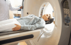
Patient lies flat on back on CT scanner in supine position. Arms above head reduce artifacts and ensure clear field view.

Coronary CT Angiography
Technique
Step 1: Contrast administration and timing:
Iodinated contrast is injected at 4 to 6 mL/sec using a power injector.
Bolus tracking initiates peak contrast scanning. Saline flush follows contrast to reduce streak artifacts in the superior vena cava.
Biphasic injection protocols prevent streak artifacts from high contrast concentrations in the right heart, using contrast followed by saline. For right heart structure imaging, triphasic protocols with sequential injections are used.
Step 2: Image Acquisition
The initial CCTA image acquisition involves obtaining scout images as low-energy scans using 120 kV tube voltage and 35 mAs.
The ‘test-bolus’ method times contrast enhancement in the ascending aorta at the carina to estimate arrival at coronary arteries.
A CT scanner with 320 detectors enhances cardiac imaging in one rotation, surpassing 64 or 128 detector systems’ capabilities.
ECG-triggered acquisition minimizes exposure by aligning X-ray activation effectively.
Retrospective ECG Gating:
Captures data throughout the cardiac cycle. It shows higher radiation allows functional assessment.
Prospective ECG Gating:
Images acquired only in mid-diastole. Lower radiation exposure but not suitable for irregular heart rhythms.
Complications:
Allergic Reactions to Iodinated Contrast
Contrast-Induced Nephropathy
Contrast Extravasation
Cumulative Radiation Exposure
Arrhythmias
Hypotension from Nitroglycerin

Coronary CTA images arteries noninvasively without discomfort.
It is used to assess coronary artery disease and detect vascular abnormalities. It has gained prominence for high sensitivity, specificity, and detailed anatomical visualization of coronary vasculature.
Multi-detector CT enhances image resolution and reduces motion artifacts.
CCTA uses contrast-enhanced CT to visualize coronary arteries.
Advantages as follows:
Non-invasive and relatively quick
High negative predictive value
To provide anatomical and functional assessment with perfusion imaging techniques
Multi-detector CT enhances image quality and minimizes motion artifacts.
Advancements in dual-source CT, AI, and reconstruction enhance image quality and diagnostic accuracy.
Modern CT scanners minimize motion artifacts by capturing images quickly. ECG gating synchronizes imaging with cardiac cycle for accuracy.
Post-processing enhances 3D images of coronary vasculature for plaque assessment.
Assessment of coronary artery disease
Evaluation of chest pain
Preoperative planning
Follow-up of coronary anomalies
Pre-Procedural Planning and Post-Intervention Assessment
Assessment of Cardiac Tumors & Masses
Pericardial Disease
Pulmonary Vein Mapping
Severe Contrast Allergy
Severe Renal Dysfunction
Dialysis patients
Pregnancy
Hemodynamic Instability
High Coronary Artery Calcification
Obesity or Large Body Habitus
Hyperthyroidism
CCTA detects significant coronary artery stenosis with high sensitivity.
Its negative predictive value makes it excellent for ruling out CAD effectively.
Non-obstructive atherosclerosis patients benefit from interventions.
FFR-CT can determine whether a stenosis is hemodynamically significant to reduce unnecessary invasive procedures.
Normal CCTA patients have excellent prognosis, with <1% risk of MI or cardiac death.
Identifies plaque traits linked to acute coronary syndrome.
CT Scanner & Imaging Equipment
Contrast Injection System
ECG Electrodes & Leads
Oxygen & Emergency Kit
Radiation Dose Optimization Tools
Image Processing & Post-Processing Software
Patient Preparation:
Pre-Scan Assessment before scheduling CCTA, assess the patient for indications, contraindications, heart rate, rhythm evaluation, and contrast allergy assessment.
Minimum 4 hours fasting before the scan to reduce motion artifacts from the diaphragm.
Caffeine or smoking should be avoided for 12 to 24 hours before the scan.
During the Scan includes IV Access for contrast injection, ECG monitoring, and breathing instructions
Informed Consent:
Explain the procedure’s risks and potential complications clearly to the patient.
Patient Positioning:
Patient lies flat on back on CT scanner in supine position. Arms above head reduce artifacts and ensure clear field view.

Coronary CT Angiography
Step 1: Contrast administration and timing:
Iodinated contrast is injected at 4 to 6 mL/sec using a power injector.
Bolus tracking initiates peak contrast scanning. Saline flush follows contrast to reduce streak artifacts in the superior vena cava.
Biphasic injection protocols prevent streak artifacts from high contrast concentrations in the right heart, using contrast followed by saline. For right heart structure imaging, triphasic protocols with sequential injections are used.
Step 2: Image Acquisition
The initial CCTA image acquisition involves obtaining scout images as low-energy scans using 120 kV tube voltage and 35 mAs.
The ‘test-bolus’ method times contrast enhancement in the ascending aorta at the carina to estimate arrival at coronary arteries.
A CT scanner with 320 detectors enhances cardiac imaging in one rotation, surpassing 64 or 128 detector systems’ capabilities.
ECG-triggered acquisition minimizes exposure by aligning X-ray activation effectively.
Retrospective ECG Gating:
Captures data throughout the cardiac cycle. It shows higher radiation allows functional assessment.
Prospective ECG Gating:
Images acquired only in mid-diastole. Lower radiation exposure but not suitable for irregular heart rhythms.
Complications:
Allergic Reactions to Iodinated Contrast
Contrast-Induced Nephropathy
Contrast Extravasation
Cumulative Radiation Exposure
Arrhythmias
Hypotension from Nitroglycerin

Both our subscription plans include Free CME/CPD AMA PRA Category 1 credits.

On course completion, you will receive a full-sized presentation quality digital certificate.
A dynamic medical simulation platform designed to train healthcare professionals and students to effectively run code situations through an immersive hands-on experience in a live, interactive 3D environment.

When you have your licenses, certificates and CMEs in one place, it's easier to track your career growth. You can easily share these with hospitals as well, using your medtigo app.


