Cricopharyngeal myotomy surgery is performed as a treatment option for patients with cricopharyngeal dysfunction (CPD) when dysphagia symptoms appear. As a portion of the upper esophageal sphincter (UES) the cricopharyngeal muscle functions to stop windpipe entry of food or liquid while forwarding food to the esophagus. The cricopharyngeal myotomy procedure weakens this muscle to create a better flow of food and liquids when swallowing occurs. The treatment procedure enables better swallowing ability and reduces dysphagia symptoms.
Indications
Chronic Dysphagia:
The patient has swallowing issues that resist treatment through swallowing therapy and dietary modifications as well as medication therapy.
The patient develops a sensation of food stuck in their throat or chest area because the cricopharyngeal muscle fails to relax correctly during swallowing.
Cricopharyngeal Spasm:
Cricopharyngeal muscle spasms or abnormal contractions that persist result in hindering swallowing ability and lead to swallowing difficulties.
Healthcare providers diagnose this condition through two tests which include manometry that measures esophageal pressure or barium swallow tests that evaluate swallowing function.
Zenker’s Diverticulum:
Zenker’s diverticulum is a pouch that can form in the upper esophagus due to pressure from the cricopharyngeal muscle. This is often associated with CPD. Cricopharyngeal myotomy can be performed in conjunction with diverticulum removal to address both the structural and functional issues.
Patients with Zenker’s diverticulum may have symptoms like regurgitation of food, aspiration, and a chronic cough.
Failed Conservative Treatments:
Patients who have undergone botulinum toxin (Botox) injections into the cricopharyngeal muscle or who have previously tried dilations to relieve muscle tension but have not achieved lasting relief from symptoms.
Cricopharyngeal myotomy can be considered after other less invasive options have failed.
Contraindications
Non-CPD Related Dysphagia:
If the dysphagia is not caused by cricopharyngeal dysfunction, i.e., structural disease of the esophagus, tumors, or neurological disease (e.g., stroke or esophageal carcinoma), cricopharyngeal myotomy would not be helpful.
Severe Esophageal Motility Disorders:
Achalasia or other severe esophageal motility disorders (in which there is an impairment of the entire esophagus, not the cricopharyngeal muscle) would also be inappropriate for cricopharyngeal myotomy. Such disorders would need alternative treatments such as pneumatic dilation or surgical myotomy of the lower esophageal sphincter (for achalasia).
Active Infections or Inflammation:
If the patient has an active infection or severe inflammation in the throat, esophagus, or surrounding areas, the surgery may be delayed until the infection resolves. Performing surgery during an active infection could increase the risk of complications like abscesses or delayed healing.
Uncontrolled Medical Conditions:
Severe uncontrolled diseases such as heart disease, diabetes, or other systemic disease may compromise the healing or tolerance capacity of the body for the surgery. The risks in such situations could potentially outweigh the benefits of surgery.
Significant Anatomical Abnormalities:
If there are significant anatomical abnormalities of the upper esophagus or cricopharyngeal area (e.g., large tumors, or extreme scarring from previous surgeries or radiation), it may be difficult to perform a myotomy safely.
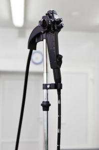
Endoscope
Video Monitor
Endoscopic Surgical Instruments
Electrocautery or Laser
Graspers/Forceps
Sutures or Staplers
Surgical Instruments for Open Surgery (if applicable):
Scalpel or Surgical Blades
Scissors
Tissue Forceps
Hemostats
Surgical Retractors
Anesthesia Equipment:
Endotracheal Tube (ET Tube)
Monitoring Devices
Sterile Drapes
Surgical Gloves
Barium Swallow or Fluoroscopy Equipment
Patient Preparation Preoperative Evaluation:
Medical History: The surgeon will review the patient’s medical history, including any existing medical conditions (such as heart disease, diabetes, or neurological conditions), previous surgeries, and allergies (especially to anesthesia).
Physical Examination: A thorough physical exam will be performed to assess overall health and identify any potential issues that may complicate surgery.
Swallowing Assessment: A speech therapist or a swallowing specialist might conduct a swallowing evaluation to confirm the diagnosis of cricopharyngeal dysfunction and determine the severity of dysphagia.
Diagnostic Tests: Barium Swallow or Fluoroscopy: This imaging test is often performed to confirm the diagnosis of cricopharyngeal dysfunction or any associated conditions (such as a Zenker’s diverticulum). It allows the surgeon to visualize the swallowing process and assess muscle function.
Manometry: An esophageal motility study might be done to assess the pressure and relaxation function of the cricopharyngeal muscle.
Preoperative Instructions:
Fasting: The patient will likely be instructed to fast for at least 6-8 hours before the surgery (usually no food or drink, including water). This is essential for reducing the risk of aspiration during anesthesia and to ensure a safe procedure.
Medications:
Some medications, especially blood thinners (like aspirin, warfarin, or clopidogrel), might need to be temporarily stopped before the surgery to reduce the risk of excessive bleeding.
Patients should discuss their regular medications (including those for chronic conditions) with their surgeon, as some may need to be adjusted.
Patient Positioning
The patient is typically placed in a supine position (lying flat on their back) on the operating table.
The patient’s head is usually extended slightly and tilted back to expose the upper esophagus and cricopharyngeal muscle.
The surgeon may use a headrest or shoulder roll to achieve this position, which provides better access to the throat and esophagus.
Technique
Step 1-Preoperative Preparation:
The patient is placed in the supine position with a slight neck extension.
The oral cavity is disinfected, and a mouth gag or oral retractor is used to keep the mouth open.
General anesthesia is administered, and the airway is secured with an endotracheal tube.
Step 2-Endoscopic Insertion:
A rigid or flexible endoscope is introduced through the mouth and passed into the hypopharynx, where the cricopharyngeal muscle is located.
The camera on the endoscope allows visualization of the cricopharyngeal region, including the upper esophagus, cricoid cartilage, and surrounding structures.
Step 3-Identification of the Cricopharyngeal Muscle:
The surgeon identifies the cricopharyngeal muscle (CPM) by visualizing the area just above the esophageal inlet (typically around the cricoid cartilage).
It may be helpful to use fluoroscopy or a barium swallow study to confirm the exact location of the muscle and determine the degree of dysfunction.
Step 4-Myotomy of the Cricopharyngeal Muscle:
The surgeon uses an electrocautery or laser device passed through the endoscope to carefully cut or divide the cricopharyngeal muscle.
The muscle is typically divided for a length of about 2-3 cm, extending from the cricoid cartilage towards the upper esophagus.
Care is taken to avoid damaging nearby structures such as the vocal cords or recurrent laryngeal nerve.
Step 5-Verification and Hemostasis:
The surgeon verifies that the muscle is sufficiently divided and checks for any signs of bleeding.
Hemostasis (stopping of bleeding) is achieved using electrocautery or other hemostatic techniques if necessary.
Step 6-Closure and Postoperative Care:
Once the muscle division is complete and bleeding controlled, the endoscope is withdrawn.
The patient is monitored postoperatively for vital signs, swallowing ability, and any signs of complications like aspiration or infection.
Step 7-Postoperative Monitoring:
After surgery, the patient is usually given a liquid or soft diet for the first few days, and swallowing therapy may be recommended to improve recovery and function.
4. Complications
Infection: As with any surgical procedure, there’s a risk of infection at the incision site (especially with open surgery) or within the deeper tissues of the neck or throat.
Bleeding: There could be bleeding during or after the surgery due to damage to small blood vessels in the area around the cricopharyngeal muscle.
Vocal Cord Paralysis: Damage to the recurrent laryngeal nerve can cause hoarseness or vocal cord paralysis, affecting speech and swallowing.
Esophageal Perforation: Accidental injury to the esophagus can lead to perforation (a hole), which is a severe complication requiring immediate intervention.
Airway Injury: The procedure involves working in the throat, so there is a small risk of damaging the airway structures, leading to difficulty breathing or other airway complications.
Persistent or New Symptoms of Dysphagia: In some cases, the surgery may not fully alleviate swallowing difficulties, or it could lead to new difficulties, such as regurgitation or difficulty swallowing solids or liquids.
Aspiration Pneumonia: There is a risk of food or liquid entering the lungs (aspiration) during or after surgery, leading to aspiration pneumonia, a potentially serious condition. This risk is particularly high in patients with preexisting swallowing difficulties.
Cricopharyngeal myotomy surgery is performed as a treatment option for patients with cricopharyngeal dysfunction (CPD) when dysphagia symptoms appear. As a portion of the upper esophageal sphincter (UES) the cricopharyngeal muscle functions to stop windpipe entry of food or liquid while forwarding food to the esophagus. The cricopharyngeal myotomy procedure weakens this muscle to create a better flow of food and liquids when swallowing occurs. The treatment procedure enables better swallowing ability and reduces dysphagia symptoms.
Chronic Dysphagia:
The patient has swallowing issues that resist treatment through swallowing therapy and dietary modifications as well as medication therapy.
The patient develops a sensation of food stuck in their throat or chest area because the cricopharyngeal muscle fails to relax correctly during swallowing.
Cricopharyngeal Spasm:
Cricopharyngeal muscle spasms or abnormal contractions that persist result in hindering swallowing ability and lead to swallowing difficulties.
Healthcare providers diagnose this condition through two tests which include manometry that measures esophageal pressure or barium swallow tests that evaluate swallowing function.
Zenker’s Diverticulum:
Zenker’s diverticulum is a pouch that can form in the upper esophagus due to pressure from the cricopharyngeal muscle. This is often associated with CPD. Cricopharyngeal myotomy can be performed in conjunction with diverticulum removal to address both the structural and functional issues.
Patients with Zenker’s diverticulum may have symptoms like regurgitation of food, aspiration, and a chronic cough.
Failed Conservative Treatments:
Patients who have undergone botulinum toxin (Botox) injections into the cricopharyngeal muscle or who have previously tried dilations to relieve muscle tension but have not achieved lasting relief from symptoms.
Cricopharyngeal myotomy can be considered after other less invasive options have failed.
Non-CPD Related Dysphagia:
If the dysphagia is not caused by cricopharyngeal dysfunction, i.e., structural disease of the esophagus, tumors, or neurological disease (e.g., stroke or esophageal carcinoma), cricopharyngeal myotomy would not be helpful.
Severe Esophageal Motility Disorders:
Achalasia or other severe esophageal motility disorders (in which there is an impairment of the entire esophagus, not the cricopharyngeal muscle) would also be inappropriate for cricopharyngeal myotomy. Such disorders would need alternative treatments such as pneumatic dilation or surgical myotomy of the lower esophageal sphincter (for achalasia).
Active Infections or Inflammation:
If the patient has an active infection or severe inflammation in the throat, esophagus, or surrounding areas, the surgery may be delayed until the infection resolves. Performing surgery during an active infection could increase the risk of complications like abscesses or delayed healing.
Uncontrolled Medical Conditions:
Severe uncontrolled diseases such as heart disease, diabetes, or other systemic disease may compromise the healing or tolerance capacity of the body for the surgery. The risks in such situations could potentially outweigh the benefits of surgery.
Significant Anatomical Abnormalities:
If there are significant anatomical abnormalities of the upper esophagus or cricopharyngeal area (e.g., large tumors, or extreme scarring from previous surgeries or radiation), it may be difficult to perform a myotomy safely.
Endoscope
Video Monitor
Endoscopic Surgical Instruments
Electrocautery or Laser
Graspers/Forceps
Sutures or Staplers
Surgical Instruments for Open Surgery (if applicable):
Scalpel or Surgical Blades
Scissors
Tissue Forceps
Hemostats
Surgical Retractors
Anesthesia Equipment:
Endotracheal Tube (ET Tube)
Monitoring Devices
Sterile Drapes
Surgical Gloves
Barium Swallow or Fluoroscopy Equipment
Patient Preparation Preoperative Evaluation:
Medical History: The surgeon will review the patient’s medical history, including any existing medical conditions (such as heart disease, diabetes, or neurological conditions), previous surgeries, and allergies (especially to anesthesia).
Physical Examination: A thorough physical exam will be performed to assess overall health and identify any potential issues that may complicate surgery.
Swallowing Assessment: A speech therapist or a swallowing specialist might conduct a swallowing evaluation to confirm the diagnosis of cricopharyngeal dysfunction and determine the severity of dysphagia.
Diagnostic Tests: Barium Swallow or Fluoroscopy: This imaging test is often performed to confirm the diagnosis of cricopharyngeal dysfunction or any associated conditions (such as a Zenker’s diverticulum). It allows the surgeon to visualize the swallowing process and assess muscle function.
Manometry: An esophageal motility study might be done to assess the pressure and relaxation function of the cricopharyngeal muscle.
Preoperative Instructions:
Fasting: The patient will likely be instructed to fast for at least 6-8 hours before the surgery (usually no food or drink, including water). This is essential for reducing the risk of aspiration during anesthesia and to ensure a safe procedure.
Medications:
Some medications, especially blood thinners (like aspirin, warfarin, or clopidogrel), might need to be temporarily stopped before the surgery to reduce the risk of excessive bleeding.
Patients should discuss their regular medications (including those for chronic conditions) with their surgeon, as some may need to be adjusted.
Patient Positioning
The patient is typically placed in a supine position (lying flat on their back) on the operating table.
The patient’s head is usually extended slightly and tilted back to expose the upper esophagus and cricopharyngeal muscle.
The surgeon may use a headrest or shoulder roll to achieve this position, which provides better access to the throat and esophagus.
Step 1-Preoperative Preparation:
The patient is placed in the supine position with a slight neck extension.
The oral cavity is disinfected, and a mouth gag or oral retractor is used to keep the mouth open.
General anesthesia is administered, and the airway is secured with an endotracheal tube.
Step 2-Endoscopic Insertion:
A rigid or flexible endoscope is introduced through the mouth and passed into the hypopharynx, where the cricopharyngeal muscle is located.
The camera on the endoscope allows visualization of the cricopharyngeal region, including the upper esophagus, cricoid cartilage, and surrounding structures.
Step 3-Identification of the Cricopharyngeal Muscle:
The surgeon identifies the cricopharyngeal muscle (CPM) by visualizing the area just above the esophageal inlet (typically around the cricoid cartilage).
It may be helpful to use fluoroscopy or a barium swallow study to confirm the exact location of the muscle and determine the degree of dysfunction.
Step 4-Myotomy of the Cricopharyngeal Muscle:
The surgeon uses an electrocautery or laser device passed through the endoscope to carefully cut or divide the cricopharyngeal muscle.
The muscle is typically divided for a length of about 2-3 cm, extending from the cricoid cartilage towards the upper esophagus.
Care is taken to avoid damaging nearby structures such as the vocal cords or recurrent laryngeal nerve.
Step 5-Verification and Hemostasis:
The surgeon verifies that the muscle is sufficiently divided and checks for any signs of bleeding.
Hemostasis (stopping of bleeding) is achieved using electrocautery or other hemostatic techniques if necessary.
Step 6-Closure and Postoperative Care:
Once the muscle division is complete and bleeding controlled, the endoscope is withdrawn.
The patient is monitored postoperatively for vital signs, swallowing ability, and any signs of complications like aspiration or infection.
Step 7-Postoperative Monitoring:
After surgery, the patient is usually given a liquid or soft diet for the first few days, and swallowing therapy may be recommended to improve recovery and function.
4. Complications
Infection: As with any surgical procedure, there’s a risk of infection at the incision site (especially with open surgery) or within the deeper tissues of the neck or throat.
Bleeding: There could be bleeding during or after the surgery due to damage to small blood vessels in the area around the cricopharyngeal muscle.
Vocal Cord Paralysis: Damage to the recurrent laryngeal nerve can cause hoarseness or vocal cord paralysis, affecting speech and swallowing.
Esophageal Perforation: Accidental injury to the esophagus can lead to perforation (a hole), which is a severe complication requiring immediate intervention.
Airway Injury: The procedure involves working in the throat, so there is a small risk of damaging the airway structures, leading to difficulty breathing or other airway complications.
Persistent or New Symptoms of Dysphagia: In some cases, the surgery may not fully alleviate swallowing difficulties, or it could lead to new difficulties, such as regurgitation or difficulty swallowing solids or liquids.
Aspiration Pneumonia: There is a risk of food or liquid entering the lungs (aspiration) during or after surgery, leading to aspiration pneumonia, a potentially serious condition. This risk is particularly high in patients with preexisting swallowing difficulties.
Both our subscription plans include Free CME/CPD AMA PRA Category 1 credits.
Digital Certificate PDF
On course completion, you will receive a full-sized presentation quality digital certificate.
medtigo Simulation
A dynamic medical simulation platform designed to train healthcare professionals and students to effectively run code situations through an immersive hands-on experience in a live, interactive 3D environment.
medtigo Points
medtigo points is our unique point redemption system created to award users for interacting on our site. These points can be redeemed for special discounts on the medtigo marketplace as well as towards the membership cost itself.
Community Forum post/reply = 5 points
*Redemption of points can occur only through the medtigo marketplace, courses, or simulation system. Money will not be credited to your bank account. 10 points = $1.
All Your Certificates in One Place
When you have your licenses, certificates and CMEs in one place, it's easier to track your career growth. You can easily share these with hospitals as well, using your medtigo app.