Digital dental radiography is the latest form of dental radiographic procedure. Instead of photographic film, digital sensors are used to obtain images of the teeth and other structures in the oral cavity. This technology benefits digital radiography and is different from conventional film-based radiography because it has high image quality, low radiation dosage, and available images.
Indications
Caries Detection and Diagnosis: To detect those areas in the teeth with a cavity and the severity of the decay.
Periodontal Disease Assessment: To assess the amount of bone resorption and other pathological conditions of the periodontium.
Endodontic Treatment: For visualizing the root canal system, for the detection of periapical lesions and, for assessing the status of working progress in root canal treatment.
Implant Planning and Placement: To determine bone density, the quantity of bone volume, and the position of structures like nerves and sinuses.
Orthodontic Assessment: This is used to control the course of orthodontic treatment and plan therapeutic orthodontic measures.
Fracture Detection: To diagnose dental and jaw traumas and determine their severity.
Contraindications
Radiation Sensitivity: Some of the patients may be sensitive to radiation or have previously been exposed to high levels of radiation hence should be taken into consideration. Alternatives or additional protective measures may be necessary.
Equipment Malfunction: It is essential to understand that there can be malfunctions associated with sensors or software in any digital equipment.
Recent Oral Surgery or Trauma: Oral surgery patients and people, who went through a recent trauma, may feel rather uncomfortable wearing intraoral sensors. Extraoral radiography could be more ideal if done until the time the area heals.
Outcomes
Equipment
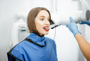
Intraoral X-ray Systems
Digital Sensors
Phosphor Plate Systems
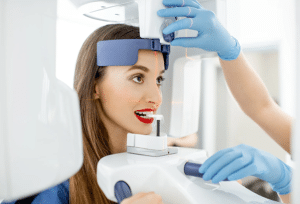
Extraoral X-ray Systems
Panoramic X-ray Machines
Cephalometric X-ray Machines
Cone Beam Computed Tomography (CBCT)
Intraoral Cameras
Patient Preparation
Before appointment
Medical History Review: Check the vital signs of the patient and get their past medical and dental history.
Patient Education: Describe the goal of the radiography and what the patient can anticipate from the process. Talk about any questions or inquiries the patient may have.
Day of the Appointment
Initial Check: Identify the patient and check whether the medical history used is the latest. Ensure to remove dental appliances, ornaments or glasses they might have been wearing.
Preparation: Check that the radiography equipment is clean, and the settings adjusted accordingly so that the equipment is in good working order. Choose the sensor size that is right for you and place it on a shielded setter cover.
Set up the computer and radiography software to the correct patient file.
Patient position
Position the patient well on the dental chair.
Secure the patient’s head with a headrest and guides to maintain the correct position and steady it.
Put on a lead apron that has a thyroid collar for the patient’s protection against radiation.
Technique
Types of Digital Radiography Sensors
Direct Digital Radiography (DDR):A digital sensor attached directly to a computer. Image taken with this sensor is available on a computer screen right after the procedure.
Indirect Digital Radiography (IDR):A phosphor plate that the image is captured on. This plate is scanned into a computer; an image is digitized and displayed.
Common Types of Digital Dental Radiographs
Intraoral Radiographs:
An X-ray was taken inside the mouth.
Bitewing: Shows the crowns of the teeth in the upper and lower arch together. Therefore, it is helpful in detecting cavities and determining bone levels.
Periapical: Shows the entire tooth from crown to root, hence it is helpful to diagnose an area that involves the tip of the root and surrounding bone.
Occlusal: Goes across the jaw and displays the entire arch of teeth on one side.
Extraoral Radiographs: Radiographs produced outside the mouth.
Panoramic: Shows the jaws, teeth, sinuses, and nasal area in a wide view.
Cephalometric: The entire side of the head is visible, useful for orthodontic planning and evaluation.
Technique and Procedure
Step 1: Preparation
Explain the procedure to the patient.
Ensure the patient removes any metallic objects that may interfere with the image quality.
Position the patient, accordingly, depending on the type of radiograph to be taken.
Step 2: Acquisition
Place the sensor or phosphor plate in the patient’s mouth for intraoral radiographs. Position the patient for extraoral radiographs according to the specific requirements of the radiograph type.
Retake the image with the software. Change the exposure settings if needed.
Step 3: Image Processing:
The image is ready to be viewed directly on the computer screen in the case of direct digital radiography.
The phosphor plate is scanned to obtain a digital image in the case of indirect digital radiography.
Change brightness, contrast, or any other setting if the image requires quality improvement.
Step 4: Review and Diagnosis
View the images for diagnosis
Measure the areas of importance and analyse it with the help of measurement tools from the software.
Store the images in the patient’s digital record for future reference.
Advantages of Digital Dental Radiography.
Reduced Radiation Exposure: Besides, the digital sensors are more sensitive to X-rays than the films to produce a desired picture at lower doses.
Enhanced Image Quality: Digital images offer the benefit of improving the images to make them clear for diagnosis through enlargement and manipulation.
Easy Storage and Sharing: Electronic images easily stored especially in the patient electronic records and can be transferred to other healthcare practitioners.
Cone Beam Computed Tomography (CBCT):Cone Beam Computed Tomography (CBCT) is X-ray imaging that has been utilized majorly in dental and orthodontic practices, of which it is variably applicable in other fields like Otolaryngology & maxillofacial surgery.
Principle: CBCT is different from conventional CT scans in that it uses a cone-shaped X-ray beam instead of a fan-shaped beam to obtain a sequence of images. At the same time, the patient’s head is being rotated 360 degrees around the area of interest.
Image Acquisition: Subsequently, the X-ray tube and a digital detector revolve around the patient to take images from various directions. They are then reconstructed into a three-dimensional picture of the treated area.
Advantages:
High Resolution: Offers high spatial resolution imaging which is very useful in imaging bone structures as well as the dental systems.
Lower Radiation Dose: Usually, CBCT produces a lower dose of radiation than the conventional CT scans do for a patient.
Quick Scanning: Most often faster than the conventional CT scans, thus saving time of both the patients and the practitioners.
Applications:
Dental: It is applied in implant planning, bone density measurement, and identification of the position of teeth and roots.
Orthodontics: They assist in treatment planning and follow-up on tooth movements.
Maxillofacial Surgery: Assists in planning surgeries and assessing anatomical structures.
Complications
Technical issues: Digital software and sensors may not work correctly, causing delays or necessitating further imaging.
Image Quality: The captured image quality can be poor because improper positioning of the sensor, low exposure rates or poor software functioning.
Radiation Exposure
Overexposure: Repeated or needless imaging can lead to considerable cumulative radiation exposure, even with lower doses.
Underexposure: Not getting enough exposure might result in low-quality pictures, so you’ll need to get more exposure by taking repeat radiographs.
Digital dental radiography is the latest form of dental radiographic procedure. Instead of photographic film, digital sensors are used to obtain images of the teeth and other structures in the oral cavity. This technology benefits digital radiography and is different from conventional film-based radiography because it has high image quality, low radiation dosage, and available images.
Caries Detection and Diagnosis: To detect those areas in the teeth with a cavity and the severity of the decay.
Periodontal Disease Assessment: To assess the amount of bone resorption and other pathological conditions of the periodontium.
Endodontic Treatment: For visualizing the root canal system, for the detection of periapical lesions and, for assessing the status of working progress in root canal treatment.
Implant Planning and Placement: To determine bone density, the quantity of bone volume, and the position of structures like nerves and sinuses.
Orthodontic Assessment: This is used to control the course of orthodontic treatment and plan therapeutic orthodontic measures.
Fracture Detection: To diagnose dental and jaw traumas and determine their severity.
Radiation Sensitivity: Some of the patients may be sensitive to radiation or have previously been exposed to high levels of radiation hence should be taken into consideration. Alternatives or additional protective measures may be necessary.
Equipment Malfunction: It is essential to understand that there can be malfunctions associated with sensors or software in any digital equipment.
Recent Oral Surgery or Trauma: Oral surgery patients and people, who went through a recent trauma, may feel rather uncomfortable wearing intraoral sensors. Extraoral radiography could be more ideal if done until the time the area heals.
Intraoral X-ray Systems
Digital Sensors
Phosphor Plate Systems
Extraoral X-ray Systems
Panoramic X-ray Machines
Cephalometric X-ray Machines
Cone Beam Computed Tomography (CBCT)
Intraoral Cameras
Before appointment
Medical History Review: Check the vital signs of the patient and get their past medical and dental history.
Patient Education: Describe the goal of the radiography and what the patient can anticipate from the process. Talk about any questions or inquiries the patient may have.
Day of the Appointment
Initial Check: Identify the patient and check whether the medical history used is the latest. Ensure to remove dental appliances, ornaments or glasses they might have been wearing.
Preparation: Check that the radiography equipment is clean, and the settings adjusted accordingly so that the equipment is in good working order. Choose the sensor size that is right for you and place it on a shielded setter cover.
Set up the computer and radiography software to the correct patient file.
Position the patient well on the dental chair.
Secure the patient’s head with a headrest and guides to maintain the correct position and steady it.
Put on a lead apron that has a thyroid collar for the patient’s protection against radiation.
Types of Digital Radiography Sensors
Direct Digital Radiography (DDR):A digital sensor attached directly to a computer. Image taken with this sensor is available on a computer screen right after the procedure.
Indirect Digital Radiography (IDR):A phosphor plate that the image is captured on. This plate is scanned into a computer; an image is digitized and displayed.
Common Types of Digital Dental Radiographs
Intraoral Radiographs:
An X-ray was taken inside the mouth.
Bitewing: Shows the crowns of the teeth in the upper and lower arch together. Therefore, it is helpful in detecting cavities and determining bone levels.
Periapical: Shows the entire tooth from crown to root, hence it is helpful to diagnose an area that involves the tip of the root and surrounding bone.
Occlusal: Goes across the jaw and displays the entire arch of teeth on one side.
Extraoral Radiographs: Radiographs produced outside the mouth.
Panoramic: Shows the jaws, teeth, sinuses, and nasal area in a wide view.
Cephalometric: The entire side of the head is visible, useful for orthodontic planning and evaluation.
Step 1: Preparation
Explain the procedure to the patient.
Ensure the patient removes any metallic objects that may interfere with the image quality.
Position the patient, accordingly, depending on the type of radiograph to be taken.
Step 2: Acquisition
Place the sensor or phosphor plate in the patient’s mouth for intraoral radiographs. Position the patient for extraoral radiographs according to the specific requirements of the radiograph type.
Retake the image with the software. Change the exposure settings if needed.
Step 3: Image Processing:
The image is ready to be viewed directly on the computer screen in the case of direct digital radiography.
The phosphor plate is scanned to obtain a digital image in the case of indirect digital radiography.
Change brightness, contrast, or any other setting if the image requires quality improvement.
Step 4: Review and Diagnosis
View the images for diagnosis
Measure the areas of importance and analyse it with the help of measurement tools from the software.
Store the images in the patient’s digital record for future reference.
Advantages of Digital Dental Radiography.
Reduced Radiation Exposure: Besides, the digital sensors are more sensitive to X-rays than the films to produce a desired picture at lower doses.
Enhanced Image Quality: Digital images offer the benefit of improving the images to make them clear for diagnosis through enlargement and manipulation.
Easy Storage and Sharing: Electronic images easily stored especially in the patient electronic records and can be transferred to other healthcare practitioners.
Cone Beam Computed Tomography (CBCT):Cone Beam Computed Tomography (CBCT) is X-ray imaging that has been utilized majorly in dental and orthodontic practices, of which it is variably applicable in other fields like Otolaryngology & maxillofacial surgery.
Principle: CBCT is different from conventional CT scans in that it uses a cone-shaped X-ray beam instead of a fan-shaped beam to obtain a sequence of images. At the same time, the patient’s head is being rotated 360 degrees around the area of interest.
Image Acquisition: Subsequently, the X-ray tube and a digital detector revolve around the patient to take images from various directions. They are then reconstructed into a three-dimensional picture of the treated area.
Advantages:
High Resolution: Offers high spatial resolution imaging which is very useful in imaging bone structures as well as the dental systems.
Lower Radiation Dose: Usually, CBCT produces a lower dose of radiation than the conventional CT scans do for a patient.
Quick Scanning: Most often faster than the conventional CT scans, thus saving time of both the patients and the practitioners.
Applications:
Dental: It is applied in implant planning, bone density measurement, and identification of the position of teeth and roots.
Orthodontics: They assist in treatment planning and follow-up on tooth movements.
Maxillofacial Surgery: Assists in planning surgeries and assessing anatomical structures.
Technical issues: Digital software and sensors may not work correctly, causing delays or necessitating further imaging.
Image Quality: The captured image quality can be poor because improper positioning of the sensor, low exposure rates or poor software functioning.
Radiation Exposure
Overexposure: Repeated or needless imaging can lead to considerable cumulative radiation exposure, even with lower doses.
Underexposure: Not getting enough exposure might result in low-quality pictures, so you’ll need to get more exposure by taking repeat radiographs.
Both our subscription plans include Free CME/CPD AMA PRA Category 1 credits.
Digital Certificate PDF
On course completion, you will receive a full-sized presentation quality digital certificate.
medtigo Simulation
A dynamic medical simulation platform designed to train healthcare professionals and students to effectively run code situations through an immersive hands-on experience in a live, interactive 3D environment.
medtigo Points
medtigo points is our unique point redemption system created to award users for interacting on our site. These points can be redeemed for special discounts on the medtigo marketplace as well as towards the membership cost itself.
Community Forum post/reply = 5 points
*Redemption of points can occur only through the medtigo marketplace, courses, or simulation system. Money will not be credited to your bank account. 10 points = $1.
All Your Certificates in One Place
When you have your licenses, certificates and CMEs in one place, it's easier to track your career growth. You can easily share these with hospitals as well, using your medtigo app.