Duraplasty is a surgical procedure that involves repairing or expanding the dura mater, the tough, outermost membrane that surrounds the brain and spinal cord. This technique is typically used to address conditions where the dura has been compromised or requires modification to relieve pressure, restore functionality, or protect the central nervous system.
Types of Duraplasty:
Primary Duraplasty: Repairing the dura directly at the site of injury or defect.
Augmentative Duraplasty: Using grafts to enlarge the dura or cover significant defects.
Reconstructive Duraplasty: In cases of repeated surgeries or complex abnormalities, where significant reconstruction is required.
Indications
Skull fractures: Dural repair is performed in open skull fractures or penetrating head injuries where there is a dural tear.
Post-traumatic cerebrospinal fluid leaks: If cerebrospinal fluid leakage persists post head-trauma surgery, it may be treated with duraplasty by closing the leak to prevent the complications of meningitis.
Surgical decompression for cranial disorders:
Chiari malformation: Duraplasty is generally performed during posterior fossa decompression to ensure the cerebellum has enough space to improve CSF flow.
Intracranial hypertension: Duraplasty may be helpful in relieving intracranial hypertension when it is refractory.
Contraindications
Active infection: Infection at the surgical site raises the risk of meningitis or wound issues.
Severe coagulopathy: Uncontrolled bleeding disorders increase hemorrhage risk.
Unstable condition: Severe systemic diseases may make surgery unsafe.
Scar tissue: Prior surgeries may heighten complication risks.
Poor healing: Conditions like diabetes can impair wound recovery.
High ICP: Elevated pressure requires careful management to avoid complications.
Contraindications
Outcomes
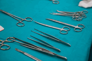
Equipment
Dural Patches
Autologous materials: (e.g., fascia lata or pericranium).
Synthetic grafts: Biocompatible materials like Gore-Tex or Dacron.
Biological grafts: Allografts (cadaver dura) or xenografts (bovine pericardium).
Microsurgical Equipment
Operating microscope
Micro-instruments
Surgical equipments
Suturing Materials
Non-absorbable sutures
Monofilament or braided sutures
Patient preparation
Preoperative Assessment
Medical History: Assess for conditions like hydrocephalus, Chiari malformation, or prior trauma.
Imaging: Conduct MRI or CT scans to evaluate the affected area.
Lab Tests: Review coagulation profile, CBC, and other baseline tests.
Neurological Evaluation: Check for signs of increased intracranial pressure or neurological deficits.
Doctor analysing the reports of patient
Patient Education
Discuss the procedure, associated risks, and potential outcomes.
Obtain informed consent after addressing patient questions.
Medications
Discontinue anticoagulants or antiplatelet drugs following protocol.
Administer prophylactic antibiotics.
Optimize the management of pre-existing medical conditions.
Patient position
Prone Position:
The patient lies face-down on a specialized operating table with support to maintain spinal alignment and reduce pressure on the chest and abdomen.
Supine Position:
The patient lies on their back with the head stabilized in a neutral or slightly extended position.
Lateral Position:
The patient is positioned on their side with proper support for the head, neck, and torso.
Step 1-Preoperative Preparation
Anesthesia: The patient is administered general anesthesia to ensure they are unconscious and pain-free during the procedure.
Positioning: The patient is positioned based on the site of the surgery (e.g., prone or supine position) to provide optimal access to the affected area.
Step 2-Incision and Exposure:
Skin Incision: A scalp or back incision is made, depending on whether the procedure is for the brain or spinal cord.
Soft Tissue Dissection: The skin, muscle, and other tissues are carefully dissected to expose the dura mater.
Step 3-Dural Exposure and Inspection:
The dura mater is gently exposed. If the procedure is related to a brain condition, the skull may be removed or partially opened (craniotomy). In spinal surgeries, the vertebral structures are carefully separated.
The dura is inspected for any tears, defects, or pathology that needs correction.
Step 4-Dural Repair:
Reconstruction: If there’s a tear, hole, or defect in the dura, it is repaired using a graft or patch. This can be an autograft (tissue taken from the patient’s body) or an allograft (tissue from a donor).
Grafting Material: Common graft materials include synthetic materials (e.g., DuraGen), biologic materials, or autologous tissues such as pericranial flaps (in cranial duraplasty).
Suturing or Stapling: The graft is carefully sutured or stapled in place, ensuring a secure and leak-proof closure.
Step 5-Hemostasis:
The surgeon ensures that there is no bleeding from the dura or surrounding tissues. Hemostasis is achieved using cautery or hemostatic agents to prevent further bleeding.
Step 6-Closure:
Dural Closure: The dura is closed once the graft or patch is in place.
Layered Closure: The muscle, fat, and skin layers are then closed in layers with sutures or staples. The closure is done carefully to minimize the risk of infection or complications.
Step 7-Postoperative Care:
Monitoring: The patient is monitored closely in the recovery room for any signs of complications, such as infection, CSF leakage, or neurological changes.
Pain Management: Pain relief is provided as needed to ensure patient comfort.
Follow-up: The patient is scheduled for follow-up visits to monitor recovery and check for any complications.
Complications
Infection:
Meningitis: Infection of the meninges (the membranes around the brain and spinal cord), which can occur due to contamination during surgery.
Wound infection: This can happen at the surgical site, leading to potential delays in recovery.
Cerebrospinal Fluid (CSF) Leaks:
CSF leakage can occur if the duraplasty site does not seal completely. This can lead to symptoms like headaches, dizziness, or even a risk of further infection.
The leak might necessitate further surgical intervention to address the issue.
Hematoma:
A hematoma (blood collection) can form in the area where the surgery was performed. This can compress brain structures and lead to neurological deficits.
Graft Failure:
If a graft (biological or synthetic) used for duraplasty fails, it can lead to complications like CSF leaks or the need for revision surgery.
Grafts can be rejected, or they may not integrate well with the surrounding tissue.
Neurological Deficits:
Damage to surrounding neural tissue during surgery may lead to new or worsening neurological deficits, such as motor weakness, sensory loss, or cognitive changes.
These can be temporary or permanent, depending on the extent of damage.
Hydrocephalus:
Hydrocephalus, or the buildup of excess CSF in the brain, can develop after duraplasty. This occurs if there is an obstruction in the normal flow of CSF due to the surgery or its aftermath.
Duraplasty is a surgical procedure that involves repairing or expanding the dura mater, the tough, outermost membrane that surrounds the brain and spinal cord. This technique is typically used to address conditions where the dura has been compromised or requires modification to relieve pressure, restore functionality, or protect the central nervous system.
Types of Duraplasty:
Primary Duraplasty: Repairing the dura directly at the site of injury or defect.
Augmentative Duraplasty: Using grafts to enlarge the dura or cover significant defects.
Reconstructive Duraplasty: In cases of repeated surgeries or complex abnormalities, where significant reconstruction is required.
Skull fractures: Dural repair is performed in open skull fractures or penetrating head injuries where there is a dural tear.
Post-traumatic cerebrospinal fluid leaks: If cerebrospinal fluid leakage persists post head-trauma surgery, it may be treated with duraplasty by closing the leak to prevent the complications of meningitis.
Surgical decompression for cranial disorders:
Chiari malformation: Duraplasty is generally performed during posterior fossa decompression to ensure the cerebellum has enough space to improve CSF flow.
Intracranial hypertension: Duraplasty may be helpful in relieving intracranial hypertension when it is refractory.
Contraindications
Active infection: Infection at the surgical site raises the risk of meningitis or wound issues.
Severe coagulopathy: Uncontrolled bleeding disorders increase hemorrhage risk.
Unstable condition: Severe systemic diseases may make surgery unsafe.
Scar tissue: Prior surgeries may heighten complication risks.
Poor healing: Conditions like diabetes can impair wound recovery.
High ICP: Elevated pressure requires careful management to avoid complications.
Dural Patches
Autologous materials: (e.g., fascia lata or pericranium).
Synthetic grafts: Biocompatible materials like Gore-Tex or Dacron.
Biological grafts: Allografts (cadaver dura) or xenografts (bovine pericardium).
Microsurgical Equipment
Operating microscope
Micro-instruments
Surgical equipments
Suturing Materials
Non-absorbable sutures
Monofilament or braided sutures
Patient preparation
Preoperative Assessment
Medical History: Assess for conditions like hydrocephalus, Chiari malformation, or prior trauma.
Imaging: Conduct MRI or CT scans to evaluate the affected area.
Lab Tests: Review coagulation profile, CBC, and other baseline tests.
Neurological Evaluation: Check for signs of increased intracranial pressure or neurological deficits.
Doctor analysing the reports of patient
Patient Education
Discuss the procedure, associated risks, and potential outcomes.
Obtain informed consent after addressing patient questions.
Medications
Discontinue anticoagulants or antiplatelet drugs following protocol.
Administer prophylactic antibiotics.
Optimize the management of pre-existing medical conditions.
Patient position
Prone Position:
The patient lies face-down on a specialized operating table with support to maintain spinal alignment and reduce pressure on the chest and abdomen.
Supine Position:
The patient lies on their back with the head stabilized in a neutral or slightly extended position.
Lateral Position:
The patient is positioned on their side with proper support for the head, neck, and torso.
Anesthesia: The patient is administered general anesthesia to ensure they are unconscious and pain-free during the procedure.
Positioning: The patient is positioned based on the site of the surgery (e.g., prone or supine position) to provide optimal access to the affected area.
Step 2-Incision and Exposure:
Skin Incision: A scalp or back incision is made, depending on whether the procedure is for the brain or spinal cord.
Soft Tissue Dissection: The skin, muscle, and other tissues are carefully dissected to expose the dura mater.
Step 3-Dural Exposure and Inspection:
The dura mater is gently exposed. If the procedure is related to a brain condition, the skull may be removed or partially opened (craniotomy). In spinal surgeries, the vertebral structures are carefully separated.
The dura is inspected for any tears, defects, or pathology that needs correction.
Step 4-Dural Repair:
Reconstruction: If there’s a tear, hole, or defect in the dura, it is repaired using a graft or patch. This can be an autograft (tissue taken from the patient’s body) or an allograft (tissue from a donor).
Grafting Material: Common graft materials include synthetic materials (e.g., DuraGen), biologic materials, or autologous tissues such as pericranial flaps (in cranial duraplasty).
Suturing or Stapling: The graft is carefully sutured or stapled in place, ensuring a secure and leak-proof closure.
Step 5-Hemostasis:
The surgeon ensures that there is no bleeding from the dura or surrounding tissues. Hemostasis is achieved using cautery or hemostatic agents to prevent further bleeding.
Step 6-Closure:
Dural Closure: The dura is closed once the graft or patch is in place.
Layered Closure: The muscle, fat, and skin layers are then closed in layers with sutures or staples. The closure is done carefully to minimize the risk of infection or complications.
Step 7-Postoperative Care:
Monitoring: The patient is monitored closely in the recovery room for any signs of complications, such as infection, CSF leakage, or neurological changes.
Pain Management: Pain relief is provided as needed to ensure patient comfort.
Follow-up: The patient is scheduled for follow-up visits to monitor recovery and check for any complications.
Complications
Infection:
Meningitis: Infection of the meninges (the membranes around the brain and spinal cord), which can occur due to contamination during surgery.
Wound infection: This can happen at the surgical site, leading to potential delays in recovery.
Cerebrospinal Fluid (CSF) Leaks:
CSF leakage can occur if the duraplasty site does not seal completely. This can lead to symptoms like headaches, dizziness, or even a risk of further infection.
The leak might necessitate further surgical intervention to address the issue.
Hematoma:
A hematoma (blood collection) can form in the area where the surgery was performed. This can compress brain structures and lead to neurological deficits.
Graft Failure:
If a graft (biological or synthetic) used for duraplasty fails, it can lead to complications like CSF leaks or the need for revision surgery.
Grafts can be rejected, or they may not integrate well with the surrounding tissue.
Neurological Deficits:
Damage to surrounding neural tissue during surgery may lead to new or worsening neurological deficits, such as motor weakness, sensory loss, or cognitive changes.
These can be temporary or permanent, depending on the extent of damage.
Hydrocephalus:
Hydrocephalus, or the buildup of excess CSF in the brain, can develop after duraplasty. This occurs if there is an obstruction in the normal flow of CSF due to the surgery or its aftermath.
Both our subscription plans include Free CME/CPD AMA PRA Category 1 credits.
Digital Certificate PDF
On course completion, you will receive a full-sized presentation quality digital certificate.
medtigo Simulation
A dynamic medical simulation platform designed to train healthcare professionals and students to effectively run code situations through an immersive hands-on experience in a live, interactive 3D environment.
medtigo Points
medtigo points is our unique point redemption system created to award users for interacting on our site. These points can be redeemed for special discounts on the medtigo marketplace as well as towards the membership cost itself.
Community Forum post/reply = 5 points
*Redemption of points can occur only through the medtigo marketplace, courses, or simulation system. Money will not be credited to your bank account. 10 points = $1.
All Your Certificates in One Place
When you have your licenses, certificates and CMEs in one place, it's easier to track your career growth. You can easily share these with hospitals as well, using your medtigo app.