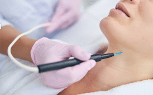
Electrocautery refers to the process in which high-frequency electrical current is used in treating tissues through cutting, coagulation, or removal of tissue. It is typically utilized in numerous surgical subfields reasonably familiar with dermatological surgery, breast reconstruction surgery, and general surgery. This involves the use of an instrument known as electrocautery and this works by passing current through the probe or electrode.
Electrocautery
There are two main types of electrocautery:
Electrosurgery: This type of electrocautery employs the use of high frequency alternation current that is passed through the patient’s body. It has been found that the current produced generates heat and this makes it possible to obtain the required result.
Thermocautery: This type of electrocautery uses direct current, through the heating of a metallic filament that has been placed within an electric circuit. The heated filament is then placed in contact with bodily tissue.
Indications
Tumor Removal: Electrocautery is used in the process of excision of tumors or any growths that are abnormal. Wound Closure: These can be employed in sealing of small vessels and tissue structures when closing the wound, and thus prevents the occurrence of bleeding after the surgery. Removal of Skin Lesions: Electrocautery is frequently applied to excision of skin lesions like papules, warts, moles or skin tags amongst others. Treatment of gynecologicalconditions: Electrocautery in gynecology may be necessary due to cervical dysplasia and other conditions like endometriosis. Dental Procedures: Dentists can employ electrocautery in different forms of surgeries for example gum surgery or extraction of teeth to facilitate coagulation. Treatment of Varicose Veins: During some of the operations you may undergo electrocautery which involves closing down the veins that are affected by varicose veins. Cosmetic Procedures: Other specialties of medical electrocautery include dermatology and cosmetic surgery; where it can be applied for surgeries like mole removal, scar correction or skin uplifting. Ear, Nose, and Throat Surgery: Electrocautery is employed during certain procedures like tonsillectomy and nasal polypectomy specifically to controlhemorrhage as well as excise tissue.
Contraindications
Electrocautery may be performed on nearly every patient because there are no definite contraindications.
Outcomes
Periprocedural care
Equipment
Electrosurgical Generator
Electrosurgical Pencil
Electrodes
Patient Plate (Grounding Pad)
Cables and Connectors
Smoke Evacuation System
Foot Pedal (Optional)
Accessories
Patient preparation
Patient Education: You must briefly describe the procedure to the patient while outlining what electrocautery is, why it is used, and what they can expect during the process and post-process. This will make it easy to address any concerns or questions that they may have regarding the procedure.
Medical History Review: Inquire for patient’s medical history to check on the presence of any past medical condition or any drug and food allergies that could be dangerous for the patient before or after the procedure.
Consent: An informed consent from the patient is required which may cover the potential risks, benefits of the procedure, and any other available choices.
Preoperative Assessment: Examine the patient for the surgical procedure to make sure they do not have any underlying medical condition. It can involve physiological assessments such as temperature, pulse, respiration and blood pressure, investigations, clinical findings, and other tests as may be relevant depending on the patient’s history.
Skin Preparation: Before conducting the electrocautery, it is important to make sure that the area of the body that will be operated on has been cleared of bacteria by washing it with an antiseptic agent.
Eye Protection: If the procedure involves the use of the equipment near the eyes such as electrocautery, ensure the patient is given protective eye gear and the same should be applicable to the healthcare team.
Monitoring: The following nursing intervention should also be implemented as a result of the procedure The patient’s vital signs should be monitored constantly during the procedure to identify any adverse reactions or complications.
Post-procedure Care: When the electrocautery is done, offer the right post-procedure care instructions especially on how to care for the wound, the number of days the patient will be restricted from certain activities and the date for check-up.
Patient position
Place the patient in a comfortable position on the surgical table or procedure chair with proper positioning that will allow the healthcare worker to easily access the specific body part that needs treatment.
It is standard practice to apply a local lidocaine injection. However, it is possible to use a solution of epinephrine and lidocaine where the latter is given within 15 minutes prior to the procedure to take leverage of the vasoconstrictive effect of epinephrine.
Technique
Pinpoint cautery
Step 1: The treatment area is regularly disinfected to prevent the spread of any contamination.
Step 2: In this case, there is no need for the patient to be administered with anesthetic drugs.
Step 3: For small and superficial lesions, a fine needle electrode tip is used while for larger lesions, a ballpoint electrode tip is used.
Step 4: The electrode tip is gently placed on the lesions, and a minimal amount of current flows through the lesion for not more than a one second.
Step 5: The treated area of skin usually forms a small scab, which, after seven to ten days, peels off spontaneously, exposing the treated skin below.
Removal of small benign lesions:
Step 1: The area around this lesion is washed and kept clean to avoid any bacteria or other microorganisms from invading the location.
Step 2: Considering the nature of the lesion, the correct electrode tip is selected depending on the size and volume.
Step 3: The electrode should be disabled when the lesion has shown a sign of necrosis or when the dermis separates from the lesion.
Step 4: The lethal tissue should naturally slough off within the first ten days or it may be removed by means of gauze or curette.
Step 5: Appearing as contours, larger and more protruding than smaller ones, the lessions may be shaved first before electrocautery is applied on it base.
Step 6: Benign lesions can be removed to improve cosmesis, reduce scarring, and provide a smooth skin surface with natural contouring.
Step 7: Regarding neovascularized skin diseases such as pyogenic granuloma, the process typically involves the use of a pre-treatment with epinephrine and lidocaine 1% solution fifteen minutes before the treatment. This makes it possible for vasoconstriction to work effectively.
Step 8: After the surgery to treat the affected area, the area can be washed, and a dressing might be applied.
Step 9: The patient is advised of the care needed in the case of an open wound, the frequency of follow-up appointments, and any other instructions that may be necessary.
Surgical punctal occlusion
Step 1: Before the procedure begins, the operative site, which is the patient’s eye and the adjacent tissue, is first cleaned.
Step 2: To minimise discomfort, local injections or topical drops are usually used to numb the affected region.
Step 3: The puncta are small openings situated on the inner corner of the upper and lower eye lids. Tear ducts are the openings through which tears enter the tear drainage system.
Step 4: An electrocautery device that has a fine tip to be used at high temperatures is introduced cold in the lacrimal punctum, the horizontal segment of the lacrimal canaliculus and the vertical segment of the lacrimal canaliculus.
Step 5: It is operated until the punctual tissue acquires a whitened state, which should not take 10 to 14 seconds.
Step 6: The device is then slowly withdrawn; the last part is removed while antibiotic ointment is then applied.
Laboratory tests
Certain tests may be needed depending on patient’s history or type of surgery. These tests could include:
Complete Blood Count: This also involves estimation of range of red and white blood cells to understand possible bleeding conditions or existence of infection.
Prothrombin time and International Normalized Ratio: These tests measure coagulation ability of the blood during the surgeries, which is crucial during procedures involving tissue manipulation. Other tests: Sometimes, the basic investigations might have to be complemented by other tests such as blood glucose and other specific infection tests depending on the presentation.
Complications
Thermal damage: Electrocautery coagulates and cuts tissue by heating it to high temperatures, and thus the heat that is produced inevitably can harm the surrounding tissue and cause burns or necrosis of tissue.
Bleeding: Failure of coagulation during electrocautery leads to bleeding at the site of operation.
Infection: In the cauterization of the tissue, there is always the risk of infection in the operative area, especially if the normalized aseptic measures are not observed.
Delayed healing: In some cases, electrocautery may also impede the process of healing or can contribute to normal wound healing or in some complicated cases, it may increase the chances of wound dehiscence.
Electrocautery refers to the process in which high-frequency electrical current is used in treating tissues through cutting, coagulation, or removal of tissue. It is typically utilized in numerous surgical subfields reasonably familiar with dermatological surgery, breast reconstruction surgery, and general surgery. This involves the use of an instrument known as electrocautery and this works by passing current through the probe or electrode.
Electrocautery
There are two main types of electrocautery:
Electrosurgery: This type of electrocautery employs the use of high frequency alternation current that is passed through the patient’s body. It has been found that the current produced generates heat and this makes it possible to obtain the required result.
Thermocautery: This type of electrocautery uses direct current, through the heating of a metallic filament that has been placed within an electric circuit. The heated filament is then placed in contact with bodily tissue.
Tumor Removal: Electrocautery is used in the process of excision of tumors or any growths that are abnormal. Wound Closure: These can be employed in sealing of small vessels and tissue structures when closing the wound, and thus prevents the occurrence of bleeding after the surgery. Removal of Skin Lesions: Electrocautery is frequently applied to excision of skin lesions like papules, warts, moles or skin tags amongst others. Treatment of gynecologicalconditions: Electrocautery in gynecology may be necessary due to cervical dysplasia and other conditions like endometriosis. Dental Procedures: Dentists can employ electrocautery in different forms of surgeries for example gum surgery or extraction of teeth to facilitate coagulation. Treatment of Varicose Veins: During some of the operations you may undergo electrocautery which involves closing down the veins that are affected by varicose veins. Cosmetic Procedures: Other specialties of medical electrocautery include dermatology and cosmetic surgery; where it can be applied for surgeries like mole removal, scar correction or skin uplifting. Ear, Nose, and Throat Surgery: Electrocautery is employed during certain procedures like tonsillectomy and nasal polypectomy specifically to controlhemorrhage as well as excise tissue.
Electrocautery may be performed on nearly every patient because there are no definite contraindications.
Equipment
Electrosurgical Generator
Electrosurgical Pencil
Electrodes
Patient Plate (Grounding Pad)
Cables and Connectors
Smoke Evacuation System
Foot Pedal (Optional)
Accessories
Patient Education: You must briefly describe the procedure to the patient while outlining what electrocautery is, why it is used, and what they can expect during the process and post-process. This will make it easy to address any concerns or questions that they may have regarding the procedure.
Medical History Review: Inquire for patient’s medical history to check on the presence of any past medical condition or any drug and food allergies that could be dangerous for the patient before or after the procedure.
Consent: An informed consent from the patient is required which may cover the potential risks, benefits of the procedure, and any other available choices.
Preoperative Assessment: Examine the patient for the surgical procedure to make sure they do not have any underlying medical condition. It can involve physiological assessments such as temperature, pulse, respiration and blood pressure, investigations, clinical findings, and other tests as may be relevant depending on the patient’s history.
Skin Preparation: Before conducting the electrocautery, it is important to make sure that the area of the body that will be operated on has been cleared of bacteria by washing it with an antiseptic agent.
Eye Protection: If the procedure involves the use of the equipment near the eyes such as electrocautery, ensure the patient is given protective eye gear and the same should be applicable to the healthcare team.
Monitoring: The following nursing intervention should also be implemented as a result of the procedure The patient’s vital signs should be monitored constantly during the procedure to identify any adverse reactions or complications.
Post-procedure Care: When the electrocautery is done, offer the right post-procedure care instructions especially on how to care for the wound, the number of days the patient will be restricted from certain activities and the date for check-up.
Patient position
Place the patient in a comfortable position on the surgical table or procedure chair with proper positioning that will allow the healthcare worker to easily access the specific body part that needs treatment.
It is standard practice to apply a local lidocaine injection. However, it is possible to use a solution of epinephrine and lidocaine where the latter is given within 15 minutes prior to the procedure to take leverage of the vasoconstrictive effect of epinephrine.
Pinpoint cautery
Step 1: The treatment area is regularly disinfected to prevent the spread of any contamination.
Step 2: In this case, there is no need for the patient to be administered with anesthetic drugs.
Step 3: For small and superficial lesions, a fine needle electrode tip is used while for larger lesions, a ballpoint electrode tip is used.
Step 4: The electrode tip is gently placed on the lesions, and a minimal amount of current flows through the lesion for not more than a one second.
Step 5: The treated area of skin usually forms a small scab, which, after seven to ten days, peels off spontaneously, exposing the treated skin below.
Removal of small benign lesions:
Step 1: The area around this lesion is washed and kept clean to avoid any bacteria or other microorganisms from invading the location.
Step 2: Considering the nature of the lesion, the correct electrode tip is selected depending on the size and volume.
Step 3: The electrode should be disabled when the lesion has shown a sign of necrosis or when the dermis separates from the lesion.
Step 4: The lethal tissue should naturally slough off within the first ten days or it may be removed by means of gauze or curette.
Step 5: Appearing as contours, larger and more protruding than smaller ones, the lessions may be shaved first before electrocautery is applied on it base.
Step 6: Benign lesions can be removed to improve cosmesis, reduce scarring, and provide a smooth skin surface with natural contouring.
Step 7: Regarding neovascularized skin diseases such as pyogenic granuloma, the process typically involves the use of a pre-treatment with epinephrine and lidocaine 1% solution fifteen minutes before the treatment. This makes it possible for vasoconstriction to work effectively.
Step 8: After the surgery to treat the affected area, the area can be washed, and a dressing might be applied.
Step 9: The patient is advised of the care needed in the case of an open wound, the frequency of follow-up appointments, and any other instructions that may be necessary.
Surgical punctal occlusion
Step 1: Before the procedure begins, the operative site, which is the patient’s eye and the adjacent tissue, is first cleaned.
Step 2: To minimise discomfort, local injections or topical drops are usually used to numb the affected region.
Step 3: The puncta are small openings situated on the inner corner of the upper and lower eye lids. Tear ducts are the openings through which tears enter the tear drainage system.
Step 4: An electrocautery device that has a fine tip to be used at high temperatures is introduced cold in the lacrimal punctum, the horizontal segment of the lacrimal canaliculus and the vertical segment of the lacrimal canaliculus.
Step 5: It is operated until the punctual tissue acquires a whitened state, which should not take 10 to 14 seconds.
Step 6: The device is then slowly withdrawn; the last part is removed while antibiotic ointment is then applied.
Laboratory tests
Certain tests may be needed depending on patient’s history or type of surgery. These tests could include:
Complete Blood Count: This also involves estimation of range of red and white blood cells to understand possible bleeding conditions or existence of infection.
Prothrombin time and International Normalized Ratio: These tests measure coagulation ability of the blood during the surgeries, which is crucial during procedures involving tissue manipulation. Other tests: Sometimes, the basic investigations might have to be complemented by other tests such as blood glucose and other specific infection tests depending on the presentation.
Thermal damage: Electrocautery coagulates and cuts tissue by heating it to high temperatures, and thus the heat that is produced inevitably can harm the surrounding tissue and cause burns or necrosis of tissue.
Bleeding: Failure of coagulation during electrocautery leads to bleeding at the site of operation.
Infection: In the cauterization of the tissue, there is always the risk of infection in the operative area, especially if the normalized aseptic measures are not observed.
Delayed healing: In some cases, electrocautery may also impede the process of healing or can contribute to normal wound healing or in some complicated cases, it may increase the chances of wound dehiscence.
Both our subscription plans include Free CME/CPD AMA PRA Category 1 credits.
Digital Certificate PDF
On course completion, you will receive a full-sized presentation quality digital certificate.
medtigo Simulation
A dynamic medical simulation platform designed to train healthcare professionals and students to effectively run code situations through an immersive hands-on experience in a live, interactive 3D environment.
medtigo Points
medtigo points is our unique point redemption system created to award users for interacting on our site. These points can be redeemed for special discounts on the medtigo marketplace as well as towards the membership cost itself.
Community Forum post/reply = 5 points
*Redemption of points can occur only through the medtigo marketplace, courses, or simulation system. Money will not be credited to your bank account. 10 points = $1.
All Your Certificates in One Place
When you have your licenses, certificates and CMEs in one place, it's easier to track your career growth. You can easily share these with hospitals as well, using your medtigo app.