Electrodiagnosis is a field which makes use of Electrophysiology in the study of human neurophysiology with the aid of electricity. Neurodiagnostics(NDS), electromyography(EMG) and evoked potentials(EPs) are some of the fundamental principles that are used in electrodiagnosis.
Electrodiagnostic testing is a critical tool in evaluating peripheral nerve or muscle injury, muscle diseases, or the localization of the problem prior to developing a treatment strategy. While electrodiagnostic tests are functional, they fundamentally differ from structures like the MRI that provides an image of the body’s anatomy. For instance, a patient may experience severe pain at the back while the MRI scan may be normal; on the other hand, individuals may have disk protrusions but have no complaints. Thus, electrodiagnostic studies can be helpful in cases where other diagnostic modalities seem insufficient. There are physiatrists with specialization and training in physical medicine and rehabilitation, neurologists, anesthesiologists among others who are qualified to perform electrodiagnostic tests.
Indications
Electromyography
The first electrodiagnostic test is Electromyography (EMG) where the needle is inserted to record the muscle activity at rest, with mild contraction, and contraction with maximum force. Usually, muscles do not have electrical activity at rest, however, myopathic fibers may exhibit spontaneous contraction during examination.
Damage to the nerve leading to neurotmetic or axonotmetic lesions causes Wallerian degeneration and produces spontaneous fibrillations and positive sharp wave activity in the period of rest. EMG can also establish primary muscle disorders.
Contractile relations in muscle fibres are determined during voluntary movement. Normal recruitment starts at 15-20 Hz as individual muscle fibers become recruited one after the other. Although identifying these patterns may sometimes be difficult and requires the use of advanced computerized equipment, they are critical during the assessment of the severity of the injuries and the prognosis since the strength of the signals does not depend on the cooperation of the patient.
In a study by Impastato et al., it was reported that voluntary motor unit recruitment at 1-9 months after trauma may be a predictor of unfavourable outcome for patients with traumatic brachial plexus injury. Overall, by 1.4 years post-injury, only 25% of muscles with severely reduced recruitment had a strength greater than 3/5.
While taking EMG can be somewhat painful due to the use of needles, there is enhanced technology in the market that involve smaller recording needles. The electromyographers should be up to date and have the right accreditation to avoid discomfort to patients. One emerging concern that has been evidenced is the expansion of EMG performance by nonphysician personnel, since the procedure involves highly technical skills and disease-specific knowledge to produce a reliable interpretation.
Nerve conduction studies
Nerve conduction studies (NCS) are mandatory for electrodiagnostic examinations which deliver electrical stimuli through peripheral nerves and evaluate muscle responses. Electrodiagnostic tests including latency, distance and nerve conduction velocity (NCV) provided information on the nerve and myelination. NCS can determine injury location, for example in carpal tunnel syndrome and can also determine other various nerve injuries and entrapments.
Studies prove that NCS outcomes may differ in diabetic peripheral neuropathy; major challenges in assessing clinical phenotypes exist. Conduction velocity correlates with nerve diameter, which suggests that only myelinated fibre’s diameter increases and reduces the capacity to measure small fibres neuropathy. Moreover, NCS can give furthermore false-positive results than ultrasonography in diagnosis of the asymptomatic carpal tunnel syndrome. However, in acute injuries, NCS continues to be useful since it can detect changes in motor unit recruitment proximal to the nerve stump in complete nerve transection.
Evoked Potentials
EPs refers to the electrical responses of the nervous system to sensory stimulation, more specifically diagnosing the CNS through electrodiagnostic approaches.
Types of EPs include:
Somatosensory EPs (SSEPs)
Visual EPs (VEPs)
Brainstem auditory EPs (BSAEPs)
Dermatomal EPs
Myotomal EPs
SSEPs, the most common, entail activating an extremity and acquiring a response from the scalp that helps in determining the site and extent of nerve root or CNS damage. They are most useful for cases such as multiple sclerosis and chronic inflammatory demyelinating polyneuropathy (CIDP). SSEPs are also useful in the evaluation of coma and brain death and general anesthesia guidelines recommend using total intravenous rather than gas anaesthesia during the procedure. VEPs elicit activity of the optic nerve and traces the responses in the visual cortex to identify damages along the optic tract.
BSAEPs work with auditory clicks to evaluate the cochlear nerve and identify the position of auditory pathway damage. In surgical scenarios, EPs are used to evaluate the condition of the spinal cord, in which intraoperative SSEPs reduce the likelihood of a permanent injury by about 50% since they offer updates to the surgeons.
Contraindications
Pacemakers or Implantable Cardioverter Defibrillators: During electrodiagnostic testing including nerve conduction studies, electrical stimulation could affect the proper working of these devices. Measures should be applied, or the test may not be conducted where it is necessary.
Severe bleeding disorders: Having hemophilia or taking anticoagulants exposes one to developing excess bleeding or hematoma when performing needle EMG. In such circumstances, diversification of methods needed for diagnosis could be required.
Active infections at the test site: Needle EMG may be contraindicated if there is a localized infection or an open wound in the area that the test is to be conducted to prevent spread of infection.
Uncooperative or noncompliant patients: This is because some of the patients may develop discomfort or may not be willing to undergo the tests and thus give inconclusive results. This can be even true in some events with use of children below certain ages or patient with certain mental diseases.
Allergies to materials used in the procedure: An important thing to note is that in case where a patient develops an allergic reaction to electrodes, gels employed in the tests, though this is hardly frequent, the patient will require to change the type of electrode to be used, or the manner of test administration will have to be altered.
Outcomes
Equipment
Electromyography (EMG) Machine: Captures electrical signals in muscles. Includes amplifiers, filters, and recording instruments to monitor and record signals picked by the needle electrodes that are inserted. Usually employed to identify muscle disorders and distinguish between neuropathies and myopathies.
Nerve Conduction Study (NCS) Equipment: Stimulates a nerve and records the velocity and amplitude of the signals sent across it. Uses surface electrodes mainly for stimulation and recording which is applied on the skin. Can evaluate the duration of nerve injury, conduction loss or demyelination.
Evoked Potentials (EP) System: Captures EEG that shows the brain’s electrical activity in response to stimuli like visual/auditory/sensory. Applied in diagnosing diseases of the central nervous system including multiple sclerosis.
Patient preparation
Patients should be informed about the procedure and mild discomfort maybe felt during electrodiagnostic testing. They should be encouraged to wear free clothing so that they don’t have to struggle to get access to the sites for tests and should not apply any lotion or creams on their skin the day they are due for testing. Jewellery and metallic should be removed. It is also important for the patient to let the physician know if they have a pacemaker, any implantable devices or if they are on any medication especially anticoagulant. The skin should be free and clear of excessive moisture.
Patient position
Patients undergoing electrodiagnostic testing are usually in a comfortable sitting or lying position on the examination table or chair. In electromyography (EMG), the patient can be told to lie down or sit depending on the muscle that is being tested. Nerve conduction studies (NCS) require that the limb being examined be relaxed and immobilized, thus there is no muscle contraction. The position should make the patient comfortable and must be able to keep still for the entire length of the process to get a correct reading.
Electrodiagnosis
Technique
Step 1-Preparation: Security of a patient as well as their understanding of the process must be guaranteed. Wipe the skin at the electrode sites with alcohol swab to reduce the amount of oil at the electrode skin interface to enhance conductivity.
Step 2-Electrode Placement:
Surface Electrodes: In nerve conduction studies (NCS), adhesive surface electrodes are fixed on the skin over the nerves’ course.
Needle Electrodes: In case of electromyography (EMG), fine needle electrodes are then inserted into muscles to record electrical signals.
Step 3-Nerve Conduction Studies (NCS): Administer a low amplitude electrical current via surface electrodes. Evaluate the signal that is generated as a result following the propagation of the signal through the nerve and calculate the conduction velocity and amplitude, among other factors.
Step 4-Electromyography (EMG): Place needle electrodes into the muscle to monitor electrical activity both at rest and contraction. Superimposed on this background, assess SA, IA, and MUAPs to determine extant muscle function.
Step 5-Evoked Potentials: When assessing sensory tracts, use proper modality (visual, auditory, or somatosensory) stimulus and record electrical activity from electrodes positioned on scalp or other locations.
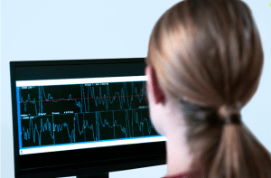
Step 6-Data Recording and Analysis: Record the signals using a computer and process them as displayed waveforms for interpretation purposes. Relative to reference values, one can note abnormalities. Step 7-Post-Procedure Care: You retire electrodes and clean the skin. Inform the patient on the instructions after the test and on the follow up depending on the results.
Complications
Discomfort or Pain: Pain sensations during needle insertions for electromyography (EMG), or when stimulating the nerve during nerve conduction studies (NCS), is moderate at worst.
Bruising or Hematoma: Percutaneous introduction of the needle can cause superficial haematomas and / or localized bleeding in patients with coagulation disorders or on oral anti-coagulant therapy.
Nerve Injury: However, there is potential harm to the nerve being tested; this is rare but more so with incorrect procedure or structural modification.
Infection: The chances of an infection at the area of needle insertion is relatively small, however, this should be compounded by poor aseptic techniques.
Allergic Reactions: There may be some complications if patients are allergic to substances like electrode gel or any local anesthetic if used.
Electrodiagnosis is a field which makes use of Electrophysiology in the study of human neurophysiology with the aid of electricity. Neurodiagnostics(NDS), electromyography(EMG) and evoked potentials(EPs) are some of the fundamental principles that are used in electrodiagnosis.
Electrodiagnostic testing is a critical tool in evaluating peripheral nerve or muscle injury, muscle diseases, or the localization of the problem prior to developing a treatment strategy. While electrodiagnostic tests are functional, they fundamentally differ from structures like the MRI that provides an image of the body’s anatomy. For instance, a patient may experience severe pain at the back while the MRI scan may be normal; on the other hand, individuals may have disk protrusions but have no complaints. Thus, electrodiagnostic studies can be helpful in cases where other diagnostic modalities seem insufficient. There are physiatrists with specialization and training in physical medicine and rehabilitation, neurologists, anesthesiologists among others who are qualified to perform electrodiagnostic tests.
Electromyography
The first electrodiagnostic test is Electromyography (EMG) where the needle is inserted to record the muscle activity at rest, with mild contraction, and contraction with maximum force. Usually, muscles do not have electrical activity at rest, however, myopathic fibers may exhibit spontaneous contraction during examination.
Damage to the nerve leading to neurotmetic or axonotmetic lesions causes Wallerian degeneration and produces spontaneous fibrillations and positive sharp wave activity in the period of rest. EMG can also establish primary muscle disorders.
Contractile relations in muscle fibres are determined during voluntary movement. Normal recruitment starts at 15-20 Hz as individual muscle fibers become recruited one after the other. Although identifying these patterns may sometimes be difficult and requires the use of advanced computerized equipment, they are critical during the assessment of the severity of the injuries and the prognosis since the strength of the signals does not depend on the cooperation of the patient.
In a study by Impastato et al., it was reported that voluntary motor unit recruitment at 1-9 months after trauma may be a predictor of unfavourable outcome for patients with traumatic brachial plexus injury. Overall, by 1.4 years post-injury, only 25% of muscles with severely reduced recruitment had a strength greater than 3/5.
While taking EMG can be somewhat painful due to the use of needles, there is enhanced technology in the market that involve smaller recording needles. The electromyographers should be up to date and have the right accreditation to avoid discomfort to patients. One emerging concern that has been evidenced is the expansion of EMG performance by nonphysician personnel, since the procedure involves highly technical skills and disease-specific knowledge to produce a reliable interpretation.
Nerve conduction studies
Nerve conduction studies (NCS) are mandatory for electrodiagnostic examinations which deliver electrical stimuli through peripheral nerves and evaluate muscle responses. Electrodiagnostic tests including latency, distance and nerve conduction velocity (NCV) provided information on the nerve and myelination. NCS can determine injury location, for example in carpal tunnel syndrome and can also determine other various nerve injuries and entrapments.
Studies prove that NCS outcomes may differ in diabetic peripheral neuropathy; major challenges in assessing clinical phenotypes exist. Conduction velocity correlates with nerve diameter, which suggests that only myelinated fibre’s diameter increases and reduces the capacity to measure small fibres neuropathy. Moreover, NCS can give furthermore false-positive results than ultrasonography in diagnosis of the asymptomatic carpal tunnel syndrome. However, in acute injuries, NCS continues to be useful since it can detect changes in motor unit recruitment proximal to the nerve stump in complete nerve transection.
Evoked Potentials
EPs refers to the electrical responses of the nervous system to sensory stimulation, more specifically diagnosing the CNS through electrodiagnostic approaches.
Types of EPs include:
Somatosensory EPs (SSEPs)
Visual EPs (VEPs)
Brainstem auditory EPs (BSAEPs)
Dermatomal EPs
Myotomal EPs
SSEPs, the most common, entail activating an extremity and acquiring a response from the scalp that helps in determining the site and extent of nerve root or CNS damage. They are most useful for cases such as multiple sclerosis and chronic inflammatory demyelinating polyneuropathy (CIDP). SSEPs are also useful in the evaluation of coma and brain death and general anesthesia guidelines recommend using total intravenous rather than gas anaesthesia during the procedure. VEPs elicit activity of the optic nerve and traces the responses in the visual cortex to identify damages along the optic tract.
BSAEPs work with auditory clicks to evaluate the cochlear nerve and identify the position of auditory pathway damage. In surgical scenarios, EPs are used to evaluate the condition of the spinal cord, in which intraoperative SSEPs reduce the likelihood of a permanent injury by about 50% since they offer updates to the surgeons.
Pacemakers or Implantable Cardioverter Defibrillators: During electrodiagnostic testing including nerve conduction studies, electrical stimulation could affect the proper working of these devices. Measures should be applied, or the test may not be conducted where it is necessary.
Severe bleeding disorders: Having hemophilia or taking anticoagulants exposes one to developing excess bleeding or hematoma when performing needle EMG. In such circumstances, diversification of methods needed for diagnosis could be required.
Active infections at the test site: Needle EMG may be contraindicated if there is a localized infection or an open wound in the area that the test is to be conducted to prevent spread of infection.
Uncooperative or noncompliant patients: This is because some of the patients may develop discomfort or may not be willing to undergo the tests and thus give inconclusive results. This can be even true in some events with use of children below certain ages or patient with certain mental diseases.
Allergies to materials used in the procedure: An important thing to note is that in case where a patient develops an allergic reaction to electrodes, gels employed in the tests, though this is hardly frequent, the patient will require to change the type of electrode to be used, or the manner of test administration will have to be altered.
Electromyography (EMG) Machine: Captures electrical signals in muscles. Includes amplifiers, filters, and recording instruments to monitor and record signals picked by the needle electrodes that are inserted. Usually employed to identify muscle disorders and distinguish between neuropathies and myopathies.
Nerve Conduction Study (NCS) Equipment: Stimulates a nerve and records the velocity and amplitude of the signals sent across it. Uses surface electrodes mainly for stimulation and recording which is applied on the skin. Can evaluate the duration of nerve injury, conduction loss or demyelination.
Evoked Potentials (EP) System: Captures EEG that shows the brain’s electrical activity in response to stimuli like visual/auditory/sensory. Applied in diagnosing diseases of the central nervous system including multiple sclerosis.
Patient preparation
Patients should be informed about the procedure and mild discomfort maybe felt during electrodiagnostic testing. They should be encouraged to wear free clothing so that they don’t have to struggle to get access to the sites for tests and should not apply any lotion or creams on their skin the day they are due for testing. Jewellery and metallic should be removed. It is also important for the patient to let the physician know if they have a pacemaker, any implantable devices or if they are on any medication especially anticoagulant. The skin should be free and clear of excessive moisture.
Patient position
Patients undergoing electrodiagnostic testing are usually in a comfortable sitting or lying position on the examination table or chair. In electromyography (EMG), the patient can be told to lie down or sit depending on the muscle that is being tested. Nerve conduction studies (NCS) require that the limb being examined be relaxed and immobilized, thus there is no muscle contraction. The position should make the patient comfortable and must be able to keep still for the entire length of the process to get a correct reading.
Electrodiagnosis
Step 1-Preparation: Security of a patient as well as their understanding of the process must be guaranteed. Wipe the skin at the electrode sites with alcohol swab to reduce the amount of oil at the electrode skin interface to enhance conductivity.
Step 2-Electrode Placement:
Surface Electrodes: In nerve conduction studies (NCS), adhesive surface electrodes are fixed on the skin over the nerves’ course.
Needle Electrodes: In case of electromyography (EMG), fine needle electrodes are then inserted into muscles to record electrical signals.
Step 3-Nerve Conduction Studies (NCS): Administer a low amplitude electrical current via surface electrodes. Evaluate the signal that is generated as a result following the propagation of the signal through the nerve and calculate the conduction velocity and amplitude, among other factors.
Step 4-Electromyography (EMG): Place needle electrodes into the muscle to monitor electrical activity both at rest and contraction. Superimposed on this background, assess SA, IA, and MUAPs to determine extant muscle function.
Step 5-Evoked Potentials: When assessing sensory tracts, use proper modality (visual, auditory, or somatosensory) stimulus and record electrical activity from electrodes positioned on scalp or other locations.
Step 6-Data Recording and Analysis: Record the signals using a computer and process them as displayed waveforms for interpretation purposes. Relative to reference values, one can note abnormalities. Step 7-Post-Procedure Care: You retire electrodes and clean the skin. Inform the patient on the instructions after the test and on the follow up depending on the results.
Complications
Discomfort or Pain: Pain sensations during needle insertions for electromyography (EMG), or when stimulating the nerve during nerve conduction studies (NCS), is moderate at worst.
Bruising or Hematoma: Percutaneous introduction of the needle can cause superficial haematomas and / or localized bleeding in patients with coagulation disorders or on oral anti-coagulant therapy.
Nerve Injury: However, there is potential harm to the nerve being tested; this is rare but more so with incorrect procedure or structural modification.
Infection: The chances of an infection at the area of needle insertion is relatively small, however, this should be compounded by poor aseptic techniques.
Allergic Reactions: There may be some complications if patients are allergic to substances like electrode gel or any local anesthetic if used.
Both our subscription plans include Free CME/CPD AMA PRA Category 1 credits.
Digital Certificate PDF
On course completion, you will receive a full-sized presentation quality digital certificate.
medtigo Simulation
A dynamic medical simulation platform designed to train healthcare professionals and students to effectively run code situations through an immersive hands-on experience in a live, interactive 3D environment.
medtigo Points
medtigo points is our unique point redemption system created to award users for interacting on our site. These points can be redeemed for special discounts on the medtigo marketplace as well as towards the membership cost itself.
Community Forum post/reply = 5 points
*Redemption of points can occur only through the medtigo marketplace, courses, or simulation system. Money will not be credited to your bank account. 10 points = $1.
All Your Certificates in One Place
When you have your licenses, certificates and CMEs in one place, it's easier to track your career growth. You can easily share these with hospitals as well, using your medtigo app.