Electroencephalogram is developed by German psychiatrist Hans Berger which is a diagnostic tool measures brain electrical activity. It has become a crucial part of neurophysiology and clinical neurology by providing insight for brainwave patterns and their connection with consciousness states and neurological disorders.
EEG uses electrodes on the scalp to detect and amplify electrical impulses from brain neurons. These signals are known as brainwaves that are categorized into different frequency bands each associated with specific mental states and activities. It is a diagnostic tool used in clinical settings to diagnose and monitor neurological conditions and in critical care to assess brain function during surgeries.
In sleep studies EEG plays a crucial role in the diagnosis of sleep disorders like insomnia and sleep apnea. Neuroscience research is largely influenced to investigate brain function and neural correlation of mental activities.
Technological advancements have improved the EEG recording techniques and are enabling the portable and ambulatory monitoring in various settings. Quantitative EEG analysis and functional connectivity mapping have expanded EEG’s utility in clinical and research.
Indications
EEG diagnose seizure disorders and identifies abnormal brain electrical patterns and seizure localization and aid in diagnosis or treatment of various seizures.
Epilepsy monitoring involves continuous EEG analysis in an epilepsy monitoring unit which helps to diagnose and locate the source of seizures over an extended period.
EEG is utilized to evaluate patients with altered consciousness or coma by providing insights into brain activity levels and determining the cause of the altered mental state. It is also used in patients with traumatic brain injuries to assess damage extent and overall function that providing prognosis insights and guiding treatment decisions.
Contraindications
Contraindications
Recent Hair Treatments
Severe scalp conditions
Patient Discomfort or Agitation
Highly Contagious Conditions
Technical considerations
Proper electrode placement is crucial for obtaining meaningful EEG signals typically positioned on specific scalp locations according to the international 10-20 system. The choice of electrode sites depends on clinical questions and brain areas which are under investigation. Before electrode application the scalp should be clean and free of substances that may hinder contact and the skin should be scrubbed with an abrasive gel.
Maintaining low electrode impedance is crucial for reliable signal quality as it measures the electrodes adhesion to the skin and conductivity of electrical signals. Impedance values are monitored and adjustments made to improve contact. Proper selection of reference and ground electrodes is essential for accurate EEG recordings.
EEG recordings minimizes the movement artifacts and ensuring patient comfort, duration of the recording lasts for 20-30 minutes. The patient state during the recording is crucial as their level of arousal can influence the EEG patterns. Continuous monitoring during the recording allows for the identification and removal of artifacts such as eye blinks, muscle movements, and electrical interference. This ensures the quality of the recordings and helps in identifying potential issues during the investigation.
An experienced EEG technologist obtains the high-quality recordings by ensuring proper electrode placement, monitoring impedance levels, and managing artifacts. The EEG signal is amplified and filtered to enhance specific frequency bands, focusing on brainwave activity. A quiet and shielded recording environment is essential to minimize external interference.
Detailed documentation of patient history, medications, and observations during the EEG is essential for proper interpretation including noting patient behavior, responses to stimuli, and clinical events.
Outcomes
EEG diagnoses and classifies the seizures in identifying abnormal electrical activity associated with different seizure types and localizing abnormalities in the brain. It also provides real-time assessments of brain function detecting abnormalities indicative of neurological disorders like encephalopathies and brain injuries.
EEG patterns helps in assessing cognitive function particularly in the assessment of cognitive disorders and dementia. They can also be used to monitor treatment response in epilepsy where changes in EEG patterns can indicate the effectiveness of medications or interventions.
Equipment
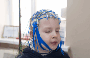
EEG electrodes are sensors attached to the scalp detecting and measuring electrical signals from brain neurons. They can be disposable or reusable and are typically made of conductive materials. To improve conductivity and reduce impedance a conductive paste or gel is applied to the electrodes. Electrode caps or headsets are used to simplify electrode placement according to the international 10-20 system. Amplifiers are part of the EEG system enhancing the strength of the electrical signals while maintaining their integrity. Modern EEG systems often use digital amplifiers.
The EEG system consists of an amplifier console, recording device, computer, and software. The amplifier console processes and records EEG signals adjusting amplification levels and filter settings. The recording device stores the amplified signals for analysis either paper-based or digital. A computer runs specialized software for real-time monitoring, recording, and analysis of EEG signals often including filtering and artifact removal tools. Impedance values are displayed on the EEG system interface. Video monitoring systems are often used to record patient behavior and clinical events during EEG recording helping correlate EEG patterns with physical movements or other observable events.
Patient preparation
Doctors provide patients with an informed consent before an EEG procedure explaining the purpose, duration, and potential discomfort. Patients are encouraged to ask questions, communication and cooperation are crucial for the success of the EEG especially during rest and activation procedures. Patients should remain still to minimize artifacts and be cooperative during recording.
The patient’s medication review ensures accurate EEG results, some medications may need to be temporarily withheld or adjusted based on healthcare provider recommendations. Caffeine should be avoided on the day of the EEG to avoid influencing brainwave patterns. Hair preparation is essential with clean hair free of oils and styling products. Electrodes are attached to specific scalp locations according to the international 10-20 system with abrasive gel and conductive gel applied to improve electrical contact.
Pre-configured electrode caps or headsets can streamline the placement process, patients should be comfortably positioned in a chair or bed allowing relaxation and minimal movement during recording.
A pillow may be used if needed, head may be supported with a pillow if needed.
EEG setups use pre-configured electrode caps or headsets to streamline the placement process ensuring consistent electrode positioning based on the patient’s head measurements. Patients are positioned comfortably in a chair or bed allowing for relaxation and minimal movement during recording.
The initial part of the EEG involves recording brainwave activity during a resting state with the patient’s eyes closed. Activation procedures may be employed to provoke specific brainwave patterns, such as opening and closing eyes, performing deep breathing, or undergoing controlled stimuli.
Video monitoring is often used in conjunction with EEG to capture the patient’s behavior and clinical events during the recording. EEG technologists continuously monitor the patient throughout the recording addressing any issues promptly and ensuring optimal electrode-skin contact.
Electroencephalogram
Step:1- Patient Preparation:
It involves explaining the procedure, ensuring patient cooperation, and obtaining a detailed medical history, including medications, sleep patterns, relevant symptoms or advising relaxation, and stillness during recording.
Step:2- Electrode Placement:
To prepare the scalp scrub it with an abrasive gel to reduce impedance. Measure the head to determine electrode placement using the international 10-20 system. Apply conductive gel to electrodes and affix them to the scalp.
Step:3- Reference and Ground Electrodes:
Choose suitable reference and ground electrodes like linked mastoids and a forehead location, ensure good skin contact to minimize artifacts.
Step:4- Monitoring Impedance:
Monitor electrode impedance levels to ensure optimal electrical contact. high impedance can cause poor signal quality. Adjust electrodes or reapply gel if impedance levels are elevated.
Step:5- Patient Positioning:
Position patient comfortably in a relaxed and semi-recumbent position, ensuring minimal movement during recording to prevent artifacts and may request specific positions or tasks based on clinical question.
Step:6- Calibration Recording:
Calibration is a process of recording standard electrical signals to ensure system accuracy identify technical issues and allow adjustments before capturing brainwave activity.
Step:7- Resting State Recording:
The brainwave activity during a resting state with closed eyes is recorded to capture spontaneous brain activity and background rhythms.
Step:8- Activation Procedures:
Clinical indications may require specific activation to trigger specific brainwave patterns like hyperventilation, photic stimulation, or eye opening and closing to identify abnormal patterns that may not present during resting state.
Electroencephalogram (EEG)
Step:9- Continuous Monitoring:
Ensure continuous patient monitoring throughout EEG recording to identify potential issues like artifacts or discomfort that address any impact on recording quality.
Step:10- Recording Duration:
EEG recording duration varies based on clinical question typically 20-30 minutes with long-term monitoring for specific conditions lasting hours or days.
Step:11- Post-Recording Analysis:
The recording process involves reviewing data for quality and artifact identification, analyzing EEG signals for specific frequency bands, patterns associated with normal or abnormal brain activity.
Step:12- Interpretation by a Neurologist:
EEG data is interpreted by a neurologist or clinical neurophysiologist, patterns, identifying abnormalities, and related findings to the patient clinical history and symptoms.
Pit-fills monitoring:
EEG involves the several challenges to ensure accurate and reliable recordings, artifacts with unwanted signals not originating from brain activity can be identified and removed during recording.
High electrode impedance can lead to poor signal quality and affect the accuracy of EEG recordings.
Continuous monitoring of electrode impedance levels is crucial and adjustments should be made promptly if impedance values are elevated. The movement during the EEG recording can introduce artifacts and affect the data quality.
Technologists should ensure patients comfortable position and informed about the importance of remaining still. Inadequate preparation of the scalp including removing dead skin cells and oils can hinder electrode-skin contact and compromise signal quality.
Incorrect electrode placement according to the international 10-20 system can lead to inaccurate localization of brainwave activity. Technologists must follow standardized electrode placement guidelines to ensure consistent and reliable recordings.
Environmental interference with electronic devices or poor wiring can introduce noise into the EEG recording. Adequate communication and efforts to make the patient comfortable can enhance cooperation and reduce the likelihood of pitfalls associated with patient movement or non-compliance.
Complications
EEG can cause mild skin irritation or anxiety in some patients, these reactions are rare and should be avoided.
EEG recordings are susceptible to artifacts which are unwanted signals not originating from brain activity. Skilled technologists work to identify and minimize these artifacts during recording.
Interpretation may sometimes lead to false positives or negatives and meaning abnormal brainwave patterns may be incorrectly identified. Clinical correlation is crucial for accurate interpretation, considering the patient’s history and other diagnostic information.
Provocation of seizures is a small risk but it is carefully weighed against the potential benefits of capturing specific EEG patterns associated with seizures.
Overinterpretation or misinterpretation of recorded patterns is a risk and the involvement of trained neurologists or clinical neurophysiologists is essential for accurate analysis.
It may also reveal asymptomatic abnormalities in brainwave patterns with raising concerns and anxiety in patients who were previously unaware of such findings.
References
Electroencephalography (EEG) and Event-Related Potentials (ERP’s) with Human Participants
Electroencephalogram – StatPearls
References
Electroencephalography (EEG) and Event-Related Potentials (ERP’s) with Human Participants
Electroencephalogram is developed by German psychiatrist Hans Berger which is a diagnostic tool measures brain electrical activity. It has become a crucial part of neurophysiology and clinical neurology by providing insight for brainwave patterns and their connection with consciousness states and neurological disorders.
EEG uses electrodes on the scalp to detect and amplify electrical impulses from brain neurons. These signals are known as brainwaves that are categorized into different frequency bands each associated with specific mental states and activities. It is a diagnostic tool used in clinical settings to diagnose and monitor neurological conditions and in critical care to assess brain function during surgeries.
In sleep studies EEG plays a crucial role in the diagnosis of sleep disorders like insomnia and sleep apnea. Neuroscience research is largely influenced to investigate brain function and neural correlation of mental activities.
Technological advancements have improved the EEG recording techniques and are enabling the portable and ambulatory monitoring in various settings. Quantitative EEG analysis and functional connectivity mapping have expanded EEG’s utility in clinical and research.
EEG diagnose seizure disorders and identifies abnormal brain electrical patterns and seizure localization and aid in diagnosis or treatment of various seizures.
Epilepsy monitoring involves continuous EEG analysis in an epilepsy monitoring unit which helps to diagnose and locate the source of seizures over an extended period.
EEG is utilized to evaluate patients with altered consciousness or coma by providing insights into brain activity levels and determining the cause of the altered mental state. It is also used in patients with traumatic brain injuries to assess damage extent and overall function that providing prognosis insights and guiding treatment decisions.
Contraindications
Recent Hair Treatments
Severe scalp conditions
Patient Discomfort or Agitation
Highly Contagious Conditions
Technical considerations
Proper electrode placement is crucial for obtaining meaningful EEG signals typically positioned on specific scalp locations according to the international 10-20 system. The choice of electrode sites depends on clinical questions and brain areas which are under investigation. Before electrode application the scalp should be clean and free of substances that may hinder contact and the skin should be scrubbed with an abrasive gel.
Maintaining low electrode impedance is crucial for reliable signal quality as it measures the electrodes adhesion to the skin and conductivity of electrical signals. Impedance values are monitored and adjustments made to improve contact. Proper selection of reference and ground electrodes is essential for accurate EEG recordings.
EEG recordings minimizes the movement artifacts and ensuring patient comfort, duration of the recording lasts for 20-30 minutes. The patient state during the recording is crucial as their level of arousal can influence the EEG patterns. Continuous monitoring during the recording allows for the identification and removal of artifacts such as eye blinks, muscle movements, and electrical interference. This ensures the quality of the recordings and helps in identifying potential issues during the investigation.
An experienced EEG technologist obtains the high-quality recordings by ensuring proper electrode placement, monitoring impedance levels, and managing artifacts. The EEG signal is amplified and filtered to enhance specific frequency bands, focusing on brainwave activity. A quiet and shielded recording environment is essential to minimize external interference.
Detailed documentation of patient history, medications, and observations during the EEG is essential for proper interpretation including noting patient behavior, responses to stimuli, and clinical events.
EEG diagnoses and classifies the seizures in identifying abnormal electrical activity associated with different seizure types and localizing abnormalities in the brain. It also provides real-time assessments of brain function detecting abnormalities indicative of neurological disorders like encephalopathies and brain injuries.
EEG patterns helps in assessing cognitive function particularly in the assessment of cognitive disorders and dementia. They can also be used to monitor treatment response in epilepsy where changes in EEG patterns can indicate the effectiveness of medications or interventions.
EEG electrodes are sensors attached to the scalp detecting and measuring electrical signals from brain neurons. They can be disposable or reusable and are typically made of conductive materials. To improve conductivity and reduce impedance a conductive paste or gel is applied to the electrodes. Electrode caps or headsets are used to simplify electrode placement according to the international 10-20 system. Amplifiers are part of the EEG system enhancing the strength of the electrical signals while maintaining their integrity. Modern EEG systems often use digital amplifiers.
The EEG system consists of an amplifier console, recording device, computer, and software. The amplifier console processes and records EEG signals adjusting amplification levels and filter settings. The recording device stores the amplified signals for analysis either paper-based or digital. A computer runs specialized software for real-time monitoring, recording, and analysis of EEG signals often including filtering and artifact removal tools. Impedance values are displayed on the EEG system interface. Video monitoring systems are often used to record patient behavior and clinical events during EEG recording helping correlate EEG patterns with physical movements or other observable events.
Doctors provide patients with an informed consent before an EEG procedure explaining the purpose, duration, and potential discomfort. Patients are encouraged to ask questions, communication and cooperation are crucial for the success of the EEG especially during rest and activation procedures. Patients should remain still to minimize artifacts and be cooperative during recording.
The patient’s medication review ensures accurate EEG results, some medications may need to be temporarily withheld or adjusted based on healthcare provider recommendations. Caffeine should be avoided on the day of the EEG to avoid influencing brainwave patterns. Hair preparation is essential with clean hair free of oils and styling products. Electrodes are attached to specific scalp locations according to the international 10-20 system with abrasive gel and conductive gel applied to improve electrical contact.
Pre-configured electrode caps or headsets can streamline the placement process, patients should be comfortably positioned in a chair or bed allowing relaxation and minimal movement during recording.
A pillow may be used if needed, head may be supported with a pillow if needed.
EEG setups use pre-configured electrode caps or headsets to streamline the placement process ensuring consistent electrode positioning based on the patient’s head measurements. Patients are positioned comfortably in a chair or bed allowing for relaxation and minimal movement during recording.
The initial part of the EEG involves recording brainwave activity during a resting state with the patient’s eyes closed. Activation procedures may be employed to provoke specific brainwave patterns, such as opening and closing eyes, performing deep breathing, or undergoing controlled stimuli.
Video monitoring is often used in conjunction with EEG to capture the patient’s behavior and clinical events during the recording. EEG technologists continuously monitor the patient throughout the recording addressing any issues promptly and ensuring optimal electrode-skin contact.
Step:1- Patient Preparation:
It involves explaining the procedure, ensuring patient cooperation, and obtaining a detailed medical history, including medications, sleep patterns, relevant symptoms or advising relaxation, and stillness during recording.
Step:2- Electrode Placement:
To prepare the scalp scrub it with an abrasive gel to reduce impedance. Measure the head to determine electrode placement using the international 10-20 system. Apply conductive gel to electrodes and affix them to the scalp.
Step:3- Reference and Ground Electrodes:
Choose suitable reference and ground electrodes like linked mastoids and a forehead location, ensure good skin contact to minimize artifacts.
Step:4- Monitoring Impedance:
Monitor electrode impedance levels to ensure optimal electrical contact. high impedance can cause poor signal quality. Adjust electrodes or reapply gel if impedance levels are elevated.
Step:5- Patient Positioning:
Position patient comfortably in a relaxed and semi-recumbent position, ensuring minimal movement during recording to prevent artifacts and may request specific positions or tasks based on clinical question.
Step:6- Calibration Recording:
Calibration is a process of recording standard electrical signals to ensure system accuracy identify technical issues and allow adjustments before capturing brainwave activity.
Step:7- Resting State Recording:
The brainwave activity during a resting state with closed eyes is recorded to capture spontaneous brain activity and background rhythms.
Step:8- Activation Procedures:
Clinical indications may require specific activation to trigger specific brainwave patterns like hyperventilation, photic stimulation, or eye opening and closing to identify abnormal patterns that may not present during resting state.
Electroencephalogram (EEG)
Step:9- Continuous Monitoring:
Ensure continuous patient monitoring throughout EEG recording to identify potential issues like artifacts or discomfort that address any impact on recording quality.
Step:10- Recording Duration:
EEG recording duration varies based on clinical question typically 20-30 minutes with long-term monitoring for specific conditions lasting hours or days.
Step:11- Post-Recording Analysis:
The recording process involves reviewing data for quality and artifact identification, analyzing EEG signals for specific frequency bands, patterns associated with normal or abnormal brain activity.
Step:12- Interpretation by a Neurologist:
EEG data is interpreted by a neurologist or clinical neurophysiologist, patterns, identifying abnormalities, and related findings to the patient clinical history and symptoms.
EEG involves the several challenges to ensure accurate and reliable recordings, artifacts with unwanted signals not originating from brain activity can be identified and removed during recording.
High electrode impedance can lead to poor signal quality and affect the accuracy of EEG recordings.
Continuous monitoring of electrode impedance levels is crucial and adjustments should be made promptly if impedance values are elevated. The movement during the EEG recording can introduce artifacts and affect the data quality.
Technologists should ensure patients comfortable position and informed about the importance of remaining still. Inadequate preparation of the scalp including removing dead skin cells and oils can hinder electrode-skin contact and compromise signal quality.
Incorrect electrode placement according to the international 10-20 system can lead to inaccurate localization of brainwave activity. Technologists must follow standardized electrode placement guidelines to ensure consistent and reliable recordings.
Environmental interference with electronic devices or poor wiring can introduce noise into the EEG recording. Adequate communication and efforts to make the patient comfortable can enhance cooperation and reduce the likelihood of pitfalls associated with patient movement or non-compliance.
EEG can cause mild skin irritation or anxiety in some patients, these reactions are rare and should be avoided.
EEG recordings are susceptible to artifacts which are unwanted signals not originating from brain activity. Skilled technologists work to identify and minimize these artifacts during recording.
Interpretation may sometimes lead to false positives or negatives and meaning abnormal brainwave patterns may be incorrectly identified. Clinical correlation is crucial for accurate interpretation, considering the patient’s history and other diagnostic information.
Provocation of seizures is a small risk but it is carefully weighed against the potential benefits of capturing specific EEG patterns associated with seizures.
Overinterpretation or misinterpretation of recorded patterns is a risk and the involvement of trained neurologists or clinical neurophysiologists is essential for accurate analysis.
It may also reveal asymptomatic abnormalities in brainwave patterns with raising concerns and anxiety in patients who were previously unaware of such findings.
Electroencephalography (EEG) and Event-Related Potentials (ERP’s) with Human Participants
Electroencephalogram – StatPearls
medtigo
Electroencephalogram (EEG)
Updated :
December 15, 2025
Electroencephalography (EEG) and Event-Related Potentials (ERP’s) with Human Participants
Electroencephalogram – StatPearls
Loading...
Free CME credits
Both our subscription plans include Free CME/CPD AMA PRA Category 1 credits.
Digital Certificate PDF
On course completion, you will receive a full-sized presentation quality digital certificate.
medtigo Simulation
A dynamic medical simulation platform designed to train healthcare professionals and students to effectively run code situations through an immersive hands-on experience in a live, interactive 3D environment.
medtigo Points
medtigo points is our unique point redemption system created to award users for interacting on our site. These points can be redeemed for special discounts on the medtigo marketplace as well as towards the membership cost itself.
Community Forum post/reply = 5 points
*Redemption of points can occur only through the medtigo marketplace, courses, or simulation system. Money will not be credited to your bank account. 10 points = $1.
All Your Certificates in One Place
When you have your licenses, certificates and CMEs in one place, it's easier to track your career growth. You can easily share these with hospitals as well, using your medtigo app.
 Electroencephalogram (EEG)


