Electromyography (EMG) and nerve conduction studies (NCS) are critical diagnostic procedures in neurology that help in the evaluation of neuromuscular diseases. These approaches assess muscle electrical activity and peripheral nerve function and provide information about a variety of neuropathological disorders. EMG and NCS are electrophysiological investigations which is used to diagnose and explain peripheral nervous system abnormalities like motor neurons, peripheral nerves, neuromuscular junctions and muscles. These approaches aid in the differentiation of myopathic and neurogenic diseases by examining the electrical characteristics of nerves and muscle.
Motor neurons control muscle contraction by transmitting electrical signals to fibers. EMG studies record the action potential generated by nerve impulses and helping diagnose disorders involving nerve dysfunction, muscle pathology or neuromuscular junction issues. Needle EMG is a method which can record electrical activity in the muscles and give detailed information on the nerve and muscle function. . It helps distinguish between myopathies and neurogenic disorders. Surface EMG uses electrodes on the skin. It is a less invasive method and is used in the rehabilitation and ergonomic studies.
NCS measures the intensity and speed of electrical impulses that go through peripheral nerves to evaluate their function. They help to determine if a disorder impacts the axons, the myelin sheath or both.
Important electrophysiological tests for evaluating peripheral nerve function are nerve conduction studies (NCS). They detect nerve injury and diagnose peripheral neuropathies, entrapment syndromes and neuromuscular illnesses by measuring the intensity and speed of electrical impulses. NCS evaluates the effectiveness of nerves in transmitting electrical impulses, revealing information on nerve integrity. Amplitude indicates the number of active axons in a nerve but conduction velocity is dependent on the integrity of the myelin sheath. Conduction speed and possible demyelination are shown by latency. NCS is able to identify anomalies in the neuromuscular junction, motor, and sensory nerves.
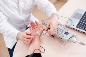
Patient nerve testing using electromyography
Motor nerve conduction study
Motor neurons are triggered in motor nerve conduction study and the muscle’s complex action potential is captured. This relates to the motor unit’s integrity. Any action that harms anterior horn cell body, axon, the neuromuscular junction, Schwann cells or muscle cell itself may have an impact on its results. The shape, size, and morphology of compound muscle impulse are also examined in order to evaluate the neuromuscular junction’s function, the number of functional muscle fibers and the myelination condition. Both postganglionic and preganglionic lesions result in aberrant motor nerve conduction because cell body of motor neurons is found in anterior horn of spinal cord.
Sensory nerve conduction study
Sensory nerve conduction study is conducted by the stimulation of the nerve at 1 point and the measurement of impulse at another point along with the course of nerve. Peripheral sensory nerve is used to locate the lesion which is related to dorsal root ganglion which contains cell body of nerve and allow to differentiate the preganglionic diseases like posterior column disease, cauda equina lesion and radiculopathy from the postganglionic diseases like plexopathy and neuropathy.
Late responses are specialized electrophysiological tests which is used in NCS to assess proximal nerve segments and long-loop reflexes that are not evaluated by standard motor and sensory conduction studies. 2 commonly studied late responses are F-waves and H-reflexes. F-waves evaluate the entire length of the motor nerve like proximal segments. It is useful in radiculopathies and demyelinating neuropathies.
F-waves are used to detect early demyelination in polyneuropathies, assess proximal nerve conduction in radiculopathies and differentiate between demyelinating and axonal neuropathies. Abnormal F-waves can result in prolonged F-wave latency, severe axonal damage or reduced F-wave persistence. The key applications of F-waves are detecting early demyelination in polyneuropathies, assessing proximal nerve conduction in radiculopathies and differentiating demyelinating and axonal neuropathies.
H-reflexes assess monosynaptic reflex arcs involving sensory and motor fibers similar to the Achilles reflex but recorded electrically. Electrical stimulation of a nerve activates Ia sensory afferent fibers which synapse at the spinal cord’s anterior horn and trigger a reflex motor response. The H-reflex is a true reflex arc making it valuable for evaluating sensory and motor pathways. The key applications of H-reflexes are diagnosing S1 radiculopathy, detecting early diabetic and inflammatory neuropathies and evaluating spinal cord lesions and myelopathies.
Insertional activity
Electromyography (EMG) is a diagnostic technique that evaluates muscle and nerve function by recording electrical activity. It consists of 2 components: insertional activity and spontaneous activity at rest. Insertional activity refers to the electrical response of a muscle when a needle electrode is inserted while spontaneous activity at rest includes abnormal electrical discharges. Abnormal insertional activity can be seen in acute denervation, myotonic disorders and inflammatory myopathies or in severe muscle atrophy or fibrosis.
Spontaneous Activity at Rest
Spontaneous activity in muscles like fibrillations, positive waves and fasciculations, can be abnormal electrical discharges without voluntary contraction. This can be seen in neurological disorders like ALS, radiculopathies, peripheral neuropathies, myopathic disorders like myotonic dystrophy, muscular dystrophies and nerve hyperexcitability syndromes.
Severity of Carpal Tunnel
Sensory Nerve Action Potential
Compound Motor Action Potential
Needle EMG Activity
Mild
Prolonged latency
Normal
Normal
Moderate
Prolonged latency and decreased amplitude
Prolonged latency
Normal
Severe
Absent
Prolonged latency and decreased amplitude
Abnormal activity
Table 1: Result of NCS and EMG in Carpal Tunnel Syndrome
Disorder
Distal Motor Latency
Distal Sensory Latency
Conduction Velocity
Amplitude of the Evoked Response
Motor neuron disease
Normal
Normal
Normal
Reduced
Axonal polyneuropathy
Normal
Normal
Slight decrease
Reduced
Demyelinating polyneuropathy
Prolonged
Prolonged
Decreased
Normal
Entrapment neuropathy
Normal (may be prolonged if this is a distal entrapment such as carpal tunnel syndrome)
Normal (May be prolonged if this is a distal entrapment such as carpal tunnel syndrome)
Decreased at the entrapment region
May be decreased when stimulating proximal to the site of entrapment
Radiculopathy
Normal
Normal
Normal
Motor response may be decreased
Myopathies
Normal
Normal
Normal
Decreased motor amplitudes
Table 2: Common diseases
Indications
EMG and NCS are vital tests to diagnose different conditions like peripheral nerve disorders, muscle disorders, radiculopathies, motor neuron diseases, injuries and toxins or metabolic conditions. EMG measures muscle activity during contraction and rest. NCS measures the speed and strength of electrical signals along nerves. These tests help diagnose nerve entrapment, damage, nerve root involvement and damage to nerve roots in conditions like herniated discs, cervical or lumbar spine herniated discs and motor nerve diseases like ALS. EMG and NCS are necessary to assess nerve damage because of trauma or surgery and identify toxins or metabolic issues that affect nerve or muscle function.
Limb temperature is crucial in electrodiagnostic studies with skin temperatures ideally between 32º C and 34º C. Cooler temperatures can cause longer-duration potentials and increased motor unit action potentials. Maintaining optimal limb temperature can be achieved using warm water, heating lamps, or hydrocollators. Age also plays a role with younger children experiencing slower conduction velocities and older individuals experiencing slower conduction velocities. Electrical noise like fluorescent lights, computers and fans can interfere with electrodiagnostic testing. To minimize disturbance, examiners should clean skin, apply electrode jelly, maintain electrode contact and place the ground between stimulators and recording electrodes.
Contraindications
Electrodiagnostic study is not performed on the patient who have external wire like guidewire, external pacing wire. Central line is not a contraindication but contralateral limb must be used, Patient who have implanted cardiac pacemaker and cardioverter defibrillator is not a contraindication to NCS. Many studies have seen no pacemake inhibition during the NCS. For patient who have IACDs, stimulation must be performed near implanted device and contralateral extremity must be used when it is possible. Stimulation intensity must be kept below 1 Hz and the pule width below 0.2 ms time. Repetition of stimulation must be avoided. Patient who has bleeding disease or anticoagulants are not contraindicated to the EMG.
Outcomes
Equipment
There are several commercial nerve conduction and EMG study devices available. Computer-based EMG equipment is produced by a number of companies. An amplifier, stimulator, speaker, preamplifier, printer, computer, and many electrodes are included.
Patient preparation
Patients should avoid applying lotions, oils, or creams to their skin on the day of the test to prevent interference with electrodes. They should inform their healthcare provider about any medications they are taking and if any changes are needed. Staying hydrated before the procedure can improve nerve conduction results. Patients should wear loose clothing that allows easy access to the test areas, and in some cases, a gown may be provided. Relaxation and calmness are essential during the test to avoid interference with results. Healthcare providers should explain the procedure, including electrodes, sensations, and expected duration, to reduce fear or confusion. No anesthesia is used for the procedure.
Patient position
The patient will must to move in various positions on the basis of on what area of body is examined.
Electromyography (EMG) Technique
Step 1: Patient preparation
The patient is positioned comfortably, cleaned and if necessary, moved or flexed to test muscles with skin over the muscle cleaned with an antiseptic wipe to prevent infection.
Step 2: Needle insertion
A sterile needle electrode is inserted into the muscle through the skin to detect electrical activity produced by muscle fibers, depending on the specific muscle or group being examined.
Step 3: Record electric activity
The needle records electrical activity in the muscle during contraction with waveforms displayed on a monitor representing the muscle’s electrical activity and the technician may ask the patient to contract the muscle in various ways.
Step 4: Muscle contraction
The patient’s voluntary muscle contraction is used by a technician to evaluate its response to nerve stimulation, assessing its response to both resting and active states.
Step 4: Data analysis
The technician analyzes recorded electrical signals detecting abnormalities that may indicate nerve damage, muscle disease or neuromuscular disorders like ALS, myopathy, or carpal tunnel syndrome.
Step 5: Needle removal
After testing the muscles, the needle is removed and a small bandage may be applied to the insertion site if necessary.
Step 6: Post care
Patients may experience mild soreness or bruising at the needle sites which usually resolves within a few days.
Nerve Conduction Study (NCS) Technique
Step 1: Patient preparation
Clean the area of the skin and remove jewelry. Make sure the patient remain still and relaxed.
Step 2: Electrode placement
Surface electrodes are placed on the skin over the nerve being tested with one at the stimulation site and the other at the recording site to measure the nerve’s response.
Step 3: Nerve stimulation
The stimulating electrode activates a nerve through a mild electrical impulse causing a brief tingling sensation without pain. The electrode can be placed on different nerve locations like wrist or ankle.
Step 4: Record nerve response
The recording electrode measures the time it takes for nerve signals to travel between electrodes displaying the response as a waveform on a monitor. Technicians assess parameters like conduction velocity and amplitude.
Step 5: Data analysis
The analysis of recorded results checks nerve signal conductance with slower conduction or reduced amplitude indicating potential nerve damage, compression or disorders like neuropathy or carpal tunnel syndrome.
Step 6: Post care
Electrodes are removed and the patient can resume normal activities without recovery time although slight discomfort from electrical stimulation usually fades quickly.
»
Home » Procedure » Electromyography and Nerve Conduction
Electromyography and Nerve Conduction
Updated :
December 15, 2025
Electromyography (EMG) and nerve conduction studies (NCS) are critical diagnostic procedures in neurology that help in the evaluation of neuromuscular diseases. These approaches assess muscle electrical activity and peripheral nerve function and provide information about a variety of neuropathological disorders. EMG and NCS are electrophysiological investigations which is used to diagnose and explain peripheral nervous system abnormalities like motor neurons, peripheral nerves, neuromuscular junctions and muscles. These approaches aid in the differentiation of myopathic and neurogenic diseases by examining the electrical characteristics of nerves and muscle.
Motor neurons control muscle contraction by transmitting electrical signals to fibers. EMG studies record the action potential generated by nerve impulses and helping diagnose disorders involving nerve dysfunction, muscle pathology or neuromuscular junction issues. Needle EMG is a method which can record electrical activity in the muscles and give detailed information on the nerve and muscle function. . It helps distinguish between myopathies and neurogenic disorders. Surface EMG uses electrodes on the skin. It is a less invasive method and is used in the rehabilitation and ergonomic studies.
NCS measures the intensity and speed of electrical impulses that go through peripheral nerves to evaluate their function. They help to determine if a disorder impacts the axons, the myelin sheath or both.
Important electrophysiological tests for evaluating peripheral nerve function are nerve conduction studies (NCS). They detect nerve injury and diagnose peripheral neuropathies, entrapment syndromes and neuromuscular illnesses by measuring the intensity and speed of electrical impulses. NCS evaluates the effectiveness of nerves in transmitting electrical impulses, revealing information on nerve integrity. Amplitude indicates the number of active axons in a nerve but conduction velocity is dependent on the integrity of the myelin sheath. Conduction speed and possible demyelination are shown by latency. NCS is able to identify anomalies in the neuromuscular junction, motor, and sensory nerves.
Patient nerve testing using electromyography
Motor nerve conduction study
Motor neurons are triggered in motor nerve conduction study and the muscle’s complex action potential is captured. This relates to the motor unit’s integrity. Any action that harms anterior horn cell body, axon, the neuromuscular junction, Schwann cells or muscle cell itself may have an impact on its results. The shape, size, and morphology of compound muscle impulse are also examined in order to evaluate the neuromuscular junction’s function, the number of functional muscle fibers and the myelination condition. Both postganglionic and preganglionic lesions result in aberrant motor nerve conduction because cell body of motor neurons is found in anterior horn of spinal cord.
Sensory nerve conduction study
Sensory nerve conduction study is conducted by the stimulation of the nerve at 1 point and the measurement of impulse at another point along with the course of nerve. Peripheral sensory nerve is used to locate the lesion which is related to dorsal root ganglion which contains cell body of nerve and allow to differentiate the preganglionic diseases like posterior column disease, cauda equina lesion and radiculopathy from the postganglionic diseases like plexopathy and neuropathy.
Late responses are specialized electrophysiological tests which is used in NCS to assess proximal nerve segments and long-loop reflexes that are not evaluated by standard motor and sensory conduction studies. 2 commonly studied late responses are F-waves and H-reflexes. F-waves evaluate the entire length of the motor nerve like proximal segments. It is useful in radiculopathies and demyelinating neuropathies.
F-waves are used to detect early demyelination in polyneuropathies, assess proximal nerve conduction in radiculopathies and differentiate between demyelinating and axonal neuropathies. Abnormal F-waves can result in prolonged F-wave latency, severe axonal damage or reduced F-wave persistence. The key applications of F-waves are detecting early demyelination in polyneuropathies, assessing proximal nerve conduction in radiculopathies and differentiating demyelinating and axonal neuropathies.
H-reflexes assess monosynaptic reflex arcs involving sensory and motor fibers similar to the Achilles reflex but recorded electrically. Electrical stimulation of a nerve activates Ia sensory afferent fibers which synapse at the spinal cord’s anterior horn and trigger a reflex motor response. The H-reflex is a true reflex arc making it valuable for evaluating sensory and motor pathways. The key applications of H-reflexes are diagnosing S1 radiculopathy, detecting early diabetic and inflammatory neuropathies and evaluating spinal cord lesions and myelopathies.
Insertional activity
Electromyography (EMG) is a diagnostic technique that evaluates muscle and nerve function by recording electrical activity. It consists of 2 components: insertional activity and spontaneous activity at rest. Insertional activity refers to the electrical response of a muscle when a needle electrode is inserted while spontaneous activity at rest includes abnormal electrical discharges. Abnormal insertional activity can be seen in acute denervation, myotonic disorders and inflammatory myopathies or in severe muscle atrophy or fibrosis.
Spontaneous Activity at Rest
Spontaneous activity in muscles like fibrillations, positive waves and fasciculations, can be abnormal electrical discharges without voluntary contraction. This can be seen in neurological disorders like ALS, radiculopathies, peripheral neuropathies, myopathic disorders like myotonic dystrophy, muscular dystrophies and nerve hyperexcitability syndromes.
Severity of Carpal Tunnel
Sensory Nerve Action Potential
Compound Motor Action Potential
Needle EMG Activity
Mild
Prolonged latency
Normal
Normal
Moderate
Prolonged latency and decreased amplitude
Prolonged latency
Normal
Severe
Absent
Prolonged latency and decreased amplitude
Abnormal activity
Table 1: Result of NCS and EMG in Carpal Tunnel Syndrome
Disorder
Distal Motor Latency
Distal Sensory Latency
Conduction Velocity
Amplitude of the Evoked Response
Motor neuron disease
Normal
Normal
Normal
Reduced
Axonal polyneuropathy
Normal
Normal
Slight decrease
Reduced
Demyelinating polyneuropathy
Prolonged
Prolonged
Decreased
Normal
Entrapment neuropathy
Normal (may be prolonged if this is a distal entrapment such as carpal tunnel syndrome)
Normal (May be prolonged if this is a distal entrapment such as carpal tunnel syndrome)
Decreased at the entrapment region
May be decreased when stimulating proximal to the site of entrapment
Radiculopathy
Normal
Normal
Normal
Motor response may be decreased
Myopathies
Normal
Normal
Normal
Decreased motor amplitudes
Table 2: Common diseases
EMG and NCS are vital tests to diagnose different conditions like peripheral nerve disorders, muscle disorders, radiculopathies, motor neuron diseases, injuries and toxins or metabolic conditions. EMG measures muscle activity during contraction and rest. NCS measures the speed and strength of electrical signals along nerves. These tests help diagnose nerve entrapment, damage, nerve root involvement and damage to nerve roots in conditions like herniated discs, cervical or lumbar spine herniated discs and motor nerve diseases like ALS. EMG and NCS are necessary to assess nerve damage because of trauma or surgery and identify toxins or metabolic issues that affect nerve or muscle function.
Limb temperature is crucial in electrodiagnostic studies with skin temperatures ideally between 32º C and 34º C. Cooler temperatures can cause longer-duration potentials and increased motor unit action potentials. Maintaining optimal limb temperature can be achieved using warm water, heating lamps, or hydrocollators. Age also plays a role with younger children experiencing slower conduction velocities and older individuals experiencing slower conduction velocities. Electrical noise like fluorescent lights, computers and fans can interfere with electrodiagnostic testing. To minimize disturbance, examiners should clean skin, apply electrode jelly, maintain electrode contact and place the ground between stimulators and recording electrodes.
Electrodiagnostic study is not performed on the patient who have external wire like guidewire, external pacing wire. Central line is not a contraindication but contralateral limb must be used, Patient who have implanted cardiac pacemaker and cardioverter defibrillator is not a contraindication to NCS. Many studies have seen no pacemake inhibition during the NCS. For patient who have IACDs, stimulation must be performed near implanted device and contralateral extremity must be used when it is possible. Stimulation intensity must be kept below 1 Hz and the pule width below 0.2 ms time. Repetition of stimulation must be avoided. Patient who has bleeding disease or anticoagulants are not contraindicated to the EMG.
There are several commercial nerve conduction and EMG study devices available. Computer-based EMG equipment is produced by a number of companies. An amplifier, stimulator, speaker, preamplifier, printer, computer, and many electrodes are included.
Patient preparation
Patients should avoid applying lotions, oils, or creams to their skin on the day of the test to prevent interference with electrodes. They should inform their healthcare provider about any medications they are taking and if any changes are needed. Staying hydrated before the procedure can improve nerve conduction results. Patients should wear loose clothing that allows easy access to the test areas, and in some cases, a gown may be provided. Relaxation and calmness are essential during the test to avoid interference with results. Healthcare providers should explain the procedure, including electrodes, sensations, and expected duration, to reduce fear or confusion. No anesthesia is used for the procedure.
Patient position
The patient will must to move in various positions on the basis of on what area of body is examined.
Step 1: Patient preparation
The patient is positioned comfortably, cleaned and if necessary, moved or flexed to test muscles with skin over the muscle cleaned with an antiseptic wipe to prevent infection.
Step 2: Needle insertion
A sterile needle electrode is inserted into the muscle through the skin to detect electrical activity produced by muscle fibers, depending on the specific muscle or group being examined.
Step 3: Record electric activity
The needle records electrical activity in the muscle during contraction with waveforms displayed on a monitor representing the muscle’s electrical activity and the technician may ask the patient to contract the muscle in various ways.
Step 4: Muscle contraction
The patient’s voluntary muscle contraction is used by a technician to evaluate its response to nerve stimulation, assessing its response to both resting and active states.
Step 4: Data analysis
The technician analyzes recorded electrical signals detecting abnormalities that may indicate nerve damage, muscle disease or neuromuscular disorders like ALS, myopathy, or carpal tunnel syndrome.
Step 5: Needle removal
After testing the muscles, the needle is removed and a small bandage may be applied to the insertion site if necessary.
Step 6: Post care
Patients may experience mild soreness or bruising at the needle sites which usually resolves within a few days.
Step 1: Patient preparation
Clean the area of the skin and remove jewelry. Make sure the patient remain still and relaxed.
Step 2: Electrode placement
Surface electrodes are placed on the skin over the nerve being tested with one at the stimulation site and the other at the recording site to measure the nerve’s response.
Step 3: Nerve stimulation
The stimulating electrode activates a nerve through a mild electrical impulse causing a brief tingling sensation without pain. The electrode can be placed on different nerve locations like wrist or ankle.
Step 4: Record nerve response
The recording electrode measures the time it takes for nerve signals to travel between electrodes displaying the response as a waveform on a monitor. Technicians assess parameters like conduction velocity and amplitude.
Step 5: Data analysis
The analysis of recorded results checks nerve signal conductance with slower conduction or reduced amplitude indicating potential nerve damage, compression or disorders like neuropathy or carpal tunnel syndrome.
Step 6: Post care
Electrodes are removed and the patient can resume normal activities without recovery time although slight discomfort from electrical stimulation usually fades quickly.
Both our subscription plans include Free CME/CPD AMA PRA Category 1 credits.
Digital Certificate PDF
On course completion, you will receive a full-sized presentation quality digital certificate.
medtigo Simulation
A dynamic medical simulation platform designed to train healthcare professionals and students to effectively run code situations through an immersive hands-on experience in a live, interactive 3D environment.
medtigo Points
medtigo points is our unique point redemption system created to award users for interacting on our site. These points can be redeemed for special discounts on the medtigo marketplace as well as towards the membership cost itself.
Community Forum post/reply = 5 points
*Redemption of points can occur only through the medtigo marketplace, courses, or simulation system. Money will not be credited to your bank account. 10 points = $1.
All Your Certificates in One Place
When you have your licenses, certificates and CMEs in one place, it's easier to track your career growth. You can easily share these with hospitals as well, using your medtigo app.