End-tidal capnography is a medical monitoring technique that measures the concentration of carbon dioxide (CO2) at the end of an exhaled breath, commonly referred to as end-tidal carbon dioxide (ETCO2).
This monitoring method provides valuable information about a patient’s respiratory status and is widely used in various medical settings, including emergency medicine, anesthesia, critical care, and respiratory therapy.
During the normal breathing cycle, the body exchanges oxygen (O2) for carbon dioxide (CO2) in the lungs. Oxygen is inhaled, and carbon dioxide, a byproduct of metabolism, is expelled during exhalation.
Capnography involves the measurement of CO2 levels in the exhaled breath using a capnograph. The capnograph produces a waveform known as the capnogram, which represents the concentration of CO2 over time.
The capnogram typically consists of three phases:
Baseline (Phase I): Represents the beginning of exhalation, where little to no CO2 is present.
Exhalation (Phase II): Reflects the rapid rise in CO2 concentration as most of the exhaled air contains CO2.
Inhalation (Phase III): Shows a gradual decrease in CO2 concentration as fresh, CO2-poor air is inhaled.
Indications
Verifying proper placement of an endotracheal tube during intubation is a critical application of capnography. A sudden increase in end-tidal CO2 confirms that the tube is in the trachea, while absence or a delayed increase may suggest esophageal intubation.
Capnography is routinely used during anesthesia to ensure proper ventilation. It helps in assessing the effectiveness of each breath and detecting issues such as hypoventilation, hyperventilation, or disconnection from the ventilator.
In emergency situations, such as cardiac arrest or respiratory failure, end-tidal capnography can be used to assess the effectiveness of cardiopulmonary resuscitation (CPR) and guide interventions.
Capnography can help identify conditions such as partial or complete airway obstruction by monitoring the waveform morphology. An abrupt increase in end-tidal CO2, accompanied by an abnormal waveform, may indicate airway obstruction.
In patients who are breathing spontaneously, end-tidal capnography provides continuous information about respiratory rate and helps detect respiratory distress or fatigue.
Contraindications
In cases where there is inadequate airflow or extremely low ventilation, such as severe respiratory distress or respiratory arrest, capnography waveforms may not be reliable or may be absent.
The quality and accuracy of capnography readings can be affected by equipment limitations. Malfunctioning or improperly calibrated equipment may provide inaccurate information.
Technical issues such as disconnected or malfunctioning sampling lines, expired mainstream or side stream CO2 sensors, or equipment failure can impact the reliability of capnography data.
Conditions that affect the collection of expired gases, such as low cardiac output, low pulmonary blood flow, or low respiratory rate, may influence the accuracy of capnography measurements.
While capnography is generally safe for use in pediatric patients, there may be considerations for infants and neonates, particularly regarding the size and placement of sensors and potential interference with proper ventilation.
Outcomes
This helps prevent complications associated with misplacement, such as inadequate ventilation or inadvertent esophageal intubation. Capnography allows for the early detection of respiratory issues, such as hypoventilation or hyperventilation, enabling prompt intervention and adjustment of ventilation parameters. This is particularly crucial in settings like anesthesia, where maintaining adequate ventilation is essential.
During cardiopulmonary resuscitation (CPR), end-tidal capnography is used to assess the quality of chest compressions and to guide interventions. In the operating room, capnography is a standard monitoring tool during anesthesia. It helps anesthesiologists ensure that patients are adequately ventilated and helps detect issues such as airway obstruction or equipment malfunction.
Capnography waveforms and numerical values can help identify airway obstructions, whether partial or complete. The information aids in the timely recognition and management of conditions such as bronchospasm or foreign body aspiration.
In spontaneously breathing patients, capnography provides continuous information about respiratory rate and the quality of each breath. This is particularly useful in monitoring patients in critical care or emergency settings. During procedural sedation, capnography helps in monitoring respiratory status and detecting hypoventilation early, ensuring patient safety.
Equipment
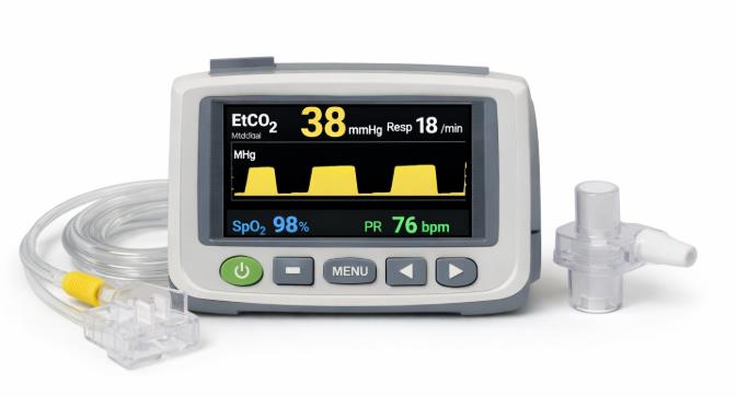
Capnograph Monitor
CO2 Sampling Line
CO2 Sensor
Airway Adapter
Patient Preparation
Informed Consent:
Before the procedure, assess the patient’s baseline respiratory status and overall health. Identify any pre-existing conditions that may affect ventilation.
Patient Positioning:
Properly position the patient for the procedure and secure the capnography equipment. Ensure that the sampling line is appropriately connected to the patient and the monitor.
TECHNIQUE
Step 1: Equipment Setup
Ensure that the capnography equipment is in good working condition.
Connect the CO2 sampling line to the patient.
Place the nasal cannula, endotracheal tube, or other airway device depending on the patient and procedure.
Step 2: Connection
Attach the CO2 sampling line to the airway adapter or the patient interface.
Step 3: Initialization
Turn on the capnograph monitor and allow it to initialize. This may involve a brief warm-up period.
Step 4: Baseline Assessment
Observe the initial baseline or Phase I of the capnogram, which represents the absence or low levels of CO2 at the beginning of exhalation.
End-Tidal Capnography equipment
COMPLICATIONS
Capnography readings may be influenced by factors that can lead to false readings, such as the presence of ambient air, water, or other contaminants in the sampling line. This can result in inaccurate measurements of end-tidal CO2.
Malfunctioning capnography equipment, including the monitor, sampling lines, or CO2 sensors, can lead to unreliable or erroneous readings.
Improper placement of the CO2 sampling line can lead to inaccurate readings. In cases of severe airway obstruction or bronchospasm, the exhalation of gases may be impeded, potentially leading to a decrease in end-tidal CO2 levels.
During cardiac arrest or situations with low cardiac output, there may be a delay in the appearance of end-tidal CO2 in the exhaled breath, as blood circulation and CO2 delivery to the lungs are compromised.
End-tidal capnography is a medical monitoring technique that measures the concentration of carbon dioxide (CO2) at the end of an exhaled breath, commonly referred to as end-tidal carbon dioxide (ETCO2).
This monitoring method provides valuable information about a patient’s respiratory status and is widely used in various medical settings, including emergency medicine, anesthesia, critical care, and respiratory therapy.
During the normal breathing cycle, the body exchanges oxygen (O2) for carbon dioxide (CO2) in the lungs. Oxygen is inhaled, and carbon dioxide, a byproduct of metabolism, is expelled during exhalation.
Capnography involves the measurement of CO2 levels in the exhaled breath using a capnograph. The capnograph produces a waveform known as the capnogram, which represents the concentration of CO2 over time.
The capnogram typically consists of three phases:
Baseline (Phase I): Represents the beginning of exhalation, where little to no CO2 is present.
Exhalation (Phase II): Reflects the rapid rise in CO2 concentration as most of the exhaled air contains CO2.
Inhalation (Phase III): Shows a gradual decrease in CO2 concentration as fresh, CO2-poor air is inhaled.
Verifying proper placement of an endotracheal tube during intubation is a critical application of capnography. A sudden increase in end-tidal CO2 confirms that the tube is in the trachea, while absence or a delayed increase may suggest esophageal intubation.
Capnography is routinely used during anesthesia to ensure proper ventilation. It helps in assessing the effectiveness of each breath and detecting issues such as hypoventilation, hyperventilation, or disconnection from the ventilator.
In emergency situations, such as cardiac arrest or respiratory failure, end-tidal capnography can be used to assess the effectiveness of cardiopulmonary resuscitation (CPR) and guide interventions.
Capnography can help identify conditions such as partial or complete airway obstruction by monitoring the waveform morphology. An abrupt increase in end-tidal CO2, accompanied by an abnormal waveform, may indicate airway obstruction.
In patients who are breathing spontaneously, end-tidal capnography provides continuous information about respiratory rate and helps detect respiratory distress or fatigue.
In cases where there is inadequate airflow or extremely low ventilation, such as severe respiratory distress or respiratory arrest, capnography waveforms may not be reliable or may be absent.
The quality and accuracy of capnography readings can be affected by equipment limitations. Malfunctioning or improperly calibrated equipment may provide inaccurate information.
Technical issues such as disconnected or malfunctioning sampling lines, expired mainstream or side stream CO2 sensors, or equipment failure can impact the reliability of capnography data.
Conditions that affect the collection of expired gases, such as low cardiac output, low pulmonary blood flow, or low respiratory rate, may influence the accuracy of capnography measurements.
While capnography is generally safe for use in pediatric patients, there may be considerations for infants and neonates, particularly regarding the size and placement of sensors and potential interference with proper ventilation.
This helps prevent complications associated with misplacement, such as inadequate ventilation or inadvertent esophageal intubation. Capnography allows for the early detection of respiratory issues, such as hypoventilation or hyperventilation, enabling prompt intervention and adjustment of ventilation parameters. This is particularly crucial in settings like anesthesia, where maintaining adequate ventilation is essential.
During cardiopulmonary resuscitation (CPR), end-tidal capnography is used to assess the quality of chest compressions and to guide interventions. In the operating room, capnography is a standard monitoring tool during anesthesia. It helps anesthesiologists ensure that patients are adequately ventilated and helps detect issues such as airway obstruction or equipment malfunction.
Capnography waveforms and numerical values can help identify airway obstructions, whether partial or complete. The information aids in the timely recognition and management of conditions such as bronchospasm or foreign body aspiration.
In spontaneously breathing patients, capnography provides continuous information about respiratory rate and the quality of each breath. This is particularly useful in monitoring patients in critical care or emergency settings. During procedural sedation, capnography helps in monitoring respiratory status and detecting hypoventilation early, ensuring patient safety.
Capnograph Monitor
CO2 Sampling Line
CO2 Sensor
Airway Adapter
Informed Consent:
Before the procedure, assess the patient’s baseline respiratory status and overall health. Identify any pre-existing conditions that may affect ventilation.
Patient Positioning:
Properly position the patient for the procedure and secure the capnography equipment. Ensure that the sampling line is appropriately connected to the patient and the monitor.
Step 1: Equipment Setup
Ensure that the capnography equipment is in good working condition.
Connect the CO2 sampling line to the patient.
Place the nasal cannula, endotracheal tube, or other airway device depending on the patient and procedure.
Step 2: Connection
Attach the CO2 sampling line to the airway adapter or the patient interface.
Step 3: Initialization
Turn on the capnograph monitor and allow it to initialize. This may involve a brief warm-up period.
Step 4: Baseline Assessment
Observe the initial baseline or Phase I of the capnogram, which represents the absence or low levels of CO2 at the beginning of exhalation.
End-Tidal Capnography equipment
Capnography readings may be influenced by factors that can lead to false readings, such as the presence of ambient air, water, or other contaminants in the sampling line. This can result in inaccurate measurements of end-tidal CO2.
Malfunctioning capnography equipment, including the monitor, sampling lines, or CO2 sensors, can lead to unreliable or erroneous readings.
Improper placement of the CO2 sampling line can lead to inaccurate readings. In cases of severe airway obstruction or bronchospasm, the exhalation of gases may be impeded, potentially leading to a decrease in end-tidal CO2 levels.
During cardiac arrest or situations with low cardiac output, there may be a delay in the appearance of end-tidal CO2 in the exhaled breath, as blood circulation and CO2 delivery to the lungs are compromised.
Both our subscription plans include Free CME/CPD AMA PRA Category 1 credits.
Digital Certificate PDF
On course completion, you will receive a full-sized presentation quality digital certificate.
medtigo Simulation
A dynamic medical simulation platform designed to train healthcare professionals and students to effectively run code situations through an immersive hands-on experience in a live, interactive 3D environment.
medtigo Points
medtigo points is our unique point redemption system created to award users for interacting on our site. These points can be redeemed for special discounts on the medtigo marketplace as well as towards the membership cost itself.
Community Forum post/reply = 5 points
*Redemption of points can occur only through the medtigo marketplace, courses, or simulation system. Money will not be credited to your bank account. 10 points = $1.
All Your Certificates in One Place
When you have your licenses, certificates and CMEs in one place, it's easier to track your career growth. You can easily share these with hospitals as well, using your medtigo app.