Endoscopic Retrograde Cholangiopancreatography (ERCP) combines luminal endoscopy and fluoroscopic imaging to diagnose and treat pancreato-biliary system conditions.
Duodenoscope passed through esophagus and stomach into second portion of duodenum for endoscopy purpose.
The hepatopancreatic ampulla is a protrusion into the duodenal lumen, where the ventral pancreatic duct and common bile duct converted into drain bile and pancreatic secretions.
The minor duodenal papilla in the second portion of the duodenum is an entry point for the dorsal pancreatic duct.
Visualized abnormalities can be treated with specialized accessories through endoscope channel with help of fluoroscopy.
ERCP has more serious complications compared to other endoscopic procedures. It required specialized training, and equipment should be used only when necessary.
Indications
Diagnostic indication:
Biliary Obstruction: It is used in evaluation of suspected obstruction in the bile duct.
Cholangitis or Pancreatitis
Biliary or Pancreatic Leaks
Biliary and Pancreatic Tumors
Therapeutic indication:
Bile Duct Stones: It is used to remove stones from the common bile duct.
Pancreatic Duct Pathologies: In treatment of pancreatic strictures or removal of intraductal stones.
Biliary Drainage: It is useful in percutaneous biliary drainage cases where it is not affordable or fails.
Stricture Management
Cholangitis
Contraindications
Absolute contraindication:
Unstable Cardiopulmonary Status
Acute Gastrointestinal Perforation
Uncorrected Coagulopathy or Bleeding Disorders
Allergy to Contrast Agents
Relative Contraindications:
Acute Pancreatitis
Severe Infection or Sepsis
Recent Upper Gastrointestinal Surgery
Severe Duodenal or Esophageal Strictures
Outcomes
Pre-procedure review of patient’s surgical history necessary to identify any anatomy contraindications for ERCP.
Deep sedation in ERCP allows for stable endoscopic position in duodenum for successful procedures.
A study found no correlation between low case volume and risk of PEP in the single endoscopist or centre.
Mucosal perforations during ERCP are usually periampullary and related to sphincterotomy are managed supportively without immediate surgery.
The duodenoscope should not be forced against resistance during insertion. The forceps elevator should be closed to prevent lacerating adjacent tissue.
Periprocedural Care
Equipment required:
Endoscope
Cannulation devices
Protective gear
Therapeutic devices
Hemostasis and Clip devices
Anesthesia equipment
Patient Preparation:
Study found no difference in papillary cannulation rates with propofol plus fentanyl compared to propofol.
Typical sedation protocol involves IV benzodiazepine and narcotic administered by nurse or physician.
Antimotility drugs help slow bowel movements during ERCP for better visualization and cannulation in patients.
Patient Positioning:
In some endoscopies, the semi prone position is preferred for better access to the small bowel despite potentially lower image quality.
Supine position is chosen based on patient comorbidities and pregnancy for closer airway monitoring.
Patient should be positioned in left lateral or semi-prone position for better accessibility.
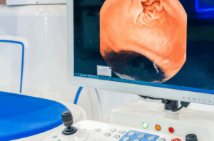
Duodenoscope used to view ampulla of Vater, biliary, and pancreatic ducts.
Endoscopic view of duodenum
Technique
Step 1: Insertion of the Endoscope
The endoscope is passed through the mouth, stomach, and duodenum to identify ampulla of Vater.
Step 2: Cannulation of the Duct
A cannula is inserted through the channel of the duodenoscope to get easy access to the duct. Put the duodenoscope in the second portion of the duodenum.
Step 3: Cannulation
Selectively cannulate the common bile duct. Cannulation of the minor duct requires a smaller cannulating device.
Step 4: Insertion of Contrast Agent:
Radiopaque contrast dye is injected into the bile or pancreatic duct.
Fluoroscopic images are taken to assess the ducts for obstructions or leaks.
Step 5: Withdrawal of wire:
Confirm guide wire with fluoroscopy in pancreatic duct. Then scope is withdrawn after completion of the procedure.
Complications:
ERCP has higher procedure-related risks compared to other endoscopic procedures due to its advanced nature.
Endoscopic Retrograde Cholangiopancreatography (ERCP) combines luminal endoscopy and fluoroscopic imaging to diagnose and treat pancreato-biliary system conditions.
Duodenoscope passed through esophagus and stomach into second portion of duodenum for endoscopy purpose.
The hepatopancreatic ampulla is a protrusion into the duodenal lumen, where the ventral pancreatic duct and common bile duct converted into drain bile and pancreatic secretions.
The minor duodenal papilla in the second portion of the duodenum is an entry point for the dorsal pancreatic duct.
Visualized abnormalities can be treated with specialized accessories through endoscope channel with help of fluoroscopy.
ERCP has more serious complications compared to other endoscopic procedures. It required specialized training, and equipment should be used only when necessary.
Diagnostic indication:
Biliary Obstruction: It is used in evaluation of suspected obstruction in the bile duct.
Cholangitis or Pancreatitis
Biliary or Pancreatic Leaks
Biliary and Pancreatic Tumors
Therapeutic indication:
Bile Duct Stones: It is used to remove stones from the common bile duct.
Pancreatic Duct Pathologies: In treatment of pancreatic strictures or removal of intraductal stones.
Biliary Drainage: It is useful in percutaneous biliary drainage cases where it is not affordable or fails.
Stricture Management
Cholangitis
Absolute contraindication:
Unstable Cardiopulmonary Status
Acute Gastrointestinal Perforation
Uncorrected Coagulopathy or Bleeding Disorders
Allergy to Contrast Agents
Relative Contraindications:
Acute Pancreatitis
Severe Infection or Sepsis
Recent Upper Gastrointestinal Surgery
Severe Duodenal or Esophageal Strictures
Pre-procedure review of patient’s surgical history necessary to identify any anatomy contraindications for ERCP.
Deep sedation in ERCP allows for stable endoscopic position in duodenum for successful procedures.
A study found no correlation between low case volume and risk of PEP in the single endoscopist or centre.
Mucosal perforations during ERCP are usually periampullary and related to sphincterotomy are managed supportively without immediate surgery.
The duodenoscope should not be forced against resistance during insertion. The forceps elevator should be closed to prevent lacerating adjacent tissue.
Equipment required:
Endoscope
Cannulation devices
Protective gear
Therapeutic devices
Hemostasis and Clip devices
Anesthesia equipment
Patient Preparation:
Study found no difference in papillary cannulation rates with propofol plus fentanyl compared to propofol.
Typical sedation protocol involves IV benzodiazepine and narcotic administered by nurse or physician.
Antimotility drugs help slow bowel movements during ERCP for better visualization and cannulation in patients.
Patient Positioning:
In some endoscopies, the semi prone position is preferred for better access to the small bowel despite potentially lower image quality.
Supine position is chosen based on patient comorbidities and pregnancy for closer airway monitoring.
Patient should be positioned in left lateral or semi-prone position for better accessibility.
Duodenoscope used to view ampulla of Vater, biliary, and pancreatic ducts.
Endoscopic view of duodenum
Step 1: Insertion of the Endoscope
The endoscope is passed through the mouth, stomach, and duodenum to identify ampulla of Vater.
Step 2: Cannulation of the Duct
A cannula is inserted through the channel of the duodenoscope to get easy access to the duct. Put the duodenoscope in the second portion of the duodenum.
Step 3: Cannulation
Selectively cannulate the common bile duct. Cannulation of the minor duct requires a smaller cannulating device.
Step 4: Insertion of Contrast Agent:
Radiopaque contrast dye is injected into the bile or pancreatic duct.
Fluoroscopic images are taken to assess the ducts for obstructions or leaks.
Step 5: Withdrawal of wire:
Confirm guide wire with fluoroscopy in pancreatic duct. Then scope is withdrawn after completion of the procedure.
Complications:
ERCP has higher procedure-related risks compared to other endoscopic procedures due to its advanced nature.
Both our subscription plans include Free CME/CPD AMA PRA Category 1 credits.
Digital Certificate PDF
On course completion, you will receive a full-sized presentation quality digital certificate.
medtigo Simulation
A dynamic medical simulation platform designed to train healthcare professionals and students to effectively run code situations through an immersive hands-on experience in a live, interactive 3D environment.
medtigo Points
medtigo points is our unique point redemption system created to award users for interacting on our site. These points can be redeemed for special discounts on the medtigo marketplace as well as towards the membership cost itself.
Community Forum post/reply = 5 points
*Redemption of points can occur only through the medtigo marketplace, courses, or simulation system. Money will not be credited to your bank account. 10 points = $1.
All Your Certificates in One Place
When you have your licenses, certificates and CMEs in one place, it's easier to track your career growth. You can easily share these with hospitals as well, using your medtigo app.