A Full-Field Electroretinogram is a diagnostic test used to measure the electrical responses generated by the retina when stimulated by light. It provides a comprehensive evaluation of the retina’s overall function, including both the rod and cone photoreceptors, as well as their associated pathways. By recording these responses, the ffERG plays a critical role in diagnosing and monitoring various retinal disorders, such as retinitis pigmentosa, cone-rod dystrophies, and retinal toxicities caused by medications. The test is non-invasive and typically involves placing electrodes on the skin or around the eyes to detect electrical activity in response to light stimuli. It is widely utilized in clinical and research settings to assess retinal health and guide treatment decisions.
Indications
Retinal Dystrophies
Retinitis Pigmentosa: To assess the extent of retinal dysfunction and monitor disease progression.
Cone-Rod Dystrophy: To differentiate between rod and cone dysfunction.
Leber Congenital Amaurosis (LCA): For diagnosis and confirmation of early-onset severe retinal dystrophy.
Stargardt Disease: To evaluate retinal function in cases with macular dystrophy.
Inherited Retinal Diseases
To confirm and classify genetic retinal disorders, especially when clinical symptoms are subtle.
Toxic Retinopathies
Drug-Induced Retinopathies: Such as chloroquine or hydroxychloroquine toxicity, to detect early retinal dysfunction before structural changes occur.
Acquired Retinal Conditions
Autoimmune Retinopathies: To diagnose immune-mediated retinal dysfunction, such as cancer-associated retinopathy (CAR) or melanoma-associated retinopathy (MAR).
Retinal Vascular Diseases: To evaluate retinal function in cases of ischemic retinopathies.
Unexplained Visual Loss
For cases where structural imaging (OCT, fundus examination) is normal, but visual complaints persist.
Night Blindness (Nyctalopia)
To evaluate rod function in conditions causing difficulty seeing in low light.
Contraindications
Severe Active Eye Infections:
Conditions like conjunctivitis, keratitis, or other ocular infections can be exacerbated by the placement of electrodes or contact lens electrodes.
Severe Ocular Surface Disorders:
Corneal injuries, severe dry eye, or other conditions affecting the ocular surface can lead to discomfort or complications during the test.
Recent Eye Surgery:
Patients who have undergone recent intraocular or corneal surgery may have fragile ocular structures, increasing the risk of complications from the procedure.
Hypersensitivity to Electrode Materials:
Allergic reactions to materials used in electrodes (e.g., gold, silver, or other contact materials) may contraindicate the use of specific types of electrodes.
Severe Photophobia:
Patients with extreme sensitivity to light may experience significant discomfort or inability to tolerate the test.
Inability to Cooperate:
Young children, individuals with cognitive impairments, or uncooperative patients may be unable to stay still or follow instructions during the procedure.
Outcomes
Equipment
Digital recording system
Ganzfield stimulator
Electrodes
Patient preparation
Schedule and Explanation: Inform the patient about the test procedure, duration, and purpose.
Eye Dilation: Administer dilating eye drops 20-30 minutes before the test.
Contact Lens: Ensure the patient is comfortable with the electrode contact lens if used.
Avoid Makeup: Advise against using eye makeup on the day of the test.
Rest and Light Exposure: Minimize exposure to bright light before testing and ensure adequate rest.
Medication Disclosure: Confirm medications or eye drops being used, as they may affect results.
Patient position
Seated or Reclined Position:
The patient is typically seated in a comfortable chair or reclined to ensure they remain still during the procedure.
A reclined position may be preferred for patients who might have difficulty holding a steady posture or for longer sessions.
Head Stabilization:
The patient’s head should be stabilized, often using a headrest, to minimize movement and maintain alignment with the recording equipment.
Technique
Step 1-Preparation:
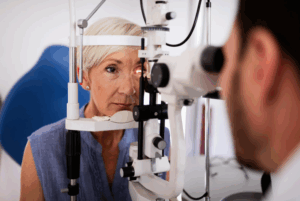
Patient Positioning: The patient should be seated comfortably in a dark room. Their head should be stabilized, often using a chin rest to minimize movement during the test.
Dilation: Pupils should be dilated using a topical ophthalmic solution (e.g., tropicamide) to allow optimal light exposure to the retina. The dilation also helps ensure accurate responses by minimizing pupil constriction during testing.
Step 2-Electrode Placement:
Corneal Electrode: A specialized electrode is placed on the cornea or close to it (often a contact lens electrode). This electrode records the electrical response generated by the retina when exposed to light stimuli.
Reference and Ground Electrodes: A reference electrode is usually placed on the skin near the outer canthus of the eye or on the forehead. A ground electrode may be placed on the earlobe or other neutral site to complete the circuit.
Step 3-Stimulus Presentation:
Flash Stimuli: The retina is stimulated with brief flashes of light, which vary in intensity. Different flash intensities are used to assess the function of both rods (which respond to dim light) and cones (which respond to bright light).
Standardized Lighting Conditions: The test is typically done in both dark-adapted and light-adapted conditions. For dark adaptation, the patient is kept in complete darkness for a period (usually 20-30 minutes), and for light adaptation, the patient may be exposed to a steady light source prior to testing.
Stimulus Duration and Intensity: Stimuli can vary in duration (e.g., milliseconds) and intensity (e.g., from very dim to bright flashes) to test different aspects of retinal function.
Step 4-Recording the Response:
The electrical responses (ERG waveforms) are recorded by the electrode. These responses include several components:
a-wave: This represents the initial negative deflection caused by the photoreceptor (rod and cone) activity.
b-wave: This positive deflection is generated by the activity of the inner retinal layers, particularly the bipolar cells.
c-wave: This may be seen in some tests, reflecting the outer retinal response.
d-wave: A later positive deflection in some conditions, reflecting further inner retinal activity.
Responses are recorded for various light intensities, and the waveform is analyzed for abnormalities in amplitude, latency, and shape.
Step 5-Data Analysis:
The results are compared to normative values for age and other factors.
Abnormalities in the waveforms (e.g., reduced amplitude, delayed latency) can indicate retinal dysfunction or damage, with potential implications for conditions like retinal degenerations, diabetic retinopathy, or optic neuropathies.
Step 6-Post-Test Considerations:
After testing, patients can usually resume normal activities, although they may experience temporary vision blurring or discomfort from the dilation.
The data are then interpreted by an ophthalmologist or a retinal specialist, who can make clinical decisions based on the findings.
Complications
Discomfort or Mild Irritation: The procedure may cause mild discomfort, as it involves the placement of electrodes on the cornea (with a contact lens electrode) or on the skin near the eye. Some patients may experience irritation or dryness from the contact lens or gel used.
Light Sensitivity: The test involves exposing the eyes to bright flashes of light, which may cause temporary discomfort or visual disturbances, especially for those sensitive to light.
Inaccurate Results: If the patient is unable to remain still or properly follow instructions during the test, the results may be inaccurate. Eye movements, blinking, or excessive noise can interfere with the measurements.
Visual Disturbances: Some individuals may experience temporary visual disturbances (e.g., afterimages or spots) due to the brightness of the test stimuli. These usually resolve shortly after the procedure.
Allergic Reactions: Although rare, some individuals may be allergic to the materials used, such as the conductive gel or materials in the contact lens electrode.
Potential for Eye Injury: While uncommon, improper handling or accidental contact with the electrode during the test may cause minor injury or irritation to the eye.
A Full-Field Electroretinogram is a diagnostic test used to measure the electrical responses generated by the retina when stimulated by light. It provides a comprehensive evaluation of the retina’s overall function, including both the rod and cone photoreceptors, as well as their associated pathways. By recording these responses, the ffERG plays a critical role in diagnosing and monitoring various retinal disorders, such as retinitis pigmentosa, cone-rod dystrophies, and retinal toxicities caused by medications. The test is non-invasive and typically involves placing electrodes on the skin or around the eyes to detect electrical activity in response to light stimuli. It is widely utilized in clinical and research settings to assess retinal health and guide treatment decisions.
Retinal Dystrophies
Retinitis Pigmentosa: To assess the extent of retinal dysfunction and monitor disease progression.
Cone-Rod Dystrophy: To differentiate between rod and cone dysfunction.
Leber Congenital Amaurosis (LCA): For diagnosis and confirmation of early-onset severe retinal dystrophy.
Stargardt Disease: To evaluate retinal function in cases with macular dystrophy.
Inherited Retinal Diseases
To confirm and classify genetic retinal disorders, especially when clinical symptoms are subtle.
Toxic Retinopathies
Drug-Induced Retinopathies: Such as chloroquine or hydroxychloroquine toxicity, to detect early retinal dysfunction before structural changes occur.
Acquired Retinal Conditions
Autoimmune Retinopathies: To diagnose immune-mediated retinal dysfunction, such as cancer-associated retinopathy (CAR) or melanoma-associated retinopathy (MAR).
Retinal Vascular Diseases: To evaluate retinal function in cases of ischemic retinopathies.
Unexplained Visual Loss
For cases where structural imaging (OCT, fundus examination) is normal, but visual complaints persist.
Night Blindness (Nyctalopia)
To evaluate rod function in conditions causing difficulty seeing in low light.
Severe Active Eye Infections:
Conditions like conjunctivitis, keratitis, or other ocular infections can be exacerbated by the placement of electrodes or contact lens electrodes.
Severe Ocular Surface Disorders:
Corneal injuries, severe dry eye, or other conditions affecting the ocular surface can lead to discomfort or complications during the test.
Recent Eye Surgery:
Patients who have undergone recent intraocular or corneal surgery may have fragile ocular structures, increasing the risk of complications from the procedure.
Hypersensitivity to Electrode Materials:
Allergic reactions to materials used in electrodes (e.g., gold, silver, or other contact materials) may contraindicate the use of specific types of electrodes.
Severe Photophobia:
Patients with extreme sensitivity to light may experience significant discomfort or inability to tolerate the test.
Inability to Cooperate:
Young children, individuals with cognitive impairments, or uncooperative patients may be unable to stay still or follow instructions during the procedure.
Digital recording system
Ganzfield stimulator
Electrodes
Schedule and Explanation: Inform the patient about the test procedure, duration, and purpose.
Eye Dilation: Administer dilating eye drops 20-30 minutes before the test.
Contact Lens: Ensure the patient is comfortable with the electrode contact lens if used.
Avoid Makeup: Advise against using eye makeup on the day of the test.
Rest and Light Exposure: Minimize exposure to bright light before testing and ensure adequate rest.
Medication Disclosure: Confirm medications or eye drops being used, as they may affect results.
Seated or Reclined Position:
The patient is typically seated in a comfortable chair or reclined to ensure they remain still during the procedure.
A reclined position may be preferred for patients who might have difficulty holding a steady posture or for longer sessions.
Head Stabilization:
The patient’s head should be stabilized, often using a headrest, to minimize movement and maintain alignment with the recording equipment.
Step 1-Preparation:
Patient Positioning: The patient should be seated comfortably in a dark room. Their head should be stabilized, often using a chin rest to minimize movement during the test.
Dilation: Pupils should be dilated using a topical ophthalmic solution (e.g., tropicamide) to allow optimal light exposure to the retina. The dilation also helps ensure accurate responses by minimizing pupil constriction during testing.
Step 2-Electrode Placement:
Corneal Electrode: A specialized electrode is placed on the cornea or close to it (often a contact lens electrode). This electrode records the electrical response generated by the retina when exposed to light stimuli.
Reference and Ground Electrodes: A reference electrode is usually placed on the skin near the outer canthus of the eye or on the forehead. A ground electrode may be placed on the earlobe or other neutral site to complete the circuit.
Step 3-Stimulus Presentation:
Flash Stimuli: The retina is stimulated with brief flashes of light, which vary in intensity. Different flash intensities are used to assess the function of both rods (which respond to dim light) and cones (which respond to bright light).
Standardized Lighting Conditions: The test is typically done in both dark-adapted and light-adapted conditions. For dark adaptation, the patient is kept in complete darkness for a period (usually 20-30 minutes), and for light adaptation, the patient may be exposed to a steady light source prior to testing.
Stimulus Duration and Intensity: Stimuli can vary in duration (e.g., milliseconds) and intensity (e.g., from very dim to bright flashes) to test different aspects of retinal function.
Step 4-Recording the Response:
The electrical responses (ERG waveforms) are recorded by the electrode. These responses include several components:
a-wave: This represents the initial negative deflection caused by the photoreceptor (rod and cone) activity.
b-wave: This positive deflection is generated by the activity of the inner retinal layers, particularly the bipolar cells.
c-wave: This may be seen in some tests, reflecting the outer retinal response.
d-wave: A later positive deflection in some conditions, reflecting further inner retinal activity.
Responses are recorded for various light intensities, and the waveform is analyzed for abnormalities in amplitude, latency, and shape.
Step 5-Data Analysis:
The results are compared to normative values for age and other factors.
Abnormalities in the waveforms (e.g., reduced amplitude, delayed latency) can indicate retinal dysfunction or damage, with potential implications for conditions like retinal degenerations, diabetic retinopathy, or optic neuropathies.
Step 6-Post-Test Considerations:
After testing, patients can usually resume normal activities, although they may experience temporary vision blurring or discomfort from the dilation.
The data are then interpreted by an ophthalmologist or a retinal specialist, who can make clinical decisions based on the findings.
Discomfort or Mild Irritation: The procedure may cause mild discomfort, as it involves the placement of electrodes on the cornea (with a contact lens electrode) or on the skin near the eye. Some patients may experience irritation or dryness from the contact lens or gel used.
Light Sensitivity: The test involves exposing the eyes to bright flashes of light, which may cause temporary discomfort or visual disturbances, especially for those sensitive to light.
Inaccurate Results: If the patient is unable to remain still or properly follow instructions during the test, the results may be inaccurate. Eye movements, blinking, or excessive noise can interfere with the measurements.
Visual Disturbances: Some individuals may experience temporary visual disturbances (e.g., afterimages or spots) due to the brightness of the test stimuli. These usually resolve shortly after the procedure.
Allergic Reactions: Although rare, some individuals may be allergic to the materials used, such as the conductive gel or materials in the contact lens electrode.
Potential for Eye Injury: While uncommon, improper handling or accidental contact with the electrode during the test may cause minor injury or irritation to the eye.
Both our subscription plans include Free CME/CPD AMA PRA Category 1 credits.
Digital Certificate PDF
On course completion, you will receive a full-sized presentation quality digital certificate.
medtigo Simulation
A dynamic medical simulation platform designed to train healthcare professionals and students to effectively run code situations through an immersive hands-on experience in a live, interactive 3D environment.
medtigo Points
medtigo points is our unique point redemption system created to award users for interacting on our site. These points can be redeemed for special discounts on the medtigo marketplace as well as towards the membership cost itself.
Community Forum post/reply = 5 points
*Redemption of points can occur only through the medtigo marketplace, courses, or simulation system. Money will not be credited to your bank account. 10 points = $1.
All Your Certificates in One Place
When you have your licenses, certificates and CMEs in one place, it's easier to track your career growth. You can easily share these with hospitals as well, using your medtigo app.