Background
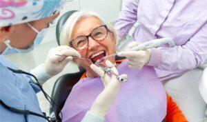
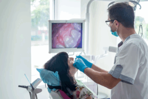
Gingivectomy and gingivoplasty are two common periodontal surgical procedures aimed at improving gum health and aesthetics. While both involve the reshaping of gum tissue, they serve distinct purposes in dental treatment.
Gingivectomy is the surgical removal of diseased or excess gum tissue to eliminate pockets between the teeth and gums, often performed to treat periodontal disease or overgrown gums due to medication or genetic conditions.
Gingivoplasty is a cosmetic or functional procedure that reshapes the gum tissue to improve its contour, often enhancing the appearance of the smile or restoring normal gum structure after disease or trauma.
Both procedures can be performed using scalpels, lasers, or electrosurgery, depending on the patient’s condition and the dentist’s approach. They contribute to better oral hygiene, improved aesthetics, and overall periodontal health.
Indications
Periodontal and Aesthetic Conditions:
Gingival Hyperplasia (Drug-Induced or Idiopathic): Overgrown gums due to medications (e.g., phenytoin, cyclosporine, calcium channel blockers) or hereditary conditions.
Chronic Inflammatory Gingival Enlargement: Due to poor oral hygiene or plaque-induced inflammation.
Functional and Restorative Indications:
Pseudopockets (False Periodontal Pockets): To eliminate excess gum tissue that traps plaque and bacteria, making oral hygiene difficult.
Pre-Prosthetic Surgery: To ensure proper fit and aesthetics of crowns, bridges, or dentures.
Post-Orthodontic Treatment:
Gingival Recontouring After Braces: To reshape thickened or irregular gum tissue after orthodontic therapy.
Contraindications
Systemic Contraindications
Uncontrolled systemic diseases (e.g., diabetes, hypertension, or cardiovascular diseases)
Bleeding disorders (e.g., hemophilia, thrombocytopenia, or patients on anticoagulants without proper medical consultation)
Immunocompromised conditions (e.g., HIV/AIDS, undergoing chemotherapy, or organ transplant recipients)
Poor wound healing conditions (e.g., uncontrolled diabetes, malnutrition)
Local Contraindications
Inadequate attached gingiva: Removing tissue could lead to mucogingival problems.
Periodontal disease with inadequate bone support: If bone loss is present, gingivectomy may worsen attachment loss.
Presence of deep periodontal pockets: If bone recontouring is required, flap surgery (not gingivectomy) is the preferred option.
Acute oral infections (e.g., herpetic gingivostomatitis, acute necrotizing ulcerative gingivitis): Treatment should be delayed until the infection resolves.
Poor oral hygiene: May lead to recurrence of the condition or post-operative infections.
High caries risk: Exposing root surfaces due to excessive tissue removal can increase susceptibility to decay.
Outcomes
Equipment’s
Basic Instruments:
Scalpel
Gingivectomy Knives – Special knives like:
Kirkland Knife (for broad incisions)
Orban Knife (for interproximal areas)
Periodontal Scissors
Tissue Forceps
Surgical Curettes
Electrosurgical & Laser Equipment:
Electrosurgical Unit
Dental Laser (Diode, CO₂, or Er:YAG)
Additional Instruments:
Periodontal Probe
Hemostatic Agents
Sutures & Needle Holders
Saliva Ejector & Suction Tips
Surgical Dressing (Periodontal Pack)
Patient Preparation
Preoperative Assessment

Medical & Dental History:
Review systemic conditions (diabetes, bleeding disorders, immunosuppression, etc.).
Check medications (anticoagulants, bisphosphonates, immunosuppressants).
Oral Examination:
Identify areas of gingival overgrowth, pocket depths, and tissue contour.
Evaluate oral hygiene and plaque levels.
Radiographic Evaluation:
Periapical or panoramic X-rays to assess bone support and root structures.
Periodontal Charting:
Measure pocket depths and gum thickness for precise surgical planning.
Patient Instructions Before Surgery
Oral Hygiene Preparation:
Advise thorough brushing and flossing.
Prescribe antimicrobial mouthwash (e.g., chlorhexidine) for pre-procedure use.
Dietary Restrictions:
Instruct fasting if sedation or general anesthesia is planned.
Medication Adjustments (if necessary):
Consult with a physician for patients on blood thinners.
Prescribe prophylactic antibiotics for immunocompromised patients.
Smoking & Alcohol:
Advise to avoid smoking and alcohol before and after surgery to promote healing.
Patient Position
Supine Position: The patient should be lying flat on their back to provide maximum visibility and accessibility to the surgical site.
Slight Head Elevation: The head should be slightly elevated to minimize blood pooling and improve the operator’s field of vision.
Comfortable Neck Support: Ensure the patient’s neck is supported to prevent strain during the procedure.
Gingivectomy
Step 1-Preoperative Preparation:
Patient Evaluation
Assess medical history, periodontal condition, and indications for gingivectomy.
Perform clinical and radiographic examinations.
Informed Consent
Explain the procedure, risks, benefits, and post-op care to the patient.
Oral Hygiene Instructions
Ensure the patient follows good oral hygiene practices before the procedure.
Anesthesia
Administer local anesthesia to numb the surgical area.
Step 2-Surgical Procedure:
Marking the Incision Line
Use a periodontal probe to measure pocket depth and determine the incision line.
Mark the incision line with a surgical pen or bleeding points using a probe.
Incision
Use a scalpel (e.g., No. 15 blade) or electrosurgery/laser to make an external bevel incision, removing excess gingival tissue.
Tissue Removal
Excise the hyperplastic or diseased gingival tissue following the incision line.
Smoothing the Margins
Use a scalpel, rotary bur, or periodontal knives (e.g., Orban knife) to shape and contour the remaining gingiva.
Hemostasis (Bleeding Control)
Achieve hemostasis using gauze pressure, electrocautery, or hemostatic agents.
Step 3-Postoperative Care:
Dressing & Protection
Apply a periodontal dressing if necessary to protect the surgical site.
Postoperative Instructions
Advise the patient on pain management, oral hygiene, diet, and activity restrictions.
Prescribe analgesics and antimicrobial mouth rinses if needed.
Follow-Up & Healing Assessment
Schedule follow-up visits to monitor healing and address any complications.
Gingivoplasty
Step 1: Patient Preparation

Gingivoplasty surgery
Obtain medical and dental history to assess contraindications.
Explain the procedure and obtain informed consent.
Perform a thorough oral examination, including periodontal probing and radiographic evaluation.
Conduct professional scaling and root planing (if necessary).
Administer local anesthesia to ensure patient comfort.
Step 2: Surgical Planning
Mark the ideal gingival contours using a periodontal probe or a surgical marker.
Define the desired gingival shape, considering aesthetic and functional factors.
Step 3: Gingival Incision
Use a scalpel (blade No. 15 or 12) or electrocautery/laser to make precise incisions.
Remove excess or hyperplastic tissue while maintaining natural scalloped contours.
Shape the gingival margins smoothly to create a natural and symmetrical appearance.
Step 4: Tissue Contouring and Refinement
Sculpt the gingival tissue carefully using gingivoplasty knives, electrosurgery, or soft tissue lasers.
Blend the tissue contours smoothly into the surrounding gingiva.
Ensure an appropriate thickness to avoid excessive exposure of underlying structures.
Step 5: Hemostasis and Post-Surgical Care
Achieve hemostasis using pressure, cautery, or hemostatic agents if necessary.
Irrigate the surgical site with sterile saline or an antiseptic solution.
Place a periodontal dressing if required to protect the surgical site.
Prescribe analgesics, antibiotics (if indicated), and an antimicrobial mouth rinse (e.g., chlorhexidine).
Step 6: Postoperative Instructions
Advise the patient to avoid hard or spicy foods and practice gentle oral hygiene.
Instruct on the use of prescribed medications and mouth rinses.
Schedule a follow-up visits to monitor healing and assess tissue response.
Complications
Pain and Discomfort
Postoperative pain is common due to exposed connective tissue and nerve endings.
The intensity varies depending on the extent of tissue removal and patient pain tolerance.
Bleeding (Hemorrhage)
Excessive bleeding can occur due to trauma to blood vessels.
Patients with clotting disorders, or those on anticoagulants, are at higher risk.
Infection
The open wound from the procedure creates a potential entry point for bacteria.
Poor oral hygiene increases the risk of postoperative infection.
Delayed Wound Healing
Healing can be prolonged in patients with systemic conditions like diabetes or smokers.
Inadequate tissue adaptation may result in slow epithelialization.
Excessive Gingival Recession
Excessive tissue removal can expose root surfaces, leading to sensitivity and aesthetic concerns.
Root exposure increases the risk of root caries and dentin hypersensitivity.
Relapse of Gingival Overgrowth
Recurrence may happen in cases of drug-induced gingival hyperplasia (e.g., phenytoin, cyclosporine, calcium channel blockers).
Poor oral hygiene and genetic predisposition can also contribute.
Scarring and Fibrosis
Irregular wound healing may lead to fibrotic tissue formation.
It can affect aesthetics and periodontal function.
References
References

Gingivectomy and gingivoplasty are two common periodontal surgical procedures aimed at improving gum health and aesthetics. While both involve the reshaping of gum tissue, they serve distinct purposes in dental treatment.
Gingivectomy is the surgical removal of diseased or excess gum tissue to eliminate pockets between the teeth and gums, often performed to treat periodontal disease or overgrown gums due to medication or genetic conditions.
Gingivoplasty is a cosmetic or functional procedure that reshapes the gum tissue to improve its contour, often enhancing the appearance of the smile or restoring normal gum structure after disease or trauma.
Both procedures can be performed using scalpels, lasers, or electrosurgery, depending on the patient’s condition and the dentist’s approach. They contribute to better oral hygiene, improved aesthetics, and overall periodontal health.
Periodontal and Aesthetic Conditions:
Gingival Hyperplasia (Drug-Induced or Idiopathic): Overgrown gums due to medications (e.g., phenytoin, cyclosporine, calcium channel blockers) or hereditary conditions.
Chronic Inflammatory Gingival Enlargement: Due to poor oral hygiene or plaque-induced inflammation.
Functional and Restorative Indications:
Pseudopockets (False Periodontal Pockets): To eliminate excess gum tissue that traps plaque and bacteria, making oral hygiene difficult.
Pre-Prosthetic Surgery: To ensure proper fit and aesthetics of crowns, bridges, or dentures.
Post-Orthodontic Treatment:
Gingival Recontouring After Braces: To reshape thickened or irregular gum tissue after orthodontic therapy.
Systemic Contraindications
Uncontrolled systemic diseases (e.g., diabetes, hypertension, or cardiovascular diseases)
Bleeding disorders (e.g., hemophilia, thrombocytopenia, or patients on anticoagulants without proper medical consultation)
Immunocompromised conditions (e.g., HIV/AIDS, undergoing chemotherapy, or organ transplant recipients)
Poor wound healing conditions (e.g., uncontrolled diabetes, malnutrition)
Local Contraindications
Inadequate attached gingiva: Removing tissue could lead to mucogingival problems.
Periodontal disease with inadequate bone support: If bone loss is present, gingivectomy may worsen attachment loss.
Presence of deep periodontal pockets: If bone recontouring is required, flap surgery (not gingivectomy) is the preferred option.
Acute oral infections (e.g., herpetic gingivostomatitis, acute necrotizing ulcerative gingivitis): Treatment should be delayed until the infection resolves.
Poor oral hygiene: May lead to recurrence of the condition or post-operative infections.
High caries risk: Exposing root surfaces due to excessive tissue removal can increase susceptibility to decay.
Basic Instruments:
Scalpel
Gingivectomy Knives – Special knives like:
Kirkland Knife (for broad incisions)
Orban Knife (for interproximal areas)
Periodontal Scissors
Tissue Forceps
Surgical Curettes
Electrosurgical & Laser Equipment:
Electrosurgical Unit
Dental Laser (Diode, CO₂, or Er:YAG)
Additional Instruments:
Periodontal Probe
Hemostatic Agents
Sutures & Needle Holders
Saliva Ejector & Suction Tips
Surgical Dressing (Periodontal Pack)
Patient Preparation
Preoperative Assessment

Medical & Dental History:
Review systemic conditions (diabetes, bleeding disorders, immunosuppression, etc.).
Check medications (anticoagulants, bisphosphonates, immunosuppressants).
Oral Examination:
Identify areas of gingival overgrowth, pocket depths, and tissue contour.
Evaluate oral hygiene and plaque levels.
Radiographic Evaluation:
Periapical or panoramic X-rays to assess bone support and root structures.
Periodontal Charting:
Measure pocket depths and gum thickness for precise surgical planning.
Patient Instructions Before Surgery
Oral Hygiene Preparation:
Advise thorough brushing and flossing.
Prescribe antimicrobial mouthwash (e.g., chlorhexidine) for pre-procedure use.
Dietary Restrictions:
Instruct fasting if sedation or general anesthesia is planned.
Medication Adjustments (if necessary):
Consult with a physician for patients on blood thinners.
Prescribe prophylactic antibiotics for immunocompromised patients.
Smoking & Alcohol:
Advise to avoid smoking and alcohol before and after surgery to promote healing.
Patient Position
Supine Position: The patient should be lying flat on their back to provide maximum visibility and accessibility to the surgical site.
Slight Head Elevation: The head should be slightly elevated to minimize blood pooling and improve the operator’s field of vision.
Comfortable Neck Support: Ensure the patient’s neck is supported to prevent strain during the procedure.
Step 1-Preoperative Preparation:
Patient Evaluation
Assess medical history, periodontal condition, and indications for gingivectomy.
Perform clinical and radiographic examinations.
Informed Consent
Explain the procedure, risks, benefits, and post-op care to the patient.
Oral Hygiene Instructions
Ensure the patient follows good oral hygiene practices before the procedure.
Anesthesia
Administer local anesthesia to numb the surgical area.
Step 2-Surgical Procedure:
Marking the Incision Line
Use a periodontal probe to measure pocket depth and determine the incision line.
Mark the incision line with a surgical pen or bleeding points using a probe.
Incision
Use a scalpel (e.g., No. 15 blade) or electrosurgery/laser to make an external bevel incision, removing excess gingival tissue.
Tissue Removal
Excise the hyperplastic or diseased gingival tissue following the incision line.
Smoothing the Margins
Use a scalpel, rotary bur, or periodontal knives (e.g., Orban knife) to shape and contour the remaining gingiva.
Hemostasis (Bleeding Control)
Achieve hemostasis using gauze pressure, electrocautery, or hemostatic agents.
Step 3-Postoperative Care:
Dressing & Protection
Apply a periodontal dressing if necessary to protect the surgical site.
Postoperative Instructions
Advise the patient on pain management, oral hygiene, diet, and activity restrictions.
Prescribe analgesics and antimicrobial mouth rinses if needed.
Follow-Up & Healing Assessment
Schedule follow-up visits to monitor healing and address any complications.
Step 1: Patient Preparation

Gingivoplasty surgery
Obtain medical and dental history to assess contraindications.
Explain the procedure and obtain informed consent.
Perform a thorough oral examination, including periodontal probing and radiographic evaluation.
Conduct professional scaling and root planing (if necessary).
Administer local anesthesia to ensure patient comfort.
Step 2: Surgical Planning
Mark the ideal gingival contours using a periodontal probe or a surgical marker.
Define the desired gingival shape, considering aesthetic and functional factors.
Step 3: Gingival Incision
Use a scalpel (blade No. 15 or 12) or electrocautery/laser to make precise incisions.
Remove excess or hyperplastic tissue while maintaining natural scalloped contours.
Shape the gingival margins smoothly to create a natural and symmetrical appearance.
Step 4: Tissue Contouring and Refinement
Sculpt the gingival tissue carefully using gingivoplasty knives, electrosurgery, or soft tissue lasers.
Blend the tissue contours smoothly into the surrounding gingiva.
Ensure an appropriate thickness to avoid excessive exposure of underlying structures.
Step 5: Hemostasis and Post-Surgical Care
Achieve hemostasis using pressure, cautery, or hemostatic agents if necessary.
Irrigate the surgical site with sterile saline or an antiseptic solution.
Place a periodontal dressing if required to protect the surgical site.
Prescribe analgesics, antibiotics (if indicated), and an antimicrobial mouth rinse (e.g., chlorhexidine).
Step 6: Postoperative Instructions
Advise the patient to avoid hard or spicy foods and practice gentle oral hygiene.
Instruct on the use of prescribed medications and mouth rinses.
Schedule a follow-up visits to monitor healing and assess tissue response.
Complications
Pain and Discomfort
Postoperative pain is common due to exposed connective tissue and nerve endings.
The intensity varies depending on the extent of tissue removal and patient pain tolerance.
Bleeding (Hemorrhage)
Excessive bleeding can occur due to trauma to blood vessels.
Patients with clotting disorders, or those on anticoagulants, are at higher risk.
Infection
The open wound from the procedure creates a potential entry point for bacteria.
Poor oral hygiene increases the risk of postoperative infection.
Delayed Wound Healing
Healing can be prolonged in patients with systemic conditions like diabetes or smokers.
Inadequate tissue adaptation may result in slow epithelialization.
Excessive Gingival Recession
Excessive tissue removal can expose root surfaces, leading to sensitivity and aesthetic concerns.
Root exposure increases the risk of root caries and dentin hypersensitivity.
Relapse of Gingival Overgrowth
Recurrence may happen in cases of drug-induced gingival hyperplasia (e.g., phenytoin, cyclosporine, calcium channel blockers).
Poor oral hygiene and genetic predisposition can also contribute.
Scarring and Fibrosis
Irregular wound healing may lead to fibrotic tissue formation.
It can affect aesthetics and periodontal function.

Both our subscription plans include Free CME/CPD AMA PRA Category 1 credits.

On course completion, you will receive a full-sized presentation quality digital certificate.
A dynamic medical simulation platform designed to train healthcare professionals and students to effectively run code situations through an immersive hands-on experience in a live, interactive 3D environment.

When you have your licenses, certificates and CMEs in one place, it's easier to track your career growth. You can easily share these with hospitals as well, using your medtigo app.


