Pulmonary embolism (PE), which is commonly caused by venous thromboembolism (VTE), continues to be a substantial and possibly preventable cause of death in hospitalised patients. While pharmacologic anticoagulation is the foundation of VTE and PE prevention and therapy, certain clinical scenarios need alternate treatments. In patients with VTE who have absolute contraindications to anticoagulation, have experienced failure or complications from anticoagulant therapy, or have progressive deep vein thrombosis (DVT) despite adequate anticoagulation, inferior vena cava (IVC) filters are an essential therapeutic option.
IVC filters, which were established in 1973, mechanically capture thrombi in the lower extremities to avoid pulmonary embolism (PE). They are classified as permanent and retrievable filters, with the latter receiving FDA approval in the early 2000s. Despite its efficiency in avoiding PE, IVC filters have not been found to improve survival rates when compared to normal anticoagulation treatment. Prophylactic IVC filters, particularly in high-risk patients without proven thrombosis, have not been linked to lower mortality and may increase the risk of recurrent DVT and other problems. An IVC filter is often inserted under imaging guidance, with access through the femoral or jugular vein. This minimally invasive treatment is appropriate for individuals with low to moderate procedural risk.
This method review focusses on the jugular approach to putting retrievable filters, such as the Gunther Tulip or Celect models, emphasising the procedural procedures and factors for safe and successful filter placement.
Indications
The placement of inferior vena cava (IVC) filters has changed considerably over the last few decades, influenced mostly by clinical necessity and the availability of newer, retrievable devices. As per guidelines issued by key organisations such as the American Heart Association (AHA), American College of Chest Physicians (ACCP), and the American College of Radiology (ACR)/Society of Interventional Radiology (SIR), IVC filter placement is recommended in patients with venous thromboembolic disease (VTE) who cannot receive anticoagulation therapy due to absolute contraindications. These include conditions in which anticoagulation must be stopped because of problems or when VTE recurs despite appropriate anticoagulant medication.
With the invention of retrievable IVC filters, their applications have grown to incorporate a variety of relative or non-traditional indicators. These include patients with substantial pulmonary embolism (PE) at high risk of recurrence, those with little cardiopulmonary reserve, poor anticoagulant adherence, big free-floating proximal DVT, and cancer-associated VTE when anticoagulation offers a considerable bleeding risk. Despite their widespread usage, a considerable number of retrievable filters are never removed, raising the possibility of long-term problems such as filter migration, fracture, or recurrent thrombosis.
Individual physician judgement is frequently employed in clinical decision-making in high-risk groups, with IVC filters used prophylactically in patients who have been immobilised for an extended period of time owing to trauma or surgery, or who have a hypercoagulable condition caused by cancer. However, evidence for prophylactic IVC filter installation is debatable. At the two-year follow-up, the filter group had a considerably greater rate of recurrent DVT.
Absolute indications for IVC filter implantation include proven VTE (DVT or PE) with an absolute contraindication to anticoagulation, recurring PE despite therapeutic anticoagulation, and serious bleeding problems associated with anticoagulation treatment.
Relative indications include free-floating thrombus in the inferior vena cava or iliofemoral veins, PE in patients with limited cardiac or pulmonary reserve, preoperative protection in patients at risk of VTE, noncompliance with anticoagulation regimens, and adjuvant protection during DVT thrombolysis.
Contraindications
Inferior vena cava (IVC) filter insertion is a safe, minimally invasive procedure, especially when performed under image guidance in appropriately selected patients. However, there are no universally recognized absolute contraindications to IVC filter placement, but lack of access to the IVC is considered an absolute technical contraindication.
Relative contraindications include uncorrected coagulopathy or severe bleeding disorders, total thrombosis of the IVC, bacteremia or sepsis, and a small IVC diameter (<15 mm). Clinical guidelines advise caution in special patient populations, such as patients with external ventricular drains (EVDs) and those with acute DVT or PE who are already receiving anticoagulation therapy.
The Neurocritical Care Society advises against routine use of IVC filters for primary VTE prophylaxis, while the American College of Chest Physicians discourages the use of IVC filters in these cases. Overuse of filters in these contexts may expose patients to complications, including filter migration, caval thrombosis, and long-term venous insufficiency.
Outcomes
Equipment
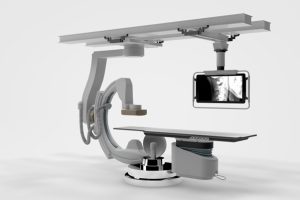
Fluoroscopy system: It is for continuous real-time imaging during catheter and filter navigation.
X-ray machine: It is used for pre-procedural and post-procedural imaging confirmation.
X-ray machine
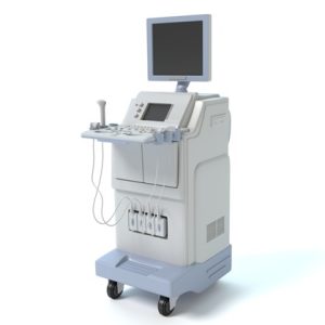
Ultrasound machine with the linear array probe: It facilitates vascular access, especially for jugular or femoral veins.
Ultrasound machine
Catheter with radiopaque calibration markers: It helps in accurate measurement of IVC diameter.
Chlorhexidine or povidone iodine solution: It is used for thorough skin disinfection prior to vascular access.
Sterile drapes and gloves: It helps to maintain a sterile field throughout the procedure.
IVC filter kit: IVC filter kit like Cook Gunther Tulip or Celect filter systems is used.
Catheter: It is used for navigating the IVC and delivering the filter.
Guide wire (0.035 inch): It provides support and direction during catheter and filter advancement.
Basic angiography set: It includes sheath introducers, syringes, and contrast injection tools.
Local anesthetic: It is typically lidocaine for numbing the vascular access site.
Sedative: Mild sedation (e.g., midazolam) may be administered for patient comfort.
Heparinized saline (1000 IU heparin in 1000 mL of 0.9% saline): It is used for flushing the catheter and preventing clot formation.
Contrast dye: It is used for visualization of venous anatomy during fluoroscopy.
Intravenous (IV) line: The IV line is used for sedation, fluid administration, and emergency medication access.
Patient Preparation
Laboratory testing: A coagulation profile (e.g., PT, aPTT, INR) is required to determine bleeding risk. Renal function tests (e.g., serum creatinine, eGFR) can assist decide if iodinated contrast is appropriate for usage.
Fasting Guidelines: Patients should be advised to avoid solid foods for at least eight hours before the procedure. Clear drinks should be avoided two hours before the operation.
Medication assessment: Conduct a thorough assessment of the patient’s medicines, making any necessary modifications for anticoagulants, antiplatelet agents, or nephrotoxic substances.
Imaging Review: Cross-sectional imaging (such as CT or MRI) is effective for assessing IVC structure, patency, diameter, and access route.
In the absence of pre-existing imaging, intra-procedural cavography should be used to look for IVC abnormalities (such as duplication), thrombus, and caval diameter.
Anesthesia: Local anesthesia is the norm for IVC filter implantation. Typically, 5 mL of 1% lidocaine is injected at the puncture site to numb the region prior to venous access. Procedural sedation may be explored for patient comfort, particularly in nervous patients or when local practices permit it. Sedation should be suited to the patient’s overall state and overseen by a competent professional.
Patient Position
The patient should be put supine on the surgery table. To expose the access site and assist cannulation with the jugular vein approach, gently shift the patient’s head to the opposite side (contralateral rotation). Padding and immobilisation can be utilised to keep the patient comfortable and restrict movement throughout the process.
Technique
Step 1: Initial Assessment and Planning
The physician reviews the patient’s medical history, including bleeding risk and renal function. Laboratory tests such as coagulation profile and serum creatinine are ordered. A review of medications is performed, with special attention to anticoagulants.
Patients are instructed to stop solid food 6–8 hours and clear liquids 2 hours before the procedure. Contrast allergy is ruled out, and consent is obtained. Some operators consider antibiotic prophylaxis.
Step 2: Pre-Procedural Imaging
Existing cross-sectional imaging (CT/MRI) is reviewed to evaluate IVC anatomy, patency, thrombus presence, and access route. If prior imaging is not available, intra-procedural cavography is planned to evaluates the patency and diameter of the IVC, presence of duplicated IVC or thrombus and location of renal veins.
Step 3: Patient Positioning and Sterile Preparation
The patient is placed in a supine position. For jugular access, the head is turned to the opposite side (contralateral rotation). The access site (neck or groin) is disinfected using chlorhexidine or povidone-iodine, and sterile drapes are applied. A sterile ultrasound probe is used to locate the target vein.
Step 4: Local Anesthesia and Venous Access
3 to 5 mL of 1% lidocaine is infiltrated for local anesthesia. A small (≤1 cm) skin incision is made at the access site. Under ultrasound guidance, an 18-gauge or micro puncture needle is used to puncture the target vein. Venous blood aspiration confirms access. A 0.035-inch guide wire is introduced into the IVC under fluoroscopy.
Step 5: Sheath and Catheter Insertion
Over the guide wire, a vascular sheath (7F for jugular or 8.5F for femoral) is advanced. A diagnostic catheter is introduced through the sheath into the IVC, usually reaching the iliac veins. The guide wire is removed, and contrast dye is injected to confirm luminal positioning.
Step 6: Cavography
A venogram is performed using hand injection or a pigtail catheter with power injection. The operator assesses IVC patency and diameter, location of the lowest renal vein and presence of thrombus or congenital anomalies. If infrarenal thrombus is present, suprarenal filter placement is considered.
Step 7: Deployment of IVC Filter
The catheter is exchanged for the filter introducer sheath. The premounted filter (e.g., Gunther Tulip or Celect) is advanced to the deployment site just below the lowest renal vein.
The sheath is retracted, allowing the filter to expand partially while still tethered to the deployment system. If the filter appears misaligned or tilted, it can be resheathed and repositioned. Once properly positioned, the filter hook is released, completing deployment.
Step 8: Final Fluoroscopic Confirmation and Closure
Filter position is confirmed on fluoroscopy. The sheath and catheter are removed. Hemostasis is achieved by applying manual pressure or using a closure device. The access site is cleaned, dressed, and the patient is monitored for immediate complications.
Step 9: Post-Procedure Monitoring
The patient is observed for bleeding, hematoma, or signs of filter-related complications. Filter type, location, and orientation are documented to facilitate future retrieval. If placed electively, follow-up is arranged to evaluate filter status and determine retrieval timing if applicable.
Complications
Complications from the use of inferior vena cava (IVC) filters can be divided into three categories: procedural, post-procedural, and retrieval related. Common complications of the insertion procedure include bleeding and thrombosis at the vascular access site. When access is acquired by the internal jugular vein, hazards include pneumothorax (rare with ultrasound guidance), access-site thrombosis (about 2%), chronic bleeding from the insertion site and potentially pulmonary embolism if the catheter passes through a thrombosed vein. Misplacement of the filter like poor alignment, severe tilt (angulation >15°), or migration (more than 2 cm displacement) might result in inadequate filtering and retrieval challenges. Operator mistake or filter deployment in the improper place or orientation increases the likelihood of difficulties.
One of the most serious post-procedure complications is IVC thrombosis which can cause bilateral lower extremities oedema and discomfort, as well as an increased risk of pulmonary embolism if thrombi expand above the filter. Renal failure may ensue if the thrombus spreads into the suprarenal IVC. Another issue is that the filter’s legs or hooks may penetrate the IVC wall, especially because hooks, which were originally inserted to prevent migration, have been linked to an increase in IVC perforation rates.
Filter fracture is also conceivable, with shards of the filter embolising important organs such as the heart and lungs. Other uncommon consequences include filter infection, penetration into nearby intestinal or skeletal structures, and retroperitoneal haematoma. Caval thrombosis occurs in around 5% of patients, while the total 30-day mortality rate from filter implantation is less than 1%.
Complications with filter retrieval are more common if the filter has been in place for a long time. The risk of fracture and IVC damage, such as dissection, rises over time. As a result, timely removal of the filter when it is no longer clinically needed is suggested. As per the studies comparing permanent and retrievable IVC filters, retrievable filters are more likely to cause complications such as fractures, whereas permanent filters are more likely to fail during initial placement. The Vascular Quality Initiative registry, which included 14,784 patients with an average follow-up of 11 months, reported immediate complications (such as venous injury, misplacement, angulation >20°, or insertion-site problems) in 1.8% of patients, while 3.1% experienced delayed complications such as filter migration, fracture, thrombus formation, or perforation.
»
Home » Procedure » Inferior Vena Cava Filter Placement
Inferior Vena Cava Filter Placement
Updated :
December 23, 2025
Pulmonary embolism (PE), which is commonly caused by venous thromboembolism (VTE), continues to be a substantial and possibly preventable cause of death in hospitalised patients. While pharmacologic anticoagulation is the foundation of VTE and PE prevention and therapy, certain clinical scenarios need alternate treatments. In patients with VTE who have absolute contraindications to anticoagulation, have experienced failure or complications from anticoagulant therapy, or have progressive deep vein thrombosis (DVT) despite adequate anticoagulation, inferior vena cava (IVC) filters are an essential therapeutic option.
IVC filters, which were established in 1973, mechanically capture thrombi in the lower extremities to avoid pulmonary embolism (PE). They are classified as permanent and retrievable filters, with the latter receiving FDA approval in the early 2000s. Despite its efficiency in avoiding PE, IVC filters have not been found to improve survival rates when compared to normal anticoagulation treatment. Prophylactic IVC filters, particularly in high-risk patients without proven thrombosis, have not been linked to lower mortality and may increase the risk of recurrent DVT and other problems. An IVC filter is often inserted under imaging guidance, with access through the femoral or jugular vein. This minimally invasive treatment is appropriate for individuals with low to moderate procedural risk.
This method review focusses on the jugular approach to putting retrievable filters, such as the Gunther Tulip or Celect models, emphasising the procedural procedures and factors for safe and successful filter placement.
The placement of inferior vena cava (IVC) filters has changed considerably over the last few decades, influenced mostly by clinical necessity and the availability of newer, retrievable devices. As per guidelines issued by key organisations such as the American Heart Association (AHA), American College of Chest Physicians (ACCP), and the American College of Radiology (ACR)/Society of Interventional Radiology (SIR), IVC filter placement is recommended in patients with venous thromboembolic disease (VTE) who cannot receive anticoagulation therapy due to absolute contraindications. These include conditions in which anticoagulation must be stopped because of problems or when VTE recurs despite appropriate anticoagulant medication.
With the invention of retrievable IVC filters, their applications have grown to incorporate a variety of relative or non-traditional indicators. These include patients with substantial pulmonary embolism (PE) at high risk of recurrence, those with little cardiopulmonary reserve, poor anticoagulant adherence, big free-floating proximal DVT, and cancer-associated VTE when anticoagulation offers a considerable bleeding risk. Despite their widespread usage, a considerable number of retrievable filters are never removed, raising the possibility of long-term problems such as filter migration, fracture, or recurrent thrombosis.
Individual physician judgement is frequently employed in clinical decision-making in high-risk groups, with IVC filters used prophylactically in patients who have been immobilised for an extended period of time owing to trauma or surgery, or who have a hypercoagulable condition caused by cancer. However, evidence for prophylactic IVC filter installation is debatable. At the two-year follow-up, the filter group had a considerably greater rate of recurrent DVT.
Absolute indications for IVC filter implantation include proven VTE (DVT or PE) with an absolute contraindication to anticoagulation, recurring PE despite therapeutic anticoagulation, and serious bleeding problems associated with anticoagulation treatment.
Relative indications include free-floating thrombus in the inferior vena cava or iliofemoral veins, PE in patients with limited cardiac or pulmonary reserve, preoperative protection in patients at risk of VTE, noncompliance with anticoagulation regimens, and adjuvant protection during DVT thrombolysis.
Inferior vena cava (IVC) filter insertion is a safe, minimally invasive procedure, especially when performed under image guidance in appropriately selected patients. However, there are no universally recognized absolute contraindications to IVC filter placement, but lack of access to the IVC is considered an absolute technical contraindication.
Relative contraindications include uncorrected coagulopathy or severe bleeding disorders, total thrombosis of the IVC, bacteremia or sepsis, and a small IVC diameter (<15 mm). Clinical guidelines advise caution in special patient populations, such as patients with external ventricular drains (EVDs) and those with acute DVT or PE who are already receiving anticoagulation therapy.
The Neurocritical Care Society advises against routine use of IVC filters for primary VTE prophylaxis, while the American College of Chest Physicians discourages the use of IVC filters in these cases. Overuse of filters in these contexts may expose patients to complications, including filter migration, caval thrombosis, and long-term venous insufficiency.
Fluoroscopy system: It is for continuous real-time imaging during catheter and filter navigation.
X-ray machine: It is used for pre-procedural and post-procedural imaging confirmation.
X-ray machine
Ultrasound machine with the linear array probe: It facilitates vascular access, especially for jugular or femoral veins.
Ultrasound machine
Catheter with radiopaque calibration markers: It helps in accurate measurement of IVC diameter.
Chlorhexidine or povidone iodine solution: It is used for thorough skin disinfection prior to vascular access.
Sterile drapes and gloves: It helps to maintain a sterile field throughout the procedure.
IVC filter kit: IVC filter kit like Cook Gunther Tulip or Celect filter systems is used.
Catheter: It is used for navigating the IVC and delivering the filter.
Guide wire (0.035 inch): It provides support and direction during catheter and filter advancement.
Basic angiography set: It includes sheath introducers, syringes, and contrast injection tools.
Local anesthetic: It is typically lidocaine for numbing the vascular access site.
Sedative: Mild sedation (e.g., midazolam) may be administered for patient comfort.
Heparinized saline (1000 IU heparin in 1000 mL of 0.9% saline): It is used for flushing the catheter and preventing clot formation.
Contrast dye: It is used for visualization of venous anatomy during fluoroscopy.
Intravenous (IV) line: The IV line is used for sedation, fluid administration, and emergency medication access.
Laboratory testing: A coagulation profile (e.g., PT, aPTT, INR) is required to determine bleeding risk. Renal function tests (e.g., serum creatinine, eGFR) can assist decide if iodinated contrast is appropriate for usage.
Fasting Guidelines: Patients should be advised to avoid solid foods for at least eight hours before the procedure. Clear drinks should be avoided two hours before the operation.
Medication assessment: Conduct a thorough assessment of the patient’s medicines, making any necessary modifications for anticoagulants, antiplatelet agents, or nephrotoxic substances.
Imaging Review: Cross-sectional imaging (such as CT or MRI) is effective for assessing IVC structure, patency, diameter, and access route.
In the absence of pre-existing imaging, intra-procedural cavography should be used to look for IVC abnormalities (such as duplication), thrombus, and caval diameter.
Anesthesia: Local anesthesia is the norm for IVC filter implantation. Typically, 5 mL of 1% lidocaine is injected at the puncture site to numb the region prior to venous access. Procedural sedation may be explored for patient comfort, particularly in nervous patients or when local practices permit it. Sedation should be suited to the patient’s overall state and overseen by a competent professional.
The patient should be put supine on the surgery table. To expose the access site and assist cannulation with the jugular vein approach, gently shift the patient’s head to the opposite side (contralateral rotation). Padding and immobilisation can be utilised to keep the patient comfortable and restrict movement throughout the process.
Step 1: Initial Assessment and Planning
The physician reviews the patient’s medical history, including bleeding risk and renal function. Laboratory tests such as coagulation profile and serum creatinine are ordered. A review of medications is performed, with special attention to anticoagulants.
Patients are instructed to stop solid food 6–8 hours and clear liquids 2 hours before the procedure. Contrast allergy is ruled out, and consent is obtained. Some operators consider antibiotic prophylaxis.
Step 2: Pre-Procedural Imaging
Existing cross-sectional imaging (CT/MRI) is reviewed to evaluate IVC anatomy, patency, thrombus presence, and access route. If prior imaging is not available, intra-procedural cavography is planned to evaluates the patency and diameter of the IVC, presence of duplicated IVC or thrombus and location of renal veins.
Step 3: Patient Positioning and Sterile Preparation
The patient is placed in a supine position. For jugular access, the head is turned to the opposite side (contralateral rotation). The access site (neck or groin) is disinfected using chlorhexidine or povidone-iodine, and sterile drapes are applied. A sterile ultrasound probe is used to locate the target vein.
Step 4: Local Anesthesia and Venous Access
3 to 5 mL of 1% lidocaine is infiltrated for local anesthesia. A small (≤1 cm) skin incision is made at the access site. Under ultrasound guidance, an 18-gauge or micro puncture needle is used to puncture the target vein. Venous blood aspiration confirms access. A 0.035-inch guide wire is introduced into the IVC under fluoroscopy.
Step 5: Sheath and Catheter Insertion
Over the guide wire, a vascular sheath (7F for jugular or 8.5F for femoral) is advanced. A diagnostic catheter is introduced through the sheath into the IVC, usually reaching the iliac veins. The guide wire is removed, and contrast dye is injected to confirm luminal positioning.
Step 6: Cavography
A venogram is performed using hand injection or a pigtail catheter with power injection. The operator assesses IVC patency and diameter, location of the lowest renal vein and presence of thrombus or congenital anomalies. If infrarenal thrombus is present, suprarenal filter placement is considered.
Step 7: Deployment of IVC Filter
The catheter is exchanged for the filter introducer sheath. The premounted filter (e.g., Gunther Tulip or Celect) is advanced to the deployment site just below the lowest renal vein.
The sheath is retracted, allowing the filter to expand partially while still tethered to the deployment system. If the filter appears misaligned or tilted, it can be resheathed and repositioned. Once properly positioned, the filter hook is released, completing deployment.
Step 8: Final Fluoroscopic Confirmation and Closure
Filter position is confirmed on fluoroscopy. The sheath and catheter are removed. Hemostasis is achieved by applying manual pressure or using a closure device. The access site is cleaned, dressed, and the patient is monitored for immediate complications.
Step 9: Post-Procedure Monitoring
The patient is observed for bleeding, hematoma, or signs of filter-related complications. Filter type, location, and orientation are documented to facilitate future retrieval. If placed electively, follow-up is arranged to evaluate filter status and determine retrieval timing if applicable.
Complications from the use of inferior vena cava (IVC) filters can be divided into three categories: procedural, post-procedural, and retrieval related. Common complications of the insertion procedure include bleeding and thrombosis at the vascular access site. When access is acquired by the internal jugular vein, hazards include pneumothorax (rare with ultrasound guidance), access-site thrombosis (about 2%), chronic bleeding from the insertion site and potentially pulmonary embolism if the catheter passes through a thrombosed vein. Misplacement of the filter like poor alignment, severe tilt (angulation >15°), or migration (more than 2 cm displacement) might result in inadequate filtering and retrieval challenges. Operator mistake or filter deployment in the improper place or orientation increases the likelihood of difficulties.
One of the most serious post-procedure complications is IVC thrombosis which can cause bilateral lower extremities oedema and discomfort, as well as an increased risk of pulmonary embolism if thrombi expand above the filter. Renal failure may ensue if the thrombus spreads into the suprarenal IVC. Another issue is that the filter’s legs or hooks may penetrate the IVC wall, especially because hooks, which were originally inserted to prevent migration, have been linked to an increase in IVC perforation rates.
Filter fracture is also conceivable, with shards of the filter embolising important organs such as the heart and lungs. Other uncommon consequences include filter infection, penetration into nearby intestinal or skeletal structures, and retroperitoneal haematoma. Caval thrombosis occurs in around 5% of patients, while the total 30-day mortality rate from filter implantation is less than 1%.
Complications with filter retrieval are more common if the filter has been in place for a long time. The risk of fracture and IVC damage, such as dissection, rises over time. As a result, timely removal of the filter when it is no longer clinically needed is suggested. As per the studies comparing permanent and retrievable IVC filters, retrievable filters are more likely to cause complications such as fractures, whereas permanent filters are more likely to fail during initial placement. The Vascular Quality Initiative registry, which included 14,784 patients with an average follow-up of 11 months, reported immediate complications (such as venous injury, misplacement, angulation >20°, or insertion-site problems) in 1.8% of patients, while 3.1% experienced delayed complications such as filter migration, fracture, thrombus formation, or perforation.
Both our subscription plans include Free CME/CPD AMA PRA Category 1 credits.
Digital Certificate PDF
On course completion, you will receive a full-sized presentation quality digital certificate.
medtigo Simulation
A dynamic medical simulation platform designed to train healthcare professionals and students to effectively run code situations through an immersive hands-on experience in a live, interactive 3D environment.
medtigo Points
medtigo points is our unique point redemption system created to award users for interacting on our site. These points can be redeemed for special discounts on the medtigo marketplace as well as towards the membership cost itself.
Community Forum post/reply = 5 points
*Redemption of points can occur only through the medtigo marketplace, courses, or simulation system. Money will not be credited to your bank account. 10 points = $1.
All Your Certificates in One Place
When you have your licenses, certificates and CMEs in one place, it's easier to track your career growth. You can easily share these with hospitals as well, using your medtigo app.