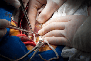
Background
Intestinal transplantation was first performed by Alexis carrel in early twentieth century experienced initial problems of organ rejection, which was sorted out with help of immunosuppressive therapies like tacrolimus. Interest in the disease re-emerged in the 1960s but reduced due to the lack of effective treatments. It was not until the year 2000 that Medicare approved coverage for intestinal transplants, which paved way to acknowledging the said procedure in managing complications arising from TPN. Despite reaching its highest level in 2007, there was a downward trend in the number of transplants, down to 91 in 2020. Survival rates differ and are higher among children who have received transplants. More emphasis in recent years have been made towards enhancing management and outcomes of patients with IF through rehabilitation and other therapies such as teduglutide limiting the need for transplants.
Indications
Indications for intestinal transplant operation also vary slightly between children and adult patient but it is indicated in case of intestinal failure- leading to multi-organ failure. Intestinal failure is defined as the inability of the physiological process to maintain adequate digestion and absorption of proteins, fats, carbohydrates, electrolytes and water and specific nutrients. The main reason for intestinal failure is short bowel syndrome which occurs in approximately 70% of transplant patients. While not all cases of short bowel syndrome require a multivisceral transplant (MVT), certain factors may make it necessary, such as:
Two or more central line infections per year or a single episode of fungemia
Parenteral nutrition, organ dysfunction such as liver dysfunction
Deep venous thrombosis or stenosis that may effectively reduce the chance for PN administration
Failure to maintain adequate hydration despite receiving parenteral nutrition and intravenous fluids
MVT in adults is used when patients have short bowel syndrome, mesenteric ischemia, abdominal tumours, inflammatory bowel disease that causes short bowel syndrome, radiation enteritis and trauma. In children, signs may include volvulus, gastroschisis, necrotizing enterocolitis, Hirschsprung’s disease, intestinal atresias, and short-bowel syndrome.
Contraindications
As with other solid organ transplants, contraindications for multivisceral transplant (MVT) may include the following:
Metastatic disease that cannot be treated by transplantation
Systemic or localized infections if they are uncontrollable or cannot be treated.
Medical issues of cardiac pulmonary nature that reduce the chances of obtaining a favourable outcome
Lack of family or social support
Substance use disorder, which includes drug or alcohol dependency
Outcomes
A multivisceral transplant entails the replacement of more than a single organ/organ system, and this is supported by a well-coordinated transdisciplinary healthcare team. From dietitians to therapists, all being involved in the healthcare delivery process contribute effectively towards improving the quality of the patient’s life after the procedure. Various medical facilities all over the world have embraced the strategy of earlier transplantation due to the benefits associated with it. In the last three decades, the results have improved more than in two times, which is attributed to the formation of specific interdisciplinary teams. Such teams must continue to be vigilant in evaluating patients even prior to the transplant as well as after the procedure. Further intensified monitoring is particularly useful in patients of the lower socio-economic status and children as it enhances treatment compliance and alerts doctors about possible side effects.
Equipment
Laparotomy sets
Hemostatic clips
Staplers and sutures
Vascular clamps
Anaesthesia machine
Ventilators
Perfusion machines
Enteral feeding pumps
Ultrasound machines
Biopsy needles
Patient preparation
The following pre-transplant evaluation should be conducted:
HLA typing and blood cross-matching
Laboratory tests: Complete blood count, comprehensive metabolic panel, prealbumin levels and coagulation studies
Serologic testing: Cytomegalovirus (CMV), Epstein-Barr virus (EBV), Hepatitis A, B and C, AIDS (HIV).
Bowel function assessment: Assessment of bowel length and functionality by CT enterography
Vascular assessment: Evaluation of the intraabdominal venous and arterial system by duplex ultrasound. In some cases, splenoportography and mesenteric angiography may be required depending on the Miami classification of the patent ductus. Additional preoperative vein or arterial angioplasty may be needed depending on classification.
Donor liver biopsy: Done in certain situations; the recipient liver biopsy may also be required if parenteral nutrition has led to liver injury.
Infectious risk assessment: Dental and possibly an ENT consultation regarding in infectious sources such as dental infections which may require extractions.
Systemic disease evaluation: This involves patient-specific interventions such as coronary angiography, pulmonary function tests, and nutrition assessment to manage avoidable risks during surgery.
Patient position
In intestinal and multivisceral transplantation patients should be positioned in a supine position where the head and the lower limbs are flat on the bed. Some positional changes like positioning the patient in a slight Trendelenburg position or a head down tilt may be employed to enhance both access and visualization of the organs. Occasionally the modified supine or the left lateral decubitus position might be used depending on the requirements of the surgery. Appropriate cushioning and support help prevent pressure ulcer formation and facilitate comfort.
Isolated Intestinal Transplantation (IITx)
Preparation:
Donor Organ Preparation:
Recipient Surgery:
Critical Steps:
Postoperative Considerations:
Recipient Surgery:
Critical Steps:
Postoperative Considerations:
Combined Liver-Intestine Transplantation
Donor Organ Preparation: Harvest the liver and the bowel together which should be either in a combined spotlight or under one surgeon. Examine and debride crucial tissue on the back table.
Recipient Surgery:
Donor Organ Preparation: Modify the allograft according to the patient’s requirements, which can be kidneys, spleen, among others.
Recipient Surgery:

Intestinal and Multivisceral Transplantation
Approach considerations
After assessing potential intestinal failure, the physician should consider:
Continued TPN: At the same time, intestinal failure should be excluded, and if this is not Done and there are no potentially lethal complications.
Isolated Intestinal Transplantation: If TPN can be discontinued, it is perhaps the simplest solution of all: with all the strictures on the use of TPN based on the fear of hyperglycemia complications, the concept of just stopping it altogether may strike as rather absurd.
Combined Liver-Small Bowel (LSB) Transplantation: Because if liver function is also affected, it complicates the dog’s condition and becomes dangerous and fatal.
Multivisceral Transplantation: In situations where the damage affects more than one organ.
Isolated Liver Transplantation: It is 90% of the capacity if only the liver is affected.
Patients who can progress to full enteral nutrition should be sent to an intestinal rehabilitation clinic for the fine tuning of TPN, control of this and other complications, and determination of the readiness for surgery or transplantation.
Teduglutide (Gattex) which was approved in adults in 2012 and in pediatric patients in 2019 helps the absorption of nutrients and fluids in the intestine and thus has the potential to decrease the patients’ requirement for transplantation.
Complications
Rejection:
Infection:
Graft Dysfunction:
Biliary Complications:
Gastrointestinal Complications:
Nutritional Complications:
Vascular Complications:

Intestinal transplantation was first performed by Alexis carrel in early twentieth century experienced initial problems of organ rejection, which was sorted out with help of immunosuppressive therapies like tacrolimus. Interest in the disease re-emerged in the 1960s but reduced due to the lack of effective treatments. It was not until the year 2000 that Medicare approved coverage for intestinal transplants, which paved way to acknowledging the said procedure in managing complications arising from TPN. Despite reaching its highest level in 2007, there was a downward trend in the number of transplants, down to 91 in 2020. Survival rates differ and are higher among children who have received transplants. More emphasis in recent years have been made towards enhancing management and outcomes of patients with IF through rehabilitation and other therapies such as teduglutide limiting the need for transplants.
Indications for intestinal transplant operation also vary slightly between children and adult patient but it is indicated in case of intestinal failure- leading to multi-organ failure. Intestinal failure is defined as the inability of the physiological process to maintain adequate digestion and absorption of proteins, fats, carbohydrates, electrolytes and water and specific nutrients. The main reason for intestinal failure is short bowel syndrome which occurs in approximately 70% of transplant patients. While not all cases of short bowel syndrome require a multivisceral transplant (MVT), certain factors may make it necessary, such as:
Two or more central line infections per year or a single episode of fungemia
Parenteral nutrition, organ dysfunction such as liver dysfunction
Deep venous thrombosis or stenosis that may effectively reduce the chance for PN administration
Failure to maintain adequate hydration despite receiving parenteral nutrition and intravenous fluids
MVT in adults is used when patients have short bowel syndrome, mesenteric ischemia, abdominal tumours, inflammatory bowel disease that causes short bowel syndrome, radiation enteritis and trauma. In children, signs may include volvulus, gastroschisis, necrotizing enterocolitis, Hirschsprung’s disease, intestinal atresias, and short-bowel syndrome.
As with other solid organ transplants, contraindications for multivisceral transplant (MVT) may include the following:
Metastatic disease that cannot be treated by transplantation
Systemic or localized infections if they are uncontrollable or cannot be treated.
Medical issues of cardiac pulmonary nature that reduce the chances of obtaining a favourable outcome
Lack of family or social support
Substance use disorder, which includes drug or alcohol dependency
A multivisceral transplant entails the replacement of more than a single organ/organ system, and this is supported by a well-coordinated transdisciplinary healthcare team. From dietitians to therapists, all being involved in the healthcare delivery process contribute effectively towards improving the quality of the patient’s life after the procedure. Various medical facilities all over the world have embraced the strategy of earlier transplantation due to the benefits associated with it. In the last three decades, the results have improved more than in two times, which is attributed to the formation of specific interdisciplinary teams. Such teams must continue to be vigilant in evaluating patients even prior to the transplant as well as after the procedure. Further intensified monitoring is particularly useful in patients of the lower socio-economic status and children as it enhances treatment compliance and alerts doctors about possible side effects.
Laparotomy sets
Hemostatic clips
Staplers and sutures
Vascular clamps
Anaesthesia machine
Ventilators
Perfusion machines
Enteral feeding pumps
Ultrasound machines
Biopsy needles
The following pre-transplant evaluation should be conducted:
HLA typing and blood cross-matching
Laboratory tests: Complete blood count, comprehensive metabolic panel, prealbumin levels and coagulation studies
Serologic testing: Cytomegalovirus (CMV), Epstein-Barr virus (EBV), Hepatitis A, B and C, AIDS (HIV).
Bowel function assessment: Assessment of bowel length and functionality by CT enterography
Vascular assessment: Evaluation of the intraabdominal venous and arterial system by duplex ultrasound. In some cases, splenoportography and mesenteric angiography may be required depending on the Miami classification of the patent ductus. Additional preoperative vein or arterial angioplasty may be needed depending on classification.
Donor liver biopsy: Done in certain situations; the recipient liver biopsy may also be required if parenteral nutrition has led to liver injury.
Infectious risk assessment: Dental and possibly an ENT consultation regarding in infectious sources such as dental infections which may require extractions.
Systemic disease evaluation: This involves patient-specific interventions such as coronary angiography, pulmonary function tests, and nutrition assessment to manage avoidable risks during surgery.
In intestinal and multivisceral transplantation patients should be positioned in a supine position where the head and the lower limbs are flat on the bed. Some positional changes like positioning the patient in a slight Trendelenburg position or a head down tilt may be employed to enhance both access and visualization of the organs. Occasionally the modified supine or the left lateral decubitus position might be used depending on the requirements of the surgery. Appropriate cushioning and support help prevent pressure ulcer formation and facilitate comfort.
Preparation:
Donor Organ Preparation:
Recipient Surgery:
Critical Steps:
Postoperative Considerations:
Recipient Surgery:
Critical Steps:
Postoperative Considerations:
Combined Liver-Intestine Transplantation
Donor Organ Preparation: Harvest the liver and the bowel together which should be either in a combined spotlight or under one surgeon. Examine and debride crucial tissue on the back table.
Recipient Surgery:
Donor Organ Preparation: Modify the allograft according to the patient’s requirements, which can be kidneys, spleen, among others.
Recipient Surgery:

Intestinal and Multivisceral Transplantation
After assessing potential intestinal failure, the physician should consider:
Continued TPN: At the same time, intestinal failure should be excluded, and if this is not Done and there are no potentially lethal complications.
Isolated Intestinal Transplantation: If TPN can be discontinued, it is perhaps the simplest solution of all: with all the strictures on the use of TPN based on the fear of hyperglycemia complications, the concept of just stopping it altogether may strike as rather absurd.
Combined Liver-Small Bowel (LSB) Transplantation: Because if liver function is also affected, it complicates the dog’s condition and becomes dangerous and fatal.
Multivisceral Transplantation: In situations where the damage affects more than one organ.
Isolated Liver Transplantation: It is 90% of the capacity if only the liver is affected.
Patients who can progress to full enteral nutrition should be sent to an intestinal rehabilitation clinic for the fine tuning of TPN, control of this and other complications, and determination of the readiness for surgery or transplantation.
Teduglutide (Gattex) which was approved in adults in 2012 and in pediatric patients in 2019 helps the absorption of nutrients and fluids in the intestine and thus has the potential to decrease the patients’ requirement for transplantation.
Rejection:
Infection:
Graft Dysfunction:
Biliary Complications:
Gastrointestinal Complications:
Nutritional Complications:
Vascular Complications:

Both our subscription plans include Free CME/CPD AMA PRA Category 1 credits.

On course completion, you will receive a full-sized presentation quality digital certificate.
A dynamic medical simulation platform designed to train healthcare professionals and students to effectively run code situations through an immersive hands-on experience in a live, interactive 3D environment.

When you have your licenses, certificates and CMEs in one place, it's easier to track your career growth. You can easily share these with hospitals as well, using your medtigo app.


