Background
Laminoplasty is performed on spine to treat spinal stenosis, herniated discs, and spinal cord compression. In laminectomy the complete removal of the lamina (the back part of the vertebra) takes place but the laminoplasty preserves the lamina while creating more space for the spinal cord and nerves. This procedure helps to relieve pressure on the spinal cord and nerves alleviating symptoms like pain, weakness, and numbness.
Anatomically, the spine is divided into cervical, thoracic, and lumbar spine. Each vertebra in the spine has a bony structure called the lamina which forms roof of the spinal canal that protect the spinal cord. In spinal stenosis the lamina can become thickened or enlarged that causing compression of the spinal cord and nerves.
During a laminoplasty an incision made in the back of neck to access the affected vertebrae. The lamina is then partially cut on one side and hinged open on the other side making a door like opening. This allows to access the spinal canal and relieve pressure on spinal cord and nerves. Metal plates or screws may be used to secure the opened lamina in its new position.
Laminoplasty is preferred over laminectomy in cases where preserving the stability of the spine is important such as in younger patients or those with certain spinal conditions. The procedure can help to restore function and mobility in patients with spinal cord compression allowing them to regain quality of life and reduce reliance on pain medications.
Indications
Cervical Spinal Stenosis
Herniated Discs
Cervical Spondylotic Myelopathy
Recurrent Disc Herniation
Traumatic Spinal Cord Compression
Tumors
Degenerative Disc Disease
Contraindications
Laminoplasty is not suitable for patients with severe spinal instability and active infections like severe osteoporosis, uncontrolled medical conditions, allergies to surgical materials, neurological deficits, personal or cultural beliefs, or previous failed laminoplasty.
Outcomes
Laminoplasty is a spine surgery that preserves the stability of the spine by creating more space within the spinal canal while maintaining the structural integrity of the posterior elements.
The choice of surgical approach depends on the patient anatomy, spinal cord compression, and the surgeon experience.
The procedure may involve implants, bone healing, or bone grafts to enhance stability.
Postoperative immobilization may be required to allow for healing and stabilization of spine.
Equipment
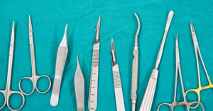
Laminoplasty is a spine surgery that involves the use of standard surgical instruments like:
scalpels
retractors
forceps
scissors
specialized instruments for cutting and hinging the laminae
High-speed drills create openings in the laminae while implants help maintain spine stability. Fluoroscopy allows real-time visualization of the spine guiding the instrument and implant placement.
Bone graft material may be used to promote bony fusion, stabilize the spine, and prevent future spinal cord compression.
Sterile drapes and supplies maintain a sterile field and neuromonitoring equipment monitors spinal cord function.
Postoperative immobilization devices may be required.
Anesthesia equipment is essential for patient monitoring.

Surgical Instruments
Patient preparation
Laminoplasty involves a preoperative evaluation, anesthesia management, patient positioning, sterile preparation, neuromonitoring setup, intraoperative imaging, and postoperative immobilization devices.
The patient is placed face down with the neck slightly extended and padded with support to comfort.
The surgical site is cleaned and prepared using antiseptic solutions and neuromonitoring equipment may be used to monitor spinal cord function.
Postoperative immobilization devices may be required to stabilize the spine and promote healing.
Monitoring and Follow-up
Postoperative care for laminoplasty involves immediate monitoring, pain management, wound care, and physical therapy to restore strength and flexibility in the neck and spine.
Patients are advised to follow-up with imaging studies to assess the spine alignment and healing process.
Regular neurological assessments are also conducted to ensure the surgery has not caused new neurological deficits and that existing deficits are improving.
These measures help to detect any immediate complications and promote healing.
Technique
Cervical laminoplasty
Cervical laminoplasty is used to decompress the spinal cord in cervical spinal stenosis where spinal canal narrows and puts pressure on spinal cord.
There are several techniques used in cervical laminoplasty like Z-laminoplasty, hinged-door laminoplasty, midline-enlargement laminoplasty, and bilateral miniplate laminoplasty.
Step 1: Patient Positioning and Anesthesia:
The patient is positioned in a prone or sitting position that depends on surgical approach.
General anesthesia is induced to make sure the patient is unconscious and free from pain during the procedure.
Step 2: Incision and Exposure:
A midline incision made over cervical spine where the skin and subcutaneous tissues are dissected to expose the posterior elements of the spine.
Fluoroscopic guidance is used to confirm the level of the laminoplasty and to ensure accurate placement of surgical instruments.
Step 3: Z-Laminoplasty:
Z-laminoplasty involves a series of Z-shaped cuts in laminae on one side of the spine.
The cuts are made by using a high speed drill or cutting instrument and the laminae are then hinged open on to the opposite side creating a Z shape.

Repaired spine
Step 4: Hinged-Door Laminoplasty:
Hinged-door laminoplasty involves a midline incision in laminae and creates a hinge on one side. The laminae are then hinged open like a door creating more space in the spinal canal.
Step 5: Midline-Enlargement Laminoplasty:
Midline-enlargement laminoplasty involves a midline incision in the laminae and removing a portion of spinous process. The remaining laminae are then hinged open which enlarges the spinal canal.
Step 6: Bilateral Miniplate Laminoplasty:
Bilateral miniplate laminoplasty involves placing a small titanium plates on both sides of hinged laminae to stabilize them in the open position.
The plates are secured with screws to maintain the desired alignment and prevent re-stenosis of the spinal canal.
Step 7: Closure and Postoperative Care:
The incision is closed and a dressing is applied to the wound. Postoperative care includes pain management, wound care, and physical therapy to restore mobility and promote healing.
Step 8: Follow-up and Monitoring:
Patients are closely monitored postoperatively for any signs of infection or neurological deficits.
Thoracolumbar laminotomy
Thoracolumbar laminotomy performed to decompress spinal cord or nerve roots in thoracic and lumbar regions of spine. It is indicated for spinal stenosis, herniated discs, and spinal cord tumors.
The goal of this surgery is to relieve pressure on spinal cord and spinal nerves to alleviate pain and improve function.
Step 1: Preoperative Planning:
Before performing thoracolumbar laminotomy a thorough preoperative evaluation is required.
This includes a review of physical examination, medical history, and imaging studies like X-rays, MRI, or CT scans.
Carefully assesses the extent and location of spinal cord compression to plan the surgical approach.
Step 2: Anesthesia and Patient Positioning:
The patient is under general anesthesia to ensure unconsciousness and pain control during the procedure.
The patient is positioned in a prone position and lying face down. Padding and supports are used to ensure proper alignment of spine to prevent pressure injuries.
Step 3: Incision and Exposure:
A midline or paramedian incision is made over the affected area of the spine. The length and location of incision depends on the number of vertebrae involved and the extent of the decompression required.
The skin and subcutaneous tissues are dissected to expose the spinous processes and laminae of the vertebrae.
Retractors are used to hold muscles and soft tissues aside providing a clear view of the surgical site.
Step 4: Laminotomy:
The facet joints and laminae are decorticated using a high-speed drill or specialized instruments. This involves removing the outer layer of bone to expose the underlying laminae.
Laminae are carefully removed using rongeurs or laminectomy punches, to create sufficient space in the spinal canal and to relieve the pressure on spinal cord and nerve roots.
If there is foraminal stenosis the surgeon may perform a foraminotomy to widen the neural foramina and relieve pressure on the exiting nerve roots. This is done using a drill or bone-cutting instruments.
Step 5: Dural Decompression:
The dura mater is the protective covering of the spinal cord and is identified and opened longitudinally. Care is taken to avoid damaging the spinal cord or nerve roots.
Any disc herniations, bone spurs, or other structures compressing the spinal cord or nerve roots are removed. This helps to relieve pressure and restore normal spinal cord function.
Step 6: Closure and Postoperative Care:
Once the decompression is complete the dura mater is closed using sutures or staples. The muscles and soft tissues are reapproximated and skin is closed with staples or sutures.
The patient is transferred to the recovery room for monitoring and pain management. Physical therapy may be initiated that may help to improve the mobility and strength.
Complications
Infection
Bleeding
Neurological Injury
Dural Tear
Pseudoarthrosis
Adjacent Segment Degeneration
Wound Complications
Persistent Pain
Urinary Retention or Incontinence

Laminoplasty is performed on spine to treat spinal stenosis, herniated discs, and spinal cord compression. In laminectomy the complete removal of the lamina (the back part of the vertebra) takes place but the laminoplasty preserves the lamina while creating more space for the spinal cord and nerves. This procedure helps to relieve pressure on the spinal cord and nerves alleviating symptoms like pain, weakness, and numbness.
Anatomically, the spine is divided into cervical, thoracic, and lumbar spine. Each vertebra in the spine has a bony structure called the lamina which forms roof of the spinal canal that protect the spinal cord. In spinal stenosis the lamina can become thickened or enlarged that causing compression of the spinal cord and nerves.
During a laminoplasty an incision made in the back of neck to access the affected vertebrae. The lamina is then partially cut on one side and hinged open on the other side making a door like opening. This allows to access the spinal canal and relieve pressure on spinal cord and nerves. Metal plates or screws may be used to secure the opened lamina in its new position.
Laminoplasty is preferred over laminectomy in cases where preserving the stability of the spine is important such as in younger patients or those with certain spinal conditions. The procedure can help to restore function and mobility in patients with spinal cord compression allowing them to regain quality of life and reduce reliance on pain medications.
Cervical Spinal Stenosis
Herniated Discs
Cervical Spondylotic Myelopathy
Recurrent Disc Herniation
Traumatic Spinal Cord Compression
Tumors
Degenerative Disc Disease
Laminoplasty is not suitable for patients with severe spinal instability and active infections like severe osteoporosis, uncontrolled medical conditions, allergies to surgical materials, neurological deficits, personal or cultural beliefs, or previous failed laminoplasty.
Laminoplasty is a spine surgery that preserves the stability of the spine by creating more space within the spinal canal while maintaining the structural integrity of the posterior elements.
The choice of surgical approach depends on the patient anatomy, spinal cord compression, and the surgeon experience.
The procedure may involve implants, bone healing, or bone grafts to enhance stability.
Postoperative immobilization may be required to allow for healing and stabilization of spine.
Laminoplasty is a spine surgery that involves the use of standard surgical instruments like:
scalpels
retractors
forceps
scissors
specialized instruments for cutting and hinging the laminae
High-speed drills create openings in the laminae while implants help maintain spine stability. Fluoroscopy allows real-time visualization of the spine guiding the instrument and implant placement.
Bone graft material may be used to promote bony fusion, stabilize the spine, and prevent future spinal cord compression.
Sterile drapes and supplies maintain a sterile field and neuromonitoring equipment monitors spinal cord function.
Postoperative immobilization devices may be required.
Anesthesia equipment is essential for patient monitoring.

Surgical Instruments
Laminoplasty involves a preoperative evaluation, anesthesia management, patient positioning, sterile preparation, neuromonitoring setup, intraoperative imaging, and postoperative immobilization devices.
The patient is placed face down with the neck slightly extended and padded with support to comfort.
The surgical site is cleaned and prepared using antiseptic solutions and neuromonitoring equipment may be used to monitor spinal cord function.
Postoperative immobilization devices may be required to stabilize the spine and promote healing.
Postoperative care for laminoplasty involves immediate monitoring, pain management, wound care, and physical therapy to restore strength and flexibility in the neck and spine.
Patients are advised to follow-up with imaging studies to assess the spine alignment and healing process.
Regular neurological assessments are also conducted to ensure the surgery has not caused new neurological deficits and that existing deficits are improving.
These measures help to detect any immediate complications and promote healing.
Cervical laminoplasty
Cervical laminoplasty is used to decompress the spinal cord in cervical spinal stenosis where spinal canal narrows and puts pressure on spinal cord.
There are several techniques used in cervical laminoplasty like Z-laminoplasty, hinged-door laminoplasty, midline-enlargement laminoplasty, and bilateral miniplate laminoplasty.
Step 1: Patient Positioning and Anesthesia:
The patient is positioned in a prone or sitting position that depends on surgical approach.
General anesthesia is induced to make sure the patient is unconscious and free from pain during the procedure.
Step 2: Incision and Exposure:
A midline incision made over cervical spine where the skin and subcutaneous tissues are dissected to expose the posterior elements of the spine.
Fluoroscopic guidance is used to confirm the level of the laminoplasty and to ensure accurate placement of surgical instruments.
Step 3: Z-Laminoplasty:
Z-laminoplasty involves a series of Z-shaped cuts in laminae on one side of the spine.
The cuts are made by using a high speed drill or cutting instrument and the laminae are then hinged open on to the opposite side creating a Z shape.

Repaired spine
Step 4: Hinged-Door Laminoplasty:
Hinged-door laminoplasty involves a midline incision in laminae and creates a hinge on one side. The laminae are then hinged open like a door creating more space in the spinal canal.
Step 5: Midline-Enlargement Laminoplasty:
Midline-enlargement laminoplasty involves a midline incision in the laminae and removing a portion of spinous process. The remaining laminae are then hinged open which enlarges the spinal canal.
Step 6: Bilateral Miniplate Laminoplasty:
Bilateral miniplate laminoplasty involves placing a small titanium plates on both sides of hinged laminae to stabilize them in the open position.
The plates are secured with screws to maintain the desired alignment and prevent re-stenosis of the spinal canal.
Step 7: Closure and Postoperative Care:
The incision is closed and a dressing is applied to the wound. Postoperative care includes pain management, wound care, and physical therapy to restore mobility and promote healing.
Step 8: Follow-up and Monitoring:
Patients are closely monitored postoperatively for any signs of infection or neurological deficits.
Thoracolumbar laminotomy performed to decompress spinal cord or nerve roots in thoracic and lumbar regions of spine. It is indicated for spinal stenosis, herniated discs, and spinal cord tumors.
The goal of this surgery is to relieve pressure on spinal cord and spinal nerves to alleviate pain and improve function.
Step 1: Preoperative Planning:
Before performing thoracolumbar laminotomy a thorough preoperative evaluation is required.
This includes a review of physical examination, medical history, and imaging studies like X-rays, MRI, or CT scans.
Carefully assesses the extent and location of spinal cord compression to plan the surgical approach.
Step 2: Anesthesia and Patient Positioning:
The patient is under general anesthesia to ensure unconsciousness and pain control during the procedure.
The patient is positioned in a prone position and lying face down. Padding and supports are used to ensure proper alignment of spine to prevent pressure injuries.
Step 3: Incision and Exposure:
A midline or paramedian incision is made over the affected area of the spine. The length and location of incision depends on the number of vertebrae involved and the extent of the decompression required.
The skin and subcutaneous tissues are dissected to expose the spinous processes and laminae of the vertebrae.
Retractors are used to hold muscles and soft tissues aside providing a clear view of the surgical site.
Step 4: Laminotomy:
The facet joints and laminae are decorticated using a high-speed drill or specialized instruments. This involves removing the outer layer of bone to expose the underlying laminae.
Laminae are carefully removed using rongeurs or laminectomy punches, to create sufficient space in the spinal canal and to relieve the pressure on spinal cord and nerve roots.
If there is foraminal stenosis the surgeon may perform a foraminotomy to widen the neural foramina and relieve pressure on the exiting nerve roots. This is done using a drill or bone-cutting instruments.
Step 5: Dural Decompression:
The dura mater is the protective covering of the spinal cord and is identified and opened longitudinally. Care is taken to avoid damaging the spinal cord or nerve roots.
Any disc herniations, bone spurs, or other structures compressing the spinal cord or nerve roots are removed. This helps to relieve pressure and restore normal spinal cord function.
Step 6: Closure and Postoperative Care:
Once the decompression is complete the dura mater is closed using sutures or staples. The muscles and soft tissues are reapproximated and skin is closed with staples or sutures.
The patient is transferred to the recovery room for monitoring and pain management. Physical therapy may be initiated that may help to improve the mobility and strength.
Infection
Bleeding
Neurological Injury
Dural Tear
Pseudoarthrosis
Adjacent Segment Degeneration
Wound Complications
Persistent Pain
Urinary Retention or Incontinence

Both our subscription plans include Free CME/CPD AMA PRA Category 1 credits.

On course completion, you will receive a full-sized presentation quality digital certificate.
A dynamic medical simulation platform designed to train healthcare professionals and students to effectively run code situations through an immersive hands-on experience in a live, interactive 3D environment.

When you have your licenses, certificates and CMEs in one place, it's easier to track your career growth. You can easily share these with hospitals as well, using your medtigo app.


