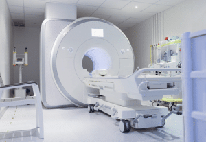
MRI is a strong medical imaging technology that uses Magnetic fields in combination with radio wave signal and a computing facility to obtain the imaging of anatomical physiological processes taking place within. MRI scanners are elongated tubelike structures, which contain a large magnet, gradient coils and a radio frequency (RF) antenna.
MRI machine
MRI stands out from other credible methods, such as X-rays or CT scans because MRI does not involve the use of ionizing radiation. This renders MRI safer for several people, in consideration of a future possibility that might require another scan.
Indications
Brain and Spinal Cord Lesions: They will be useful for diagnosing tumor, bleeding, infection, inflammation or present at birth factors which are congenital.
Multiple Sclerosis: It’s suitable for the careful diagnosis of the changes that may result in developing plaques in the brain and spinal cord.
Stroke: In order to differentiate the type and the area of the brain that was injured and the degree of injury as well.
Traumatic Brain Injury: This shall help in assessing the magnitude of damage and regular bleeding at the concerned area.
Epilepsy: To locate specific areas that might be likely to be involved in seizures.
Contraindications
Implanted Cardiac Devices:
Unless they are MRI-compatible, implanted cardioverter-defibrillators (ICDs) and permanent pacemakers are generally not advised for MRIs.
Cochlear Implants:
Staying on the dependency of the strength and style of the setting of the cochlear implants, some of them are not MRI Safe.
Certain Metal Implants:
Usually, ferromagnetic devices that are non-MRI compatible used in neurosurgery operations for instance, aneurism clips, some metal stents and other implants pose such risks.
Metallic Foreign Bodies:
During the scan, metallic foreign bodies in sensitive areas (such as close to the brain or eyes) may move or heat up.
Neurostimulators:
Certain neurostimulators are affected by magnetic field but none of them are designed to work with MRI.
Outcomes
Equipment
Magnet
Gradient Coils
Radiofrequency (RF) Coils
Computer System
Patient Table
Shielding
Patient preparation
Medical History and Screening:
The patient will have to fill forms which will contain general information about their health.
Any internal medical device such as pacemaker, cochlear implant, or special types of metal implant, when present, claustrophobia, and, finally, an allergy to the contrast agents to be used during the procedure.
Patient Instructions:
Normally, it is advised that the patients should not take food or liquids before the scan specifically if a contrast material is to be administered in the scan.
Clothing and Accessories:
It is expected that the patients would come wearing loose attires so as to accommodate the probe and these should not come with metallic parts like zippers, snaps and buttons. Occasionally, the patients are provided with hospital gowns.
Communication:
However, to ensure all inconveniences have been corrected, the patient being assessed can use a call button to talk to the technologist.
To counter this, patients are provided with earplugs or headphones to reduce on the noise made by the MRI machine.
Immobility:
The patient has to be as still as they could, for these types of images to work at their optimal best.
Patient position:
The position of the patient shall be sitting or lying on MRI table which is fitted into the MRI machine.
Accessory details may include padding and straps which may be used to restrain the patient and ensure maximal comfort.
Approach considerations
Screening: At the present, it is considerable important to do the scan for relevant metallic articles that includes implant surgical used equipment, metal fragment, skin tattoo.
Gadolinium Contrast: It should not be used in patients who have kidney diseases because they experience more NSF after being exposed to products containing gadolinium.
Magnet Strength: It is significant to mention that there some benefits accruable to having higher field strength magnets 3T as against 1.5T as it aids in getting a better definition of the body part but at the same time is likely to produce artifacts and Specific Absorption Rates (SAR).
Coil Selection: Use appropriate coils to obtain a good picture of a certain part of the body.
Pulse Sequences: Time the specific pulse sequences to get adequate signals from the target organ or area with pathological changes.
MRI machine technique
Step 1: The patients are advised to lie stationary on the back and as much as possible, they should avoid any form of movement. At some point during the process there will be brief occasions where the patient will be asked to hold their breath briefly. Also, short intervals might be provided between scans in which the patient will be engaged in scan activities.
Step 2: The scans are at most 3 minutes long, whereas port-based scans on average may take an hour.
Step 3: The head-coil helmet is then placed around the patient’s head before the scan. The helmet enables the patient to cover his head, but one is able to see outside thus it reduces claustrophobia.
Step 4: A two-way intercom facility is available to the patient and through this, the patient can call anyone outside the room.
Step 5: Music or TV is also available in some machines and hence, symptoms such as claustrophobia are reduced as well as the loud noises of the scanner.
Step 6: In addition to the scanner’s sounds, vibrations may also be sensed.
Step 7: There is also a panic button throughout the procedure for the patient; in case something that the patient is not comfortable with happens, then the patient drops the procedure.
Complications
Claustrophobia: MRI scans may be uncomfortable for some patients because of the enclosed MRI machine.
Metal Implants and Devices: MRI can weaken or harm metal implants, pacemakers, cochlear implants, and other similar medical devices. These electrodes require precautions before an MRI is performed on the patient.
Allergic Reactions: It should be noted that some of the patients may develop an acute allergic-like reaction to the compounds (gadolinium) used in some MRI procedures.
Thermal Injuries: Since the radiofrequency energy is utilized during the scan, a patient may be subjected to heating at especially those points where the patient’s body has a direct interaction with the scanner.
Noise: The MRI machine gives out loud sounds when undertaking the scan and this may be painful to the patient. This is generally managed by offering here protection which may include usage of ear plugs.
MRI is a strong medical imaging technology that uses Magnetic fields in combination with radio wave signal and a computing facility to obtain the imaging of anatomical physiological processes taking place within. MRI scanners are elongated tubelike structures, which contain a large magnet, gradient coils and a radio frequency (RF) antenna.
MRI machine
MRI stands out from other credible methods, such as X-rays or CT scans because MRI does not involve the use of ionizing radiation. This renders MRI safer for several people, in consideration of a future possibility that might require another scan.
Brain and Spinal Cord Lesions: They will be useful for diagnosing tumor, bleeding, infection, inflammation or present at birth factors which are congenital.
Multiple Sclerosis: It’s suitable for the careful diagnosis of the changes that may result in developing plaques in the brain and spinal cord.
Stroke: In order to differentiate the type and the area of the brain that was injured and the degree of injury as well.
Traumatic Brain Injury: This shall help in assessing the magnitude of damage and regular bleeding at the concerned area.
Epilepsy: To locate specific areas that might be likely to be involved in seizures.
Implanted Cardiac Devices:
Unless they are MRI-compatible, implanted cardioverter-defibrillators (ICDs) and permanent pacemakers are generally not advised for MRIs.
Cochlear Implants:
Staying on the dependency of the strength and style of the setting of the cochlear implants, some of them are not MRI Safe.
Certain Metal Implants:
Usually, ferromagnetic devices that are non-MRI compatible used in neurosurgery operations for instance, aneurism clips, some metal stents and other implants pose such risks.
Metallic Foreign Bodies:
During the scan, metallic foreign bodies in sensitive areas (such as close to the brain or eyes) may move or heat up.
Neurostimulators:
Certain neurostimulators are affected by magnetic field but none of them are designed to work with MRI.
Magnet
Gradient Coils
Radiofrequency (RF) Coils
Computer System
Patient Table
Shielding
Medical History and Screening:
The patient will have to fill forms which will contain general information about their health.
Any internal medical device such as pacemaker, cochlear implant, or special types of metal implant, when present, claustrophobia, and, finally, an allergy to the contrast agents to be used during the procedure.
Patient Instructions:
Normally, it is advised that the patients should not take food or liquids before the scan specifically if a contrast material is to be administered in the scan.
Clothing and Accessories:
It is expected that the patients would come wearing loose attires so as to accommodate the probe and these should not come with metallic parts like zippers, snaps and buttons. Occasionally, the patients are provided with hospital gowns.
Communication:
However, to ensure all inconveniences have been corrected, the patient being assessed can use a call button to talk to the technologist.
To counter this, patients are provided with earplugs or headphones to reduce on the noise made by the MRI machine.
Immobility:
The patient has to be as still as they could, for these types of images to work at their optimal best.
Patient position:
The position of the patient shall be sitting or lying on MRI table which is fitted into the MRI machine.
Accessory details may include padding and straps which may be used to restrain the patient and ensure maximal comfort.
Screening: At the present, it is considerable important to do the scan for relevant metallic articles that includes implant surgical used equipment, metal fragment, skin tattoo.
Gadolinium Contrast: It should not be used in patients who have kidney diseases because they experience more NSF after being exposed to products containing gadolinium.
Magnet Strength: It is significant to mention that there some benefits accruable to having higher field strength magnets 3T as against 1.5T as it aids in getting a better definition of the body part but at the same time is likely to produce artifacts and Specific Absorption Rates (SAR).
Coil Selection: Use appropriate coils to obtain a good picture of a certain part of the body.
Pulse Sequences: Time the specific pulse sequences to get adequate signals from the target organ or area with pathological changes.
Step 1: The patients are advised to lie stationary on the back and as much as possible, they should avoid any form of movement. At some point during the process there will be brief occasions where the patient will be asked to hold their breath briefly. Also, short intervals might be provided between scans in which the patient will be engaged in scan activities.
Step 2: The scans are at most 3 minutes long, whereas port-based scans on average may take an hour.
Step 3: The head-coil helmet is then placed around the patient’s head before the scan. The helmet enables the patient to cover his head, but one is able to see outside thus it reduces claustrophobia.
Step 4: A two-way intercom facility is available to the patient and through this, the patient can call anyone outside the room.
Step 5: Music or TV is also available in some machines and hence, symptoms such as claustrophobia are reduced as well as the loud noises of the scanner.
Step 6: In addition to the scanner’s sounds, vibrations may also be sensed.
Step 7: There is also a panic button throughout the procedure for the patient; in case something that the patient is not comfortable with happens, then the patient drops the procedure.
Claustrophobia: MRI scans may be uncomfortable for some patients because of the enclosed MRI machine.
Metal Implants and Devices: MRI can weaken or harm metal implants, pacemakers, cochlear implants, and other similar medical devices. These electrodes require precautions before an MRI is performed on the patient.
Allergic Reactions: It should be noted that some of the patients may develop an acute allergic-like reaction to the compounds (gadolinium) used in some MRI procedures.
Thermal Injuries: Since the radiofrequency energy is utilized during the scan, a patient may be subjected to heating at especially those points where the patient’s body has a direct interaction with the scanner.
Noise: The MRI machine gives out loud sounds when undertaking the scan and this may be painful to the patient. This is generally managed by offering here protection which may include usage of ear plugs.
Both our subscription plans include Free CME/CPD AMA PRA Category 1 credits.
Digital Certificate PDF
On course completion, you will receive a full-sized presentation quality digital certificate.
medtigo Simulation
A dynamic medical simulation platform designed to train healthcare professionals and students to effectively run code situations through an immersive hands-on experience in a live, interactive 3D environment.
medtigo Points
medtigo points is our unique point redemption system created to award users for interacting on our site. These points can be redeemed for special discounts on the medtigo marketplace as well as towards the membership cost itself.
Community Forum post/reply = 5 points
*Redemption of points can occur only through the medtigo marketplace, courses, or simulation system. Money will not be credited to your bank account. 10 points = $1.
All Your Certificates in One Place
When you have your licenses, certificates and CMEs in one place, it's easier to track your career growth. You can easily share these with hospitals as well, using your medtigo app.