This procedure of median nerve block is easy, reliable and safe option for doing surgeries related to thumb, index, middle, radial palm and inferior half of the ring finger. Such methods have been effectively being employed for many years now that are based on landmarking. Nevertheless, due to advancements in technology and availability of ultrasound, anaesthesia is more often now effective with less doses of medicines and fewer side effects. Peripheral nerve blocks for the hand are widely applied in anaesthesiology practices and hand surgeries during and after operations. In the recent past, emergency physicians and trauma specialists have also employed these blocks in treating acute pain as well as injuries.
Indications
In minor wounds, wrist nerve blocks may take longer and are not as effective as local infiltration or digital blocks. However, they are particularly beneficial in the emergency department (ED) in the following scenarios:
Injuries where more than one finger is affected at the same time
Severe injuries of the hand that required washing or removal of debris and dirt.
Preventing distortion of anatomy in areas of thin skin or where tissues are edematous
Administering anaesthesia to multiple lacerations given that they are adjacent or are near one another.
In these cases, median nerve block can be supplementary by ulnar or radial nerve block proximal to the wrist to make sure that the wrist, hand and fingers are sufficiently anesthetized.
Contraindications
Nerve blocks should not be performed in the following situations:
For instance, if the patient refuses to undergo the procedure
Cellulitis or abscess at the site of injection
In cases with a higher risk for compartment syndrome such as high energy injuries, it is a relative contraindication; seek consensus with the other specialities involved.
Outcomes
Equipment
This area should be cleaned in a sterile fashion and there are many people who use chlorhexidine solution for cleaning as suggested by the CDC for any sterility purposes.
The size of the needle that is commonly used in the procedure is the 25 to 27-gauge needle. Reducing the needle size aids in preventing direct injury on the nerve fibres and minimizes pain when administering the injection. However, there are some drawbacks to smaller needles: They render aspiration more difficult to confirm intravascular placement, and they also may decrease the chances of eliciting paraesthesia if the needle is within the nerve.
Patient preparation
The following preparatory steps should be taken:
Relatedly, get the patient’s informed consent of the nerve block procedure through explaining the risks, benefits, and other treatment options available.Ensure the identity of the patient and the exact area to be closed.
Perform and document a detailed neurovascular assessment of the injured extremity.
Place the involved extremity on a stand with the elbow joint locked and the forearm pronated.
Wash the area from the elbow crease towards the forearm or the area exposed from the clothing down to the middle forearm either with chlorhexidine 2% or povidone iodine and allow the solution to dry.
If possible, make sure that the ultrasound machine is in a sight line of the employee so that it does not pose a problem to access it every now and then.
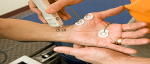
Place a high-frequency linear ultrasound probe in front of the patient and cover it with a sterile cover and apply sterile gel.
The local anesthetic solution should be filled a sterile syringe.
Median Nerve Block
Patient position
Patient position plays an integral role in all the procedures performed in the emergency department since positioning affects both the patient’s and the provider’s comfort, potentially influencing the probability of the procedure’s success.
Elevate the bed to an appropriate level and position the patients arm on a mayo stand or other flat surface. Place a small roll underneath the wrist to introduce mild wrist extension.
Technique
Step 1-Place the ultrasound probe at the level of the elbow crease to ensure that it is parallel to the proximal radius.
Step 2-Identify the radius and notice the pronator teres muscle located medially.
Step 3-Locate the two muscles flexor digitorum superficialis (FDS) and the flexor digitorum profundus (FDP) muscles which are medial to the pronator teres.
Step 4-Identify the median nerve between the FDS FDP it will be revealed as hyperechoic which has an appearance of a honey comb.
Step 5-Place the block needle alongside the probe direction so that the needle is parallel to the probe surface with the tip always in sight.
Step 6-Bring the needle tip to the superficial layer of the fascia that lies between the FDS and FDP muscles.
Step 7-Aspirate to confirm that the tip of the needle is out of the blood vessel.
Step 8-Aspirate a little of local anesthetic to ensure that the needle is on the right place. If needed, reposition the needle tip to be slightly further within the fascial plane to ensure that the anesthetic is around the nerve.
Step 9-If the location of the anesthetic spread coincides with the fascial plane of the median nerve, inject 1-2 mL of the local anesthetic in small increments while aspirating before achieving the desired distribution of the anesthetic solution.
This procedure of median nerve block is easy, reliable and safe option for doing surgeries related to thumb, index, middle, radial palm and inferior half of the ring finger. Such methods have been effectively being employed for many years now that are based on landmarking. Nevertheless, due to advancements in technology and availability of ultrasound, anaesthesia is more often now effective with less doses of medicines and fewer side effects. Peripheral nerve blocks for the hand are widely applied in anaesthesiology practices and hand surgeries during and after operations. In the recent past, emergency physicians and trauma specialists have also employed these blocks in treating acute pain as well as injuries.
In minor wounds, wrist nerve blocks may take longer and are not as effective as local infiltration or digital blocks. However, they are particularly beneficial in the emergency department (ED) in the following scenarios:
Injuries where more than one finger is affected at the same time
Severe injuries of the hand that required washing or removal of debris and dirt.
Preventing distortion of anatomy in areas of thin skin or where tissues are edematous
Administering anaesthesia to multiple lacerations given that they are adjacent or are near one another.
In these cases, median nerve block can be supplementary by ulnar or radial nerve block proximal to the wrist to make sure that the wrist, hand and fingers are sufficiently anesthetized.
Nerve blocks should not be performed in the following situations:
For instance, if the patient refuses to undergo the procedure
Cellulitis or abscess at the site of injection
In cases with a higher risk for compartment syndrome such as high energy injuries, it is a relative contraindication; seek consensus with the other specialities involved.
This area should be cleaned in a sterile fashion and there are many people who use chlorhexidine solution for cleaning as suggested by the CDC for any sterility purposes.
The size of the needle that is commonly used in the procedure is the 25 to 27-gauge needle. Reducing the needle size aids in preventing direct injury on the nerve fibres and minimizes pain when administering the injection. However, there are some drawbacks to smaller needles: They render aspiration more difficult to confirm intravascular placement, and they also may decrease the chances of eliciting paraesthesia if the needle is within the nerve.
The following preparatory steps should be taken:
Relatedly, get the patient’s informed consent of the nerve block procedure through explaining the risks, benefits, and other treatment options available.Ensure the identity of the patient and the exact area to be closed.
Perform and document a detailed neurovascular assessment of the injured extremity.
Place the involved extremity on a stand with the elbow joint locked and the forearm pronated.
Wash the area from the elbow crease towards the forearm or the area exposed from the clothing down to the middle forearm either with chlorhexidine 2% or povidone iodine and allow the solution to dry.
If possible, make sure that the ultrasound machine is in a sight line of the employee so that it does not pose a problem to access it every now and then.
Place a high-frequency linear ultrasound probe in front of the patient and cover it with a sterile cover and apply sterile gel.
The local anesthetic solution should be filled a sterile syringe.
Median Nerve Block
Patient position plays an integral role in all the procedures performed in the emergency department since positioning affects both the patient’s and the provider’s comfort, potentially influencing the probability of the procedure’s success.
Elevate the bed to an appropriate level and position the patients arm on a mayo stand or other flat surface. Place a small roll underneath the wrist to introduce mild wrist extension.
Step 1-Place the ultrasound probe at the level of the elbow crease to ensure that it is parallel to the proximal radius.
Step 2-Identify the radius and notice the pronator teres muscle located medially.
Step 3-Locate the two muscles flexor digitorum superficialis (FDS) and the flexor digitorum profundus (FDP) muscles which are medial to the pronator teres.
Step 4-Identify the median nerve between the FDS FDP it will be revealed as hyperechoic which has an appearance of a honey comb.
Step 5-Place the block needle alongside the probe direction so that the needle is parallel to the probe surface with the tip always in sight.
Step 6-Bring the needle tip to the superficial layer of the fascia that lies between the FDS and FDP muscles.
Step 7-Aspirate to confirm that the tip of the needle is out of the blood vessel.
Step 8-Aspirate a little of local anesthetic to ensure that the needle is on the right place. If needed, reposition the needle tip to be slightly further within the fascial plane to ensure that the anesthetic is around the nerve.
Step 9-If the location of the anesthetic spread coincides with the fascial plane of the median nerve, inject 1-2 mL of the local anesthetic in small increments while aspirating before achieving the desired distribution of the anesthetic solution.
Both our subscription plans include Free CME/CPD AMA PRA Category 1 credits.
Digital Certificate PDF
On course completion, you will receive a full-sized presentation quality digital certificate.
medtigo Simulation
A dynamic medical simulation platform designed to train healthcare professionals and students to effectively run code situations through an immersive hands-on experience in a live, interactive 3D environment.
medtigo Points
medtigo points is our unique point redemption system created to award users for interacting on our site. These points can be redeemed for special discounts on the medtigo marketplace as well as towards the membership cost itself.
Community Forum post/reply = 5 points
*Redemption of points can occur only through the medtigo marketplace, courses, or simulation system. Money will not be credited to your bank account. 10 points = $1.
All Your Certificates in One Place
When you have your licenses, certificates and CMEs in one place, it's easier to track your career growth. You can easily share these with hospitals as well, using your medtigo app.