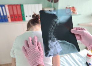
Myelography is a procedure in which the spinal cord, nerve roots, and other structures are seen using a contrast agent. It consists of administering a contrast dye into the subarachnoid space, which often involves a lumbar puncture. This dye increases the contrast of the spine and nerve structures on the X-ray, CT scan, or MRI.
Indications
Herniated Discs: To identify bulging and slipped discs that may be applying pressure on the nerves.
Spinal Stenosis: To evaluate the degree of spinal stenosis which leads to compression of the spinal nerves and causing symptoms such as pain.
Spinal Tumors: To diagnose tumors involving the spinal cord and nerve roots or structures that are in the surrounding structure.
Infections or Inflammatory Conditions: To detect neoplastic diseases such as meningitis or any other infection or inflammatory conditions like arachnoiditis.
Spinal Cord Injuries: To determine the severity of damage in the spinal cord and the adjacent structures.
Congenital Abnormalities: For abnormalities of the spinal column at birth like spina bifida.
Persistent Pain or Neurological Symptoms: When MRI or CT scan has not given enough information on the patients’ health condition.
Spinal Cysts: For the diagnosis of cysts nestled in or around the spinal cord.
Contraindications
Allergy to Contrast Medium: Patients should not have myelography if they have a known allergy to the iodinated contrast chemicals used in the technique unless premedication guidelines are followed and there are no other choices for imaging.
Acute Infection: Localization of the process in the active phase of the disease, as well as in the field of around the spinal column, is strictly prohibited due to the possibility of infection spread.
Coagulopathy and Anticoagulant Therapy: This is mainly so because the patient may be on drugs that predispose them to bleeding or even have a bleeding disorder.
Recent Lumbar Puncture: Cortical stimulation in these circumstances could raise the risk of post procedure complications such as infection or leakage of CSF in the lumbar punctured area.
Outcomes
Equipment
X-ray or Fluoroscopy Machine
CT Scanner
Contrast Dye
Needles and Catheters
Tilt Table
Monitoring Equipment
Image Processing and Viewing Workstations
Sterile Drapes and Tools
Patient Preparation
Patient History and Consent:
Take a comprehensive case history on the patient medical history in terms of their disease, allergies to any compounds such as contrast media or iodine, current and past medicines as well as past procedures done.
Discuss the procedure, risks, benefits and any side effects that might be associated with genomic sequencing should be explained to the patient.
Obtain written informed consent.
Medication Review:
Evaluate the current medications the patient is currently using. Anticoagulants and some other medicines may be taken a few days prior to the procedure.
Because one is diabetic, the patient should consult their physician about how the fasting will impact the insulin level.
Fasting:
The patient can only take food or drink after the procedure in 4 to 8 hours.
Hydration:
Remind the patient to intake sufficient fluids before arriving when they cannot consume food or fluids before the surgery.
Clothing and Personal Items:
Loose and comfortable clothing should be worn by the patient.
All jewellery, watches, and other metal items should be taken off for these can affect the images taken.
Premedication:
Give any preoperative medications, usually a mild sedative, before the procedure to help calm the patient.
Technique
Step 1-Positioning the Patient:
Place the patient on the X-ray table laying on their side or entirely on their abdomen.
Step 2-Sterilization and Local Anesthesia:
Wipe the lumbar area of the patient with an appropriate antiseptic agent.
Cover the environment around the patient with sterile towels.
Give an injection of lidocaine to help local anesthetic for the puncture.
Step 3-Lumbar Puncture:
Puncture the subarachnoid space using a spinal needle, commonly between intervertebral space.
Assure the correct position of the needle by checking the drip of the cerebrospinal fluid (CSF).
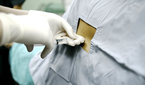
Step 4-Contrast Medium Injection:
Connect a syringe with the contrast medium to the spinal needle.
Administer the contrast medium into the subarachnoid space gradually.
Take out the spinal needle once the injection has been made.
Step 5-Imaging:
Position the patient as required for imaging. Usually, it is used to take several views of the spine where many positions are involved.
Assess the flow of the contrast medium through X-ray images or CT scans.
The possible side effects of the contrast medium should also be observed in the patient.
Step 6-Post-procedure
Observation:
Keep the patient in a supine position for a few hours to prevent headaches caused by CSF leakage.
Observe for the manifestations of allergy or any complication that may occur in the patient.
Post-procedure Care:
For elimination of the contrast medium from the body, the patient should be encouraged to take plenty of fluids.
Avoid strenuous activities for at least 24 hours.
Laboratory tests
Blood Tests:
Complete Blood Count (CBC): To ensure no infections or blood disorders that might complicate the procedure.
Coagulation Profile: To screen for any coagulopathies that prevent normal coagulation and might cause bleeding during the procedure.
Allergy Tests:
Allergy History: A patient’s history of allergies, particularly iodine or contrast dyes, is needed to avoid close reactions to the substance.
Complications
Nausea and Vomiting: This may be due to the contrast dye or an allergic reaction to the procedure
Infection: There is always some chance of infection at the puncture site; however, this is very rare
Bleeding: There can be minor bleeding at the site of injection. Significant bleeding can also occur, which may subsequently develop into a hematoma.
Allergic Reaction: The patient may develop a mild to severe allergic reaction to the contrast dye.
Neurological Symptoms: Temporary symptoms like vertigo, tingling, or even muscle weakness can be experienced by the patient due to irritation or damage to his spinal cord or nerves.
Seizures: The contrast dye can cause seizures in very rare instances.
Myelography is a procedure in which the spinal cord, nerve roots, and other structures are seen using a contrast agent. It consists of administering a contrast dye into the subarachnoid space, which often involves a lumbar puncture. This dye increases the contrast of the spine and nerve structures on the X-ray, CT scan, or MRI.
Herniated Discs: To identify bulging and slipped discs that may be applying pressure on the nerves.
Spinal Stenosis: To evaluate the degree of spinal stenosis which leads to compression of the spinal nerves and causing symptoms such as pain.
Spinal Tumors: To diagnose tumors involving the spinal cord and nerve roots or structures that are in the surrounding structure.
Infections or Inflammatory Conditions: To detect neoplastic diseases such as meningitis or any other infection or inflammatory conditions like arachnoiditis.
Spinal Cord Injuries: To determine the severity of damage in the spinal cord and the adjacent structures.
Congenital Abnormalities: For abnormalities of the spinal column at birth like spina bifida.
Persistent Pain or Neurological Symptoms: When MRI or CT scan has not given enough information on the patients’ health condition.
Spinal Cysts: For the diagnosis of cysts nestled in or around the spinal cord.
Allergy to Contrast Medium: Patients should not have myelography if they have a known allergy to the iodinated contrast chemicals used in the technique unless premedication guidelines are followed and there are no other choices for imaging.
Acute Infection: Localization of the process in the active phase of the disease, as well as in the field of around the spinal column, is strictly prohibited due to the possibility of infection spread.
Coagulopathy and Anticoagulant Therapy: This is mainly so because the patient may be on drugs that predispose them to bleeding or even have a bleeding disorder.
Recent Lumbar Puncture: Cortical stimulation in these circumstances could raise the risk of post procedure complications such as infection or leakage of CSF in the lumbar punctured area.
X-ray or Fluoroscopy Machine
CT Scanner
Contrast Dye
Needles and Catheters
Tilt Table
Monitoring Equipment
Image Processing and Viewing Workstations
Sterile Drapes and Tools
Patient History and Consent:
Take a comprehensive case history on the patient medical history in terms of their disease, allergies to any compounds such as contrast media or iodine, current and past medicines as well as past procedures done.
Discuss the procedure, risks, benefits and any side effects that might be associated with genomic sequencing should be explained to the patient.
Obtain written informed consent.
Medication Review:
Evaluate the current medications the patient is currently using. Anticoagulants and some other medicines may be taken a few days prior to the procedure.
Because one is diabetic, the patient should consult their physician about how the fasting will impact the insulin level.
Fasting:
The patient can only take food or drink after the procedure in 4 to 8 hours.
Hydration:
Remind the patient to intake sufficient fluids before arriving when they cannot consume food or fluids before the surgery.
Clothing and Personal Items:
Loose and comfortable clothing should be worn by the patient.
All jewellery, watches, and other metal items should be taken off for these can affect the images taken.
Premedication:
Give any preoperative medications, usually a mild sedative, before the procedure to help calm the patient.
Step 1-Positioning the Patient:
Place the patient on the X-ray table laying on their side or entirely on their abdomen.
Step 2-Sterilization and Local Anesthesia:
Wipe the lumbar area of the patient with an appropriate antiseptic agent.
Cover the environment around the patient with sterile towels.
Give an injection of lidocaine to help local anesthetic for the puncture.
Step 3-Lumbar Puncture:
Puncture the subarachnoid space using a spinal needle, commonly between intervertebral space.
Assure the correct position of the needle by checking the drip of the cerebrospinal fluid (CSF).
Step 4-Contrast Medium Injection:
Connect a syringe with the contrast medium to the spinal needle.
Administer the contrast medium into the subarachnoid space gradually.
Take out the spinal needle once the injection has been made.
Step 5-Imaging:
Position the patient as required for imaging. Usually, it is used to take several views of the spine where many positions are involved.
Assess the flow of the contrast medium through X-ray images or CT scans.
The possible side effects of the contrast medium should also be observed in the patient.
Step 6-Post-procedure
Observation:
Keep the patient in a supine position for a few hours to prevent headaches caused by CSF leakage.
Observe for the manifestations of allergy or any complication that may occur in the patient.
Post-procedure Care:
For elimination of the contrast medium from the body, the patient should be encouraged to take plenty of fluids.
Avoid strenuous activities for at least 24 hours.
Blood Tests:
Complete Blood Count (CBC): To ensure no infections or blood disorders that might complicate the procedure.
Coagulation Profile: To screen for any coagulopathies that prevent normal coagulation and might cause bleeding during the procedure.
Allergy Tests:
Allergy History: A patient’s history of allergies, particularly iodine or contrast dyes, is needed to avoid close reactions to the substance.
Nausea and Vomiting: This may be due to the contrast dye or an allergic reaction to the procedure
Infection: There is always some chance of infection at the puncture site; however, this is very rare
Bleeding: There can be minor bleeding at the site of injection. Significant bleeding can also occur, which may subsequently develop into a hematoma.
Allergic Reaction: The patient may develop a mild to severe allergic reaction to the contrast dye.
Neurological Symptoms: Temporary symptoms like vertigo, tingling, or even muscle weakness can be experienced by the patient due to irritation or damage to his spinal cord or nerves.
Seizures: The contrast dye can cause seizures in very rare instances.
Both our subscription plans include Free CME/CPD AMA PRA Category 1 credits.
Digital Certificate PDF
On course completion, you will receive a full-sized presentation quality digital certificate.
medtigo Simulation
A dynamic medical simulation platform designed to train healthcare professionals and students to effectively run code situations through an immersive hands-on experience in a live, interactive 3D environment.
medtigo Points
medtigo points is our unique point redemption system created to award users for interacting on our site. These points can be redeemed for special discounts on the medtigo marketplace as well as towards the membership cost itself.
Community Forum post/reply = 5 points
*Redemption of points can occur only through the medtigo marketplace, courses, or simulation system. Money will not be credited to your bank account. 10 points = $1.
All Your Certificates in One Place
When you have your licenses, certificates and CMEs in one place, it's easier to track your career growth. You can easily share these with hospitals as well, using your medtigo app.