Neuroimaging refers to methods employed to visualize the anatomy, function, or activity of the brain and nervous system. These techniques play a vital role in the diagnosis, treatment planning, and monitoring of neurological diseases.
Neuroimaging has transformed the practice of neurology, providing information on brain function and assisting in the treatment of diseases like Alzheimer’s disease, epilepsy, brain tumors, stroke, and psychiatric illness. It has also played a crucial role in research, allowing for a better understanding of how the brain functions and how neurological disease arises.
Indications
Acute Neurological Symptoms:
Sudden onset of severe headache, especially in cases of suspected subarachnoid hemorrhage (e.g., CT or MRI for ruling out hemorrhage).
New-onset seizures (to evaluate for underlying brain abnormalities like tumors, infarcts, or structural malformations).
Sudden neurological deficit (e.g., weakness, sensory loss) suggestive of acute stroke (CT or MRI to confirm).
Confusion or altered mental status (MRI/CT to rule out mass lesions, ischemic stroke, or acute metabolic changes).
Chronic or Progressive Neurological Symptoms:
Progressive memory loss or cognitive decline (MRI to evaluate for conditions like Alzheimer’s disease, frontotemporal dementia, or other neurodegenerative disorders).
Chronic headaches with warning signs (e.g., nausea, visual disturbances) or a change in pattern, indicating the need for imaging to rule out intracranial masses or other pathology.
Progressive motor symptoms (e.g., tremor, gait instability) where neurodegenerative diseases like Parkinson’s disease or multiple system atrophy need to be evaluated.
Trauma or Injury:
Head trauma with loss of consciousness, confusion, or focal neurological signs (CT or MRI to detect fractures, hemorrhage, or brain contusions).
Suspected spinal cord injury or vertebral fractures (CT or MRI to assess structural damage and spinal cord integrity).
Vascular Pathologies:
Suspected stroke or transient ischemic attack (TIA) (CT or MRI to detect acute ischemia, infarction, or hemorrhage).
Suspected aneurysm, arteriovenous malformation, or other vascular malformations (CT angiography or MR angiography).
Evaluation of venous sinus thrombosis (CT venography or MR venography).
Tumors and Mass Lesions:
Patients with a clinical suspicion of brain tumors, metastases, or benign lesions (MRI is the gold standard for brain tumor detection).
Suspected pituitary or spinal cord tumors (MRI or CT depending on the location).
Infectious Diseases and Inflammatory Conditions:
Suspected brain abscess, encephalitis, or meningitis (MRI or CT to assess the extent of infection or inflammation).
Inflammatory conditions like multiple sclerosis (MRI is the primary imaging modality for detecting plaques and disease progression).
Contraindications
Pacemakers or Implantable Cardiac Devices: The intense magnetic field may disrupt the operation of pacemakers, defibrillators, and other implantable devices, resulting in malfunction or patient injury.
Cochlear Implants: The magnetic field may cause damage to cochlear implants, resulting in failure or malfunction.
Metallic Foreign Bodies: Patients with metallic foreign bodies, including shrapnel, bullet fragments, or retained surgical instruments, in critical locations (e.g., the eye) can be at risk from magnetic forces.
Pregnancy: CT scans are associated with radiation, which could be harmful to a developing fetus, especially in early pregnancy. MRI is used in pregnant patients when brain or spine imaging is required.
Severe Renal Impairment: Administration of iodinated contrast media in CT can aggravate kidney function in patients with established renal insufficiency.
Allergy to Contrast Media: Known allergy to iodinated contrast agents can result in reactions ranging from mild dermatitis to life-threatening anaphylactic reaction.
Uncontrolled Hypertension or Cardiac Instability: PET imaging necessitates some degree of patient cooperation, and unstable cardiovascular patients may not be able to tolerate the procedure.
Age-Related Factors: In extremely young or old patients, handling and positioning for imaging might require extra care. Neonates, especially, might need special procedures or imaging protocols.
Patient Preparation Informed Consent:
Patients should be informed about the type of neuroimaging, its purpose, potential risks, and necessary preparations before giving consent.
Medical History:
A detailed history should be taken, including claustrophobia, allergies, existing conditions (e.g., kidney disease, pregnancy), and implanted devices like pacemakers or stents to determine suitability and identify contraindications.
Medication Review:
Patients should disclose any medications, especially sedatives, anticoagulants, or drugs affecting imaging agents. Adjustments may be needed before the procedure.
Metal and Implants:
Patients must remove metal items (jewelry, hearing aids, eyeglasses, dentures) before an MRI. Implanted devices should be checked for MRI compatibility.
Claustrophobia Considerations:
Patients with claustrophobia should be informed about the MRI’s enclosed space. If needed, a mild sedative can be prescribed to ease anxiety.
Contrast Agent Precautions:
Before CT with contrast, patients should be screened for iodine allergies and kidney or thyroid conditions. Blood tests may be required to assess kidney function.
Fasting and Hydration:
For PET scans, fasting for 4-6 hours helps improve image quality. Water is generally allowed in limited amounts.
Substance Restrictions:
Before an EEG, patients should avoid caffeine, nicotine, and stimulants as they can affect brain activity.
Pediatric Considerations:
Children may need extra preparation, with parents involved in explanations. Depending on age, sedation or anesthesia may be required for cooperation.
Patient position
For most brain MRI scans, the patient typically lies in a supine (on their back) position. The head is placed in a dedicated coil or headrest, and the head is aligned with the center of the scanner’s magnetic field. This ensures high-quality images of the brain.
The head must be placed symmetrically in the scanner’s gantry (the circular opening of the machine) to ensure the brain is imaged properly.
PET and SPECT are usually performed with the patient in a supine position. The head is aligned in the center of the scanner, with the patient’s body positioned so that the imaging field of view covers the entire brain.
Magnetic Resonance Imaging (MRI)
MRI Machine
MRI is a widely used technique for obtaining high-resolution images of brain structures. It uses magnetic fields and radio waves to create detailed images.
Step 1-Preparation:
The patient may be asked to change into a gown and remove any metal objects (jewellery, hearing aids, etc.).
The patient lies down on the MRI table, and a coil is placed around the head.
If contrast is needed (for enhanced image quality), a contrast agent (gadolinium) is injected intravenously.
Step 2-Scanning:
The MRI machine creates a magnetic field, causing hydrogen atoms in the body to align.
Radiofrequency pulses are applied, and when the pulse stops, the hydrogen atoms emit signals.
These signals are captured by the MRI detectors and used to create an image.
Step 3-Image Reconstruction:
The signals are processed by a computer to generate a high-resolution 3D image of the brain.
Images can be in multiple orientations: axial, sagittal, and coronal.
Functional Magnetic Resonance Imaging (fMRI)
fMRI measures brain activity by detecting changes in blood flow, which is related to neuronal activity.
Step 1-Preparation:
Like standard MRI, the patient is asked to lie down and remove metal objects.
If required, a task or stimulus (visual, auditory, or motor) may be presented to the patient during the scan.
Step 2-Scanning:
fMRI uses the BOLD (Blood Oxygen Level Dependent) signal, which detects changes in the ratio of oxygenated to deoxygenated blood in the brain.
As neurons become active, the demand for oxygen increases, leading to changes in blood flow, which can be detected by the MRI scanner.
Step 3-Image Reconstruction:
The data is processed to create functional images that show brain activity. The resulting images are overlaid on the structural MRI to identify active brain regions.
Positron Emission Tomography (PET)
PET Scan Machine
PET is a functional imaging technique that measures metabolic activity, such as glucose consumption in the brain, to assess brain function.
Step 1-Preparation:
A radiolabelled tracer (such as fluorodeoxyglucose, FDG) is injected intravenously. This tracer is typically glucose or a glucose analog that emits positrons as it decays.
The patient is usually asked to rest quietly for a period after the injection.
Step 2-Scanning:
The PET scanner detects the gamma rays produced when positrons emitted by the tracer collide with electrons in the body.
These collisions produce detectable photons, which are captured by the PET scanner.
Step 3-Image Reconstruction:
The data collected by the scanner is processed to create 3D images showing the distribution of the tracer in the brain, revealing areas of high or low metabolic activity.
Computed Tomography (CT) Scan
CT uses X-rays to create cross-sectional images of the brain, often used in emergency situations to quickly assess brain structure and detect abnormalities.
Step 1-Preparation:
The patient is asked to lie on the CT scanner table.
Metal objects are removed, and contrast may be injected if needed.
Step 2-Scanning:
The CT scanner takes a series of X-ray images from different angles around the head.
The patient may be asked to hold their breath briefly during the scan to minimize movement.
Step 3-Image Reconstruction:
The X-ray images are processed by a computer to produce detailed cross-sectional images (slices) of the brain.
These slices can be reconstructed into 3D images for a more comprehensive view.
Electroencephalography (EEG)
EEG measures electrical activity in the brain through electrodes placed on the scalp.
Step 1-Preparation:
Electrodes are placed on the scalp using a conductive gel or paste.
The patient is typically asked to remain still, and the EEG technician ensures that the electrodes are securely attached.
Step 2-Recording:
The EEG records electrical activity from the brain, detecting brain waves (e.g., alpha, beta, delta, theta).
The activity is recorded continuously, usually for 20 to 60 minutes.
Step 3-Data Analysis:
The recorded brain wave patterns are analysed for abnormalities that could indicate seizures, sleep disorders, or other neurological conditions.
Magnetoencephalography (MEG)
MEG measures the magnetic fields produced by neuronal activity, offering high temporal resolution.
Step 1 – Preparation:
The patient is fitted with a helmet-like device that holds superconducting sensors (SQUIDs), which can pick up magnetic fields.
The patient is instructed to relax and remain still.
Step 2 – Recording:
MEG detects the weak magnetic fields generated by the brain’s electrical activity.
The data is recorded and monitored continuously for abnormal or specific brain patterns.
Step 3 – Data Analysis:
MEG allows for high temporal resolution, making it suitable for examining fast brain activity, such as task-related cognitive function or epilepsy occurrence.
Diffusion Tensor Imaging (DTI)
DTI is a type of MRI that specifically measures the diffusion of water molecules in the brain, particularly useful for imaging white matter tracts.
Step 1-Preparation:
As with standard MRI, the patient lies in the MRI scanner, and the procedure is explained.
Step 2-Scanning:
DTI applies the same fundamental principles as MRI but is directed at quantifying the direction of motion (diffusion) of water molecules.
Water molecules spread more freely in the direction of white matter tracts, permitting tracing of such tracts.
Step 3-Image Reconstruction:
DTI creates images that display the orientation and integrity of white matter tracts, which are vital to know for brain connectivity.
Complications Allergic Reactions:
Mild to severe allergic reactions might occur in some patients, such as itching, rash, or anaphylaxis. They are infrequent but can happen, especially with iodinated contrast in CT or gadolinium in MRI.
Nephrotoxicity:
Gadolinium (used in MRIs) and iodinated contrast (used in CT scans) can cause kidney damage, especially in patients with existing kidney disease. Contrast-induced nephropathy (CIN) may lead to acute kidney injury, requiring careful monitoring.
Extravasation:
If contrast agents are injected incorrectly, they may leak into surrounding tissues, causing swelling, pain, or tissue damage. This is more common with intravenous injections.
Thyroid Dysfunction:
Iodine-based contrast agents (used in CT scans) can affect thyroid function, particularly in individuals with thyroid disorders.
Pregnancy Risks:
Radiation exposure during pregnancy, especially in the early stages, may increase the risk of fetal development issues. For this reason, non-emergency CT, PET, and SPECT scans are generally avoided during pregnancy.
Neurological Side Effects:
Some patients may experience mild symptoms like dizziness, headache, or nausea after receiving radioactive tracers. Higher doses, though rare, can cause more severe effects.
Claustrophobia:
The enclosed space of an MRI machine can trigger anxiety or panic in some patients, potentially making the procedure difficult. Sedation or alternative imaging methods may be needed.
Sedation Risks:
Patients who require sedation, such as children or those with severe anxiety, face risks including breathing difficulties, allergic reactions, or cardiovascular issues.
Metal Artifacts:
MRI scans can be distorted by metal objects in or on the body, such as pacemakers, implants, or jewellery, sometimes making the images unusable.
Skin Irritation:
Electrodes placed on the scalp for imaging can cause mild irritation, redness, or itching, particularly in patients with sensitive skin or during prolonged procedures.
Stress and Anxiety:
The findings may influence long-term treatment plans, adding to anxiety about potential diagnoses like tumors or neurodegenerative conditions.
Post-Procedure Psychological Impact:
A serious diagnosis following a scan, such as a brain tumor or dementia, can lead to emotional distress, including anxiety or depression.
Injection Site Complications:
Pain, swelling, bruising, or infection may occur at the injection site after receiving contrast agents or radiotracers.
Cardiovascular Complications:
Though rare, the injection of radiotracers can cause heart-related issues like palpitations or fainting, particularly in patients with pre-existing heart conditions.
Neuroimaging refers to methods employed to visualize the anatomy, function, or activity of the brain and nervous system. These techniques play a vital role in the diagnosis, treatment planning, and monitoring of neurological diseases.
Neuroimaging has transformed the practice of neurology, providing information on brain function and assisting in the treatment of diseases like Alzheimer’s disease, epilepsy, brain tumors, stroke, and psychiatric illness. It has also played a crucial role in research, allowing for a better understanding of how the brain functions and how neurological disease arises.
Acute Neurological Symptoms:
Sudden onset of severe headache, especially in cases of suspected subarachnoid hemorrhage (e.g., CT or MRI for ruling out hemorrhage).
New-onset seizures (to evaluate for underlying brain abnormalities like tumors, infarcts, or structural malformations).
Sudden neurological deficit (e.g., weakness, sensory loss) suggestive of acute stroke (CT or MRI to confirm).
Confusion or altered mental status (MRI/CT to rule out mass lesions, ischemic stroke, or acute metabolic changes).
Chronic or Progressive Neurological Symptoms:
Progressive memory loss or cognitive decline (MRI to evaluate for conditions like Alzheimer’s disease, frontotemporal dementia, or other neurodegenerative disorders).
Chronic headaches with warning signs (e.g., nausea, visual disturbances) or a change in pattern, indicating the need for imaging to rule out intracranial masses or other pathology.
Progressive motor symptoms (e.g., tremor, gait instability) where neurodegenerative diseases like Parkinson’s disease or multiple system atrophy need to be evaluated.
Trauma or Injury:
Head trauma with loss of consciousness, confusion, or focal neurological signs (CT or MRI to detect fractures, hemorrhage, or brain contusions).
Suspected spinal cord injury or vertebral fractures (CT or MRI to assess structural damage and spinal cord integrity).
Vascular Pathologies:
Suspected stroke or transient ischemic attack (TIA) (CT or MRI to detect acute ischemia, infarction, or hemorrhage).
Suspected aneurysm, arteriovenous malformation, or other vascular malformations (CT angiography or MR angiography).
Evaluation of venous sinus thrombosis (CT venography or MR venography).
Tumors and Mass Lesions:
Patients with a clinical suspicion of brain tumors, metastases, or benign lesions (MRI is the gold standard for brain tumor detection).
Suspected pituitary or spinal cord tumors (MRI or CT depending on the location).
Infectious Diseases and Inflammatory Conditions:
Suspected brain abscess, encephalitis, or meningitis (MRI or CT to assess the extent of infection or inflammation).
Inflammatory conditions like multiple sclerosis (MRI is the primary imaging modality for detecting plaques and disease progression).
Pacemakers or Implantable Cardiac Devices: The intense magnetic field may disrupt the operation of pacemakers, defibrillators, and other implantable devices, resulting in malfunction or patient injury.
Cochlear Implants: The magnetic field may cause damage to cochlear implants, resulting in failure or malfunction.
Metallic Foreign Bodies: Patients with metallic foreign bodies, including shrapnel, bullet fragments, or retained surgical instruments, in critical locations (e.g., the eye) can be at risk from magnetic forces.
Pregnancy: CT scans are associated with radiation, which could be harmful to a developing fetus, especially in early pregnancy. MRI is used in pregnant patients when brain or spine imaging is required.
Severe Renal Impairment: Administration of iodinated contrast media in CT can aggravate kidney function in patients with established renal insufficiency.
Allergy to Contrast Media: Known allergy to iodinated contrast agents can result in reactions ranging from mild dermatitis to life-threatening anaphylactic reaction.
Uncontrolled Hypertension or Cardiac Instability: PET imaging necessitates some degree of patient cooperation, and unstable cardiovascular patients may not be able to tolerate the procedure.
Age-Related Factors: In extremely young or old patients, handling and positioning for imaging might require extra care. Neonates, especially, might need special procedures or imaging protocols.
Patient Preparation Informed Consent:
Patients should be informed about the type of neuroimaging, its purpose, potential risks, and necessary preparations before giving consent.
Medical History:
A detailed history should be taken, including claustrophobia, allergies, existing conditions (e.g., kidney disease, pregnancy), and implanted devices like pacemakers or stents to determine suitability and identify contraindications.
Medication Review:
Patients should disclose any medications, especially sedatives, anticoagulants, or drugs affecting imaging agents. Adjustments may be needed before the procedure.
Metal and Implants:
Patients must remove metal items (jewelry, hearing aids, eyeglasses, dentures) before an MRI. Implanted devices should be checked for MRI compatibility.
Claustrophobia Considerations:
Patients with claustrophobia should be informed about the MRI’s enclosed space. If needed, a mild sedative can be prescribed to ease anxiety.
Contrast Agent Precautions:
Before CT with contrast, patients should be screened for iodine allergies and kidney or thyroid conditions. Blood tests may be required to assess kidney function.
Fasting and Hydration:
For PET scans, fasting for 4-6 hours helps improve image quality. Water is generally allowed in limited amounts.
Substance Restrictions:
Before an EEG, patients should avoid caffeine, nicotine, and stimulants as they can affect brain activity.
Pediatric Considerations:
Children may need extra preparation, with parents involved in explanations. Depending on age, sedation or anesthesia may be required for cooperation.
Patient position
For most brain MRI scans, the patient typically lies in a supine (on their back) position. The head is placed in a dedicated coil or headrest, and the head is aligned with the center of the scanner’s magnetic field. This ensures high-quality images of the brain.
The head must be placed symmetrically in the scanner’s gantry (the circular opening of the machine) to ensure the brain is imaged properly.
PET and SPECT are usually performed with the patient in a supine position. The head is aligned in the center of the scanner, with the patient’s body positioned so that the imaging field of view covers the entire brain.
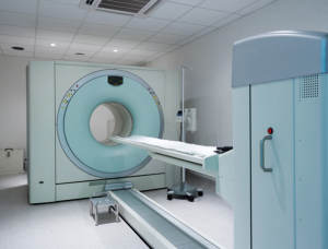
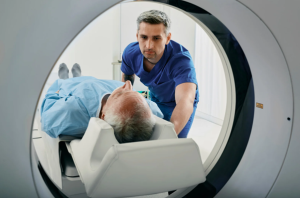
MRI Machine
MRI is a widely used technique for obtaining high-resolution images of brain structures. It uses magnetic fields and radio waves to create detailed images.
Step 1-Preparation:
The patient may be asked to change into a gown and remove any metal objects (jewellery, hearing aids, etc.).
The patient lies down on the MRI table, and a coil is placed around the head.
If contrast is needed (for enhanced image quality), a contrast agent (gadolinium) is injected intravenously.
Step 2-Scanning:
The MRI machine creates a magnetic field, causing hydrogen atoms in the body to align.
Radiofrequency pulses are applied, and when the pulse stops, the hydrogen atoms emit signals.
These signals are captured by the MRI detectors and used to create an image.
Step 3-Image Reconstruction:
The signals are processed by a computer to generate a high-resolution 3D image of the brain.
Images can be in multiple orientations: axial, sagittal, and coronal.
fMRI measures brain activity by detecting changes in blood flow, which is related to neuronal activity.
Step 1-Preparation:
Like standard MRI, the patient is asked to lie down and remove metal objects.
If required, a task or stimulus (visual, auditory, or motor) may be presented to the patient during the scan.
Step 2-Scanning:
fMRI uses the BOLD (Blood Oxygen Level Dependent) signal, which detects changes in the ratio of oxygenated to deoxygenated blood in the brain.
As neurons become active, the demand for oxygen increases, leading to changes in blood flow, which can be detected by the MRI scanner.
Step 3-Image Reconstruction:
The data is processed to create functional images that show brain activity. The resulting images are overlaid on the structural MRI to identify active brain regions.
PET Scan Machine
PET is a functional imaging technique that measures metabolic activity, such as glucose consumption in the brain, to assess brain function.
Step 1-Preparation:
A radiolabelled tracer (such as fluorodeoxyglucose, FDG) is injected intravenously. This tracer is typically glucose or a glucose analog that emits positrons as it decays.
The patient is usually asked to rest quietly for a period after the injection.
Step 2-Scanning:
The PET scanner detects the gamma rays produced when positrons emitted by the tracer collide with electrons in the body.
These collisions produce detectable photons, which are captured by the PET scanner.
Step 3-Image Reconstruction:
The data collected by the scanner is processed to create 3D images showing the distribution of the tracer in the brain, revealing areas of high or low metabolic activity.
CT uses X-rays to create cross-sectional images of the brain, often used in emergency situations to quickly assess brain structure and detect abnormalities.
Step 1-Preparation:
The patient is asked to lie on the CT scanner table.
Metal objects are removed, and contrast may be injected if needed.
Step 2-Scanning:
The CT scanner takes a series of X-ray images from different angles around the head.
The patient may be asked to hold their breath briefly during the scan to minimize movement.
Step 3-Image Reconstruction:
The X-ray images are processed by a computer to produce detailed cross-sectional images (slices) of the brain.
These slices can be reconstructed into 3D images for a more comprehensive view.
EEG measures electrical activity in the brain through electrodes placed on the scalp.
Step 1-Preparation:
Electrodes are placed on the scalp using a conductive gel or paste.
The patient is typically asked to remain still, and the EEG technician ensures that the electrodes are securely attached.
Step 2-Recording:
The EEG records electrical activity from the brain, detecting brain waves (e.g., alpha, beta, delta, theta).
The activity is recorded continuously, usually for 20 to 60 minutes.
Step 3-Data Analysis:
The recorded brain wave patterns are analysed for abnormalities that could indicate seizures, sleep disorders, or other neurological conditions.
MEG measures the magnetic fields produced by neuronal activity, offering high temporal resolution.
Step 1 – Preparation:
The patient is fitted with a helmet-like device that holds superconducting sensors (SQUIDs), which can pick up magnetic fields.
The patient is instructed to relax and remain still.
Step 2 – Recording:
MEG detects the weak magnetic fields generated by the brain’s electrical activity.
The data is recorded and monitored continuously for abnormal or specific brain patterns.
Step 3 – Data Analysis:
MEG allows for high temporal resolution, making it suitable for examining fast brain activity, such as task-related cognitive function or epilepsy occurrence.
DTI is a type of MRI that specifically measures the diffusion of water molecules in the brain, particularly useful for imaging white matter tracts.
Step 1-Preparation:
As with standard MRI, the patient lies in the MRI scanner, and the procedure is explained.
Step 2-Scanning:
DTI applies the same fundamental principles as MRI but is directed at quantifying the direction of motion (diffusion) of water molecules.
Water molecules spread more freely in the direction of white matter tracts, permitting tracing of such tracts.
Step 3-Image Reconstruction:
DTI creates images that display the orientation and integrity of white matter tracts, which are vital to know for brain connectivity.
Complications Allergic Reactions:
Mild to severe allergic reactions might occur in some patients, such as itching, rash, or anaphylaxis. They are infrequent but can happen, especially with iodinated contrast in CT or gadolinium in MRI.
Nephrotoxicity:
Gadolinium (used in MRIs) and iodinated contrast (used in CT scans) can cause kidney damage, especially in patients with existing kidney disease. Contrast-induced nephropathy (CIN) may lead to acute kidney injury, requiring careful monitoring.
Extravasation:
If contrast agents are injected incorrectly, they may leak into surrounding tissues, causing swelling, pain, or tissue damage. This is more common with intravenous injections.
Thyroid Dysfunction:
Iodine-based contrast agents (used in CT scans) can affect thyroid function, particularly in individuals with thyroid disorders.
Pregnancy Risks:
Radiation exposure during pregnancy, especially in the early stages, may increase the risk of fetal development issues. For this reason, non-emergency CT, PET, and SPECT scans are generally avoided during pregnancy.
Neurological Side Effects:
Some patients may experience mild symptoms like dizziness, headache, or nausea after receiving radioactive tracers. Higher doses, though rare, can cause more severe effects.
Claustrophobia:
The enclosed space of an MRI machine can trigger anxiety or panic in some patients, potentially making the procedure difficult. Sedation or alternative imaging methods may be needed.
Sedation Risks:
Patients who require sedation, such as children or those with severe anxiety, face risks including breathing difficulties, allergic reactions, or cardiovascular issues.
Metal Artifacts:
MRI scans can be distorted by metal objects in or on the body, such as pacemakers, implants, or jewellery, sometimes making the images unusable.
Skin Irritation:
Electrodes placed on the scalp for imaging can cause mild irritation, redness, or itching, particularly in patients with sensitive skin or during prolonged procedures.
Stress and Anxiety:
The findings may influence long-term treatment plans, adding to anxiety about potential diagnoses like tumors or neurodegenerative conditions.
Post-Procedure Psychological Impact:
A serious diagnosis following a scan, such as a brain tumor or dementia, can lead to emotional distress, including anxiety or depression.
Injection Site Complications:
Pain, swelling, bruising, or infection may occur at the injection site after receiving contrast agents or radiotracers.
Cardiovascular Complications:
Though rare, the injection of radiotracers can cause heart-related issues like palpitations or fainting, particularly in patients with pre-existing heart conditions.
Both our subscription plans include Free CME/CPD AMA PRA Category 1 credits.
Digital Certificate PDF
On course completion, you will receive a full-sized presentation quality digital certificate.
medtigo Simulation
A dynamic medical simulation platform designed to train healthcare professionals and students to effectively run code situations through an immersive hands-on experience in a live, interactive 3D environment.
medtigo Points
medtigo points is our unique point redemption system created to award users for interacting on our site. These points can be redeemed for special discounts on the medtigo marketplace as well as towards the membership cost itself.
Community Forum post/reply = 5 points
*Redemption of points can occur only through the medtigo marketplace, courses, or simulation system. Money will not be credited to your bank account. 10 points = $1.
All Your Certificates in One Place
When you have your licenses, certificates and CMEs in one place, it's easier to track your career growth. You can easily share these with hospitals as well, using your medtigo app.