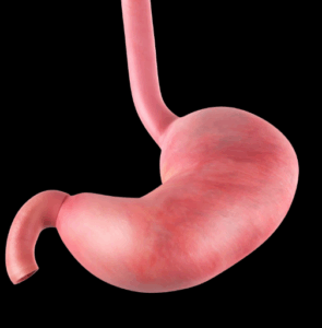
Partial Gastrectomy, often known as stomach resection, is a medical intervention in which surgeons choose to resect a part of the stomach to treat several diseases of this essential organ.
Partial gastrectomy
By altering the structure of the stomach through surgery, Partial gastrectomy improved the health and overall well-being of the affected patients, with attempts made to ease the symptoms instead of causing additional complications.
Indications
From the management of gastric conditions, it is proved that partial gastrectomy might be used as method of dealing with some gastric conditions. For instance, it may be required in cases of gastric cancer that has not spread to other areas of the body for a localized form of gastric cancer.
Additional serious problems associated with peptic ulcer disease, including perforation, bleeding or obstruction might require partial gastrectomy to resect the compromised segments of stomach.
For chronic or major resistant gastric bleeding, this surgery can be used to excise the expected bleeding cause such as taking out a part of the stomach, partial gastrectomy.
Gastroparesis starts with a specific issue of slowing down and in most cases, one may ever experience some stages of bloating, nausea, or vomiting. If necessary, it is possible to attempt to partially remove the stomach, which may help to improve emptying and minimize pain.
Contraindications
Advanced cancer: Surgery might not be feasible or effective if the cancer has progressed far beyond the stomach to other organs.
Severe malnutrition: Some patient may have severe malnourishment; they are at higher risk of not surviving the surgery. Feeding support can be required prior to the decision on surgery.
Uncontrolled medical conditions: It is contraindicated in critically ill patient or any patient who has serious complicating disease like chronic obstructive pulmonary disease.
History of surgery: Unstable diabetes or severe mental illness because they are likely to undertake surgical operations and it slows down their recovery period.
Outcomes
Equipment
Basic surgical instruments:
Scalpels
Scissors
Hemostats for clamping blood vessels.
Forceps for grasping and manipulating tissues.
Needle drivers and sutures for closing incisions and anastomoses.
Suction and irrigation equipment.
For open surgery: Retractors, abdominal retractors.
Use a laparoscope, trocars, insufflator, or energy devices for laparoscopic surgery.
Staplers: Linear staplers, Circular staplers.
Patient preparation
Consume lots of nutrient rich and vitamin packed foods to make more progress in minimising inflammation and assisting in speedy recovery.
Fluid intake should be adequate to avoid the dehydrated state of the body. Obesity can be a predictor of poor surgical outcomes and slow recovery thus the need to exercise and take a proper meal is advised. Furthermore, ordinarily, standard medical procedure includes the use of general anesthesia and endotracheal intubation.
Patient position
Reverse Trendelenburg
The operating table’s head end is lowered 15 to 30 degrees below the foot end. This pushes the abdominal contents downward, allowing for enhanced visibility, especially in the stomach area.
Staging laparoscopy
Laparoscopy for staging before partial gastrectomy is another surgical process used to check the extension of the cancer to other parts of the stomach before the partial gastrectomy. This is usually done where there is a suspicion of malignancies commonly, gastric cancer or other malignancies in the stomach.
Partial Gastrectomy
Step 1: Anesthesia
General anesthesia is used to make sure the patient doesn’t feel any discomfort and stays asleep during the treatment. Additionally, the surgical team will clean and sterilise the abdomen of the patient.
Step 2: Incision
From the incision made on the abdomen, the surgeon is in a position of accessing an entry point into the stomach. It may also depend on the specifics of the position and sizes of the proposed incision as related to the mode of the minimally invasive surgery to be used.
Essentially the incision from the xiphoid process down to and below the umbilicus or otherwise inferior to an umbilicus requires significantly less blood loss to create an efficient opening into the abdominal cavity.
After the stomach is made visible, the surgeon carefully inspects it to evaluate the severity of the illness and determine the amount of stomach that must be removed.
Step 3: Duodenal mobilization Mobilization of the duodenum can also be made through operation that require an incision to the side of peritoneum. Next, the duodenum is shifted to the left side gently, thereby exposing the inferior vena cava.
Step 4: Omental mobilization By dividing the omentum anterior leaf from the transverse colon and separating both by an avascular plane, a large omentum detaches from the transverse colon.
This is true if the avascular window is located on the left side of the transverse colon. Omental reflection is divided additionally & omental expansion to the right further.
This procedure has one disadvantage in that could harm the middle colic artery. In doing so, the transverse colon is mobilized to obtain an optimal view of the vascular pattern of the transverse colon and the gastrocolic omentum is reflected along the greater curvature to visually display and the vascular pattern of gastroepiploic vessels which should prevent such injury. An abdominal posterior wall is liberated by making an incision obliterating the peritoneal folds which are referred to as gastropancreatic folds. The lesser omentum is peeled off from the underside of the hepatic for malignancy and lesser curvature for ulcer associated condition.
Step 5: Duodenal division The right gastric artery arises from the gastroduodenal or the proximal hepatic artery and runs along the lesser curvature, and it occupies the inferior aspect. It has two ligations & two divisions. Similarly, in greater curvature a gastroepiploic artery is present at the lower end, is divided & the ligament is doubly ligated. At this point, the duodenum’s 1 to 2 cm next to the pylorus is evidently devoid of fat deposits and vascular attachments.
Special attention is paid to the protection of the pancreatic tissues when duodenum clearance is performed. At this point, the duodenum is divided using linear stapler.
Step 6: Gastric division More possible abdominal movement is gained after the division of the duodenum. An additional part of the mobilization of the greater curvature is done through the gastrosplenic ligament. Again, the manipulation of the greater curvature during the gastrectomy differs based on the level of operation to be performed. It implies a change in the location of the greater curvature in a way that the position of the gastroepiploic artery more to the stomach wall or in the vicinity of the second short abdominal artery. The choice of the first short gastric artery is left intact because it is used to supply the blood to the rest of an abdomen.
Also, the position at which the abdomen is divided beside the least curvature is in line with a third eminent vein or slightly lesser to the intersection between the esophagus & stomach.
The selected site is to be prepped for the smaller & greater curvatures & omental fat is to be taken for a few cm along the section line.
Approximately at the area of proximal resection, the linear stapler is used to divide the stomach. At this point the specimen is disposed off.
Step 7: Lymph node dissection
D1 Dissection: Dissection entails the removal of lymph nodes, particularly the perigastric lymph nodes, from the stomach’s greater and lesser curvatures.
D2 Dissection: Involves dissection of D1 stations and others from left gastric, hepatic common, celiac and splenic arteries. D3 Dissection: Involved removal of the D2 stations and other together with extra lymph nodes beneath the pancreatic head, in the hepatoduodenal ligament, and surrounding the portal vein.
D4 Dissection: This is the most extensive type of lymph node dissection that involves lymph nodes even beyond the D3, and the areas include retropancreatic, around superior mesenteric artery and paraaortic regions.
Step 8: Reconstruction
Through Billroth I
The bottom part of the gastric staple line is slashed to match the duodenum end’s diameter, with the orientation towards the greater curvature. Delayed interrupted absorbable sutures removal is also expected in another surgical site, which is the centre of an abdominal posterior wall & duodenum. All the sutures are put in place alongside a posterior layer that must be tied as soon as possible, starting from the lesser curvature’s side. Similarly, the anterior layer is also closed in layers using sutures like its counterpart, the posterior layer. It is sutured with proper care and a suitable thread without adding any pressure to the tumor.
Partial gastrectomy with stapled anastomosis
Partial gastrectomy with stapled anastomosis that requires areas of the stomach to be severed and cleared and the remaining parts of the stomach to be stapled together. Partial gastrectomy with stapled anastomosis requires that part of the stomach is removed by severing the stomach and then joining the incised surfaces with the help of a staplers. It is often used to treat stomach cancer, severe ulceration and other conditions affecting the normal alteration of the stomach.
Complications
Bleeding: There may be hematomas and other injuries requiring treatment in subsequent days after surgery.
Anastomotic leak: The anastomosis established between the rest of the stomach and small intestine can be incompetent, which can cause infection or any other severe complications.
Dumping syndrome: It results to early and frequent emptying of the stomach and the first part of the small intestine and comes with symptoms like nausea and vomiting soon after eating foods or undergoing other activities.
Marginal ulcer: In areas where a stomach reducing operation was done after removal of a portion of the stomach lining, the ragged incised surface of the stomach becomes predisposed to ulcer formation.
Nutritional deficiencies: Dietary changes and supplements may be necessary due to reduced stomach size and function, which can make it difficult to absorb vital nutrients.
Partial Gastrectomy, often known as stomach resection, is a medical intervention in which surgeons choose to resect a part of the stomach to treat several diseases of this essential organ.
Partial gastrectomy
By altering the structure of the stomach through surgery, Partial gastrectomy improved the health and overall well-being of the affected patients, with attempts made to ease the symptoms instead of causing additional complications.
From the management of gastric conditions, it is proved that partial gastrectomy might be used as method of dealing with some gastric conditions. For instance, it may be required in cases of gastric cancer that has not spread to other areas of the body for a localized form of gastric cancer.
Additional serious problems associated with peptic ulcer disease, including perforation, bleeding or obstruction might require partial gastrectomy to resect the compromised segments of stomach.
For chronic or major resistant gastric bleeding, this surgery can be used to excise the expected bleeding cause such as taking out a part of the stomach, partial gastrectomy.
Gastroparesis starts with a specific issue of slowing down and in most cases, one may ever experience some stages of bloating, nausea, or vomiting. If necessary, it is possible to attempt to partially remove the stomach, which may help to improve emptying and minimize pain.
Advanced cancer: Surgery might not be feasible or effective if the cancer has progressed far beyond the stomach to other organs.
Severe malnutrition: Some patient may have severe malnourishment; they are at higher risk of not surviving the surgery. Feeding support can be required prior to the decision on surgery.
Uncontrolled medical conditions: It is contraindicated in critically ill patient or any patient who has serious complicating disease like chronic obstructive pulmonary disease.
History of surgery: Unstable diabetes or severe mental illness because they are likely to undertake surgical operations and it slows down their recovery period.
Basic surgical instruments:
Scalpels
Scissors
Hemostats for clamping blood vessels.
Forceps for grasping and manipulating tissues.
Needle drivers and sutures for closing incisions and anastomoses.
Suction and irrigation equipment.
For open surgery: Retractors, abdominal retractors.
Use a laparoscope, trocars, insufflator, or energy devices for laparoscopic surgery.
Staplers: Linear staplers, Circular staplers.
Consume lots of nutrient rich and vitamin packed foods to make more progress in minimising inflammation and assisting in speedy recovery.
Fluid intake should be adequate to avoid the dehydrated state of the body. Obesity can be a predictor of poor surgical outcomes and slow recovery thus the need to exercise and take a proper meal is advised. Furthermore, ordinarily, standard medical procedure includes the use of general anesthesia and endotracheal intubation.
Reverse Trendelenburg
The operating table’s head end is lowered 15 to 30 degrees below the foot end. This pushes the abdominal contents downward, allowing for enhanced visibility, especially in the stomach area.
Laparoscopy for staging before partial gastrectomy is another surgical process used to check the extension of the cancer to other parts of the stomach before the partial gastrectomy. This is usually done where there is a suspicion of malignancies commonly, gastric cancer or other malignancies in the stomach.
Step 1: Anesthesia
General anesthesia is used to make sure the patient doesn’t feel any discomfort and stays asleep during the treatment. Additionally, the surgical team will clean and sterilise the abdomen of the patient.
Step 2: Incision
From the incision made on the abdomen, the surgeon is in a position of accessing an entry point into the stomach. It may also depend on the specifics of the position and sizes of the proposed incision as related to the mode of the minimally invasive surgery to be used.
Essentially the incision from the xiphoid process down to and below the umbilicus or otherwise inferior to an umbilicus requires significantly less blood loss to create an efficient opening into the abdominal cavity.
After the stomach is made visible, the surgeon carefully inspects it to evaluate the severity of the illness and determine the amount of stomach that must be removed.
Step 3: Duodenal mobilization Mobilization of the duodenum can also be made through operation that require an incision to the side of peritoneum. Next, the duodenum is shifted to the left side gently, thereby exposing the inferior vena cava.
Step 4: Omental mobilization By dividing the omentum anterior leaf from the transverse colon and separating both by an avascular plane, a large omentum detaches from the transverse colon.
This is true if the avascular window is located on the left side of the transverse colon. Omental reflection is divided additionally & omental expansion to the right further.
This procedure has one disadvantage in that could harm the middle colic artery. In doing so, the transverse colon is mobilized to obtain an optimal view of the vascular pattern of the transverse colon and the gastrocolic omentum is reflected along the greater curvature to visually display and the vascular pattern of gastroepiploic vessels which should prevent such injury. An abdominal posterior wall is liberated by making an incision obliterating the peritoneal folds which are referred to as gastropancreatic folds. The lesser omentum is peeled off from the underside of the hepatic for malignancy and lesser curvature for ulcer associated condition.
Step 5: Duodenal division The right gastric artery arises from the gastroduodenal or the proximal hepatic artery and runs along the lesser curvature, and it occupies the inferior aspect. It has two ligations & two divisions. Similarly, in greater curvature a gastroepiploic artery is present at the lower end, is divided & the ligament is doubly ligated. At this point, the duodenum’s 1 to 2 cm next to the pylorus is evidently devoid of fat deposits and vascular attachments.
Special attention is paid to the protection of the pancreatic tissues when duodenum clearance is performed. At this point, the duodenum is divided using linear stapler.
Step 6: Gastric division More possible abdominal movement is gained after the division of the duodenum. An additional part of the mobilization of the greater curvature is done through the gastrosplenic ligament. Again, the manipulation of the greater curvature during the gastrectomy differs based on the level of operation to be performed. It implies a change in the location of the greater curvature in a way that the position of the gastroepiploic artery more to the stomach wall or in the vicinity of the second short abdominal artery. The choice of the first short gastric artery is left intact because it is used to supply the blood to the rest of an abdomen.
Also, the position at which the abdomen is divided beside the least curvature is in line with a third eminent vein or slightly lesser to the intersection between the esophagus & stomach.
The selected site is to be prepped for the smaller & greater curvatures & omental fat is to be taken for a few cm along the section line.
Approximately at the area of proximal resection, the linear stapler is used to divide the stomach. At this point the specimen is disposed off.
Step 7: Lymph node dissection
D1 Dissection: Dissection entails the removal of lymph nodes, particularly the perigastric lymph nodes, from the stomach’s greater and lesser curvatures.
D2 Dissection: Involves dissection of D1 stations and others from left gastric, hepatic common, celiac and splenic arteries. D3 Dissection: Involved removal of the D2 stations and other together with extra lymph nodes beneath the pancreatic head, in the hepatoduodenal ligament, and surrounding the portal vein.
D4 Dissection: This is the most extensive type of lymph node dissection that involves lymph nodes even beyond the D3, and the areas include retropancreatic, around superior mesenteric artery and paraaortic regions.
Step 8: Reconstruction
Through Billroth I
The bottom part of the gastric staple line is slashed to match the duodenum end’s diameter, with the orientation towards the greater curvature. Delayed interrupted absorbable sutures removal is also expected in another surgical site, which is the centre of an abdominal posterior wall & duodenum. All the sutures are put in place alongside a posterior layer that must be tied as soon as possible, starting from the lesser curvature’s side. Similarly, the anterior layer is also closed in layers using sutures like its counterpart, the posterior layer. It is sutured with proper care and a suitable thread without adding any pressure to the tumor.
Partial gastrectomy with stapled anastomosis that requires areas of the stomach to be severed and cleared and the remaining parts of the stomach to be stapled together. Partial gastrectomy with stapled anastomosis requires that part of the stomach is removed by severing the stomach and then joining the incised surfaces with the help of a staplers. It is often used to treat stomach cancer, severe ulceration and other conditions affecting the normal alteration of the stomach.
Bleeding: There may be hematomas and other injuries requiring treatment in subsequent days after surgery.
Anastomotic leak: The anastomosis established between the rest of the stomach and small intestine can be incompetent, which can cause infection or any other severe complications.
Dumping syndrome: It results to early and frequent emptying of the stomach and the first part of the small intestine and comes with symptoms like nausea and vomiting soon after eating foods or undergoing other activities.
Marginal ulcer: In areas where a stomach reducing operation was done after removal of a portion of the stomach lining, the ragged incised surface of the stomach becomes predisposed to ulcer formation.
Nutritional deficiencies: Dietary changes and supplements may be necessary due to reduced stomach size and function, which can make it difficult to absorb vital nutrients.
Both our subscription plans include Free CME/CPD AMA PRA Category 1 credits.
Digital Certificate PDF
On course completion, you will receive a full-sized presentation quality digital certificate.
medtigo Simulation
A dynamic medical simulation platform designed to train healthcare professionals and students to effectively run code situations through an immersive hands-on experience in a live, interactive 3D environment.
medtigo Points
medtigo points is our unique point redemption system created to award users for interacting on our site. These points can be redeemed for special discounts on the medtigo marketplace as well as towards the membership cost itself.
Community Forum post/reply = 5 points
*Redemption of points can occur only through the medtigo marketplace, courses, or simulation system. Money will not be credited to your bank account. 10 points = $1.
All Your Certificates in One Place
When you have your licenses, certificates and CMEs in one place, it's easier to track your career growth. You can easily share these with hospitals as well, using your medtigo app.