The pericardial window is used for both diagnostic and therapeutic purposes to drain accumulated pericardial fluid. It is a condition which arises following heart surgery but may have many reasons.
The pericardium surrounds the heart like a cocoon, once this cavity fills with too much fluid, then cardiac filling is compromised. Heart compression and tamponade may occur when the small gap between the heart and the noncompliant pericardium suddenly filled with blood or fluid.
In patients with neoplastic pericardial illness, the pericardial window combined with systemic chemotherapy which help to avoid the buildup of high fluid volumes.
The pericardial window permits the effusion to constantly drain into the chest or peritoneum by removing a piece of the pericardium. Procedure including thoracotomy, thoracoscopic, and subxiphoid incision can be used to remove the fluid.
When a patient is hemodynamically stable, ultrasound can be used as a primary screening method to identify hidden penetrating lesions.
Indications
Symptomatic pericardial effusions
Asymptomatic pericardial effusions that warrant a pericardial window for diagnosis
Hemodynamically stable patients with an undiagnosed pericardial effusion
Coexisting pericardial, pleural, or pulmonary pathology that requires diagnosis
Drainage of a purulent pericardial effusion
Early fungal or tuberculous pericarditis in which resection of the pericardium is required to stop future pericardial constriction
Loculated effusions situated unilaterally or posteriorly
Contraindications
Uncorrectable coagulopathy
Hemodynamic instability unsuitable for surgery
Localized pericardial effusion not amenable to drainage
Suspected or confirmed purulent pericarditis
Previous sternotomy or thoracic surgery
Pericardial effusion resolving with medical therapy
Malignancy with widespread metastasis
Outcomes
A pericardial window surgery usually has a positive outcome, especially when it is done to treat cardiac tamponade or pericardial effusion.
It creates a continuous drainage pathway into the peritoneal or pleural cavity to prevent fluid from returning.
Assisting in the identification of underlying reasons including infection, cancer, or autoimmune illness, the process also permits diagnostic sample of pericardial fluid and tissue.
In patients with malignant pericardial effusion symptoms may minimize, but the primary disease shows a poor long-term prognosis.
Equipment
For the subxiphoid and thoracotomy approaches includes standard thoracic instruments:
Long curved Allis clamp
Aspirating needle used for mediastinoscopy
Thoracoscopic scissors
Endoscopic gastrointestinal anastomosis (GIA) stapler with vascular staple load
Peanut dissector and tonsil sponge
Yankauer sucker
External defibrillator pads
Equipment required for the thoracoscopic approach includes:
30º thoracoscope
Ring forceps
Patient Preparation
General anesthesia is ideal and preferable for the subxiphoid technique.
If an arrhythmia occurs during surgery, external defibrillator pads may be attached to a defibrillator and put to the chest and back before to preparation.
The thoracotomy and thoracoscopic procedures need single-lung ventilation and general anaesthesia. External defibrillator pads and arterial and central venous pressure monitoring may be required, much like with the subxiphoid technique.
Patient Positioning
The patient should be positioned in supine position for the subxiphoid technique.
Anterolateral or posterolateral thoracotomy positions are used for the thoracotomy and thoracoscopic procedures depending on the pericardium’s exposure and approach.
If the patient’s hemodynamic stability or capacity to withstand single-lung anaesthesia are in question, they can be put in a supine position with the side they want to access raised using a pressure bag or jelly roll.
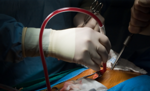
Figure 1: Pericardial window surgery
Technique
For Subxiphoid approach:
An incision is created above the xiphoid, reaching over the midline of the belly, and it is brief, around 5-8 cm long. In many cases, the xiphoid is removed entirely, and the linea alba is cut. The dissection of the finger is used to enter the retrosternal region. The pericardium’s diaphragmatic aspect can be seen with upward retraction.
Using the hook or an Adson or Allis clamp, the pericardium is grasped; it can also be cut directly. By cutting the pericardium carefully, the aperture is made larger. A sucker is placed into the pericardial region. This sucker or finger is frequently utilized to further dissect any adhesions.
The pericardium is also used to get a biopsy specimen. The epicardium is examined following the aspiration of all the fluid. Surgeon should insert a finger into the pericardial space to check for nodules and other adhesions in the pericardium. Finally, the incision is closed in layers after a tube is attached to the pericardial area.
For Thoracotomy approach:
For a little anterior thoracotomy, the fourth or fifth intercostal spaces are used. The selected intercostal space can be seen by dividing the pectoralis muscle by an inframammary skin incision (6-8 cm long).
For entering the pleural cavity, the intercostal gap should be opened over the superior border of the rib. After inserting a retractor, the pericardium region is visible.
With help of scalpel or pair of scissors cut the pericardium prior to the phrenic nerve. The pericardium is sent for pathologic examination, a generous window is made, and samples of the pleural effusion are taken. A biopsy can be easily performed if necessary, and the adjacent lung is palpated. A chest tube is inserted into the pleura or pericardium and sealed or suctioned in place. Layers are used to seal the incision.
For Thoracoscopic approach:
The thoracoscope is positioned in the seventh intercostal space, either in line with the anterior superior iliac spine on the right or in the midaxillary line. The incision is made on the left, immediately behind this line. An incision is made in the fifth intercostal space, in the posterior axillary line, or just above the pericardium. When the lung is deflated, the phrenic nerve may be seen running along the pericardium vertically; this nerve should be always seen during the procedure.
A sharp mobilization off the pericardium is necessary to enable tension-free retraction and broader access to the pericardial surface on the left, where the nerve passes through the center of the lateral surface. This is carried out in cases where a left-side pericardiectomy is planned.
The phrenic nerve on the right is located directly in front of the lung’s hilum and does not obstruct pericardial resection. The lower lobe is grabbed and retracted superiorly, followed by an electrocautery to mobilize the pulmonary ligament if the posterior pericardium is to be cut.
From the level of the inferior pulmonary vein to the mainstem bronchus, the posterior mediastinal pleura is opened to reach the posterior pericardial area. To increase exposure, the esophagus on the right must be mobilized. By using a tonsil sponge stick for blunt dissection, the pericardium may be isolated from the surrounding soft tissue.
If the pericardium is too enlarged to permit this, a spinal needle inserted through the chest wall while under thoracoscopic vision is used to aspirate the effusion.
The pericardium is cut and opened using scissors or electrocautery. After the initial pericardial opening is made, the incision can be extended using an endoscopic gastrointestinal anastomosis (GIA) vascular stapler if the pericardium is extremely thickened or vascular.
Depending on their density or tenacity, adhesions between the heart and the pericardium can be cut with scissors or bluntly using a peanut dissector or Yankauer sucker.
Electrocautery dissection of the substernal plane can be done to the contralateral chest or across the midline, depending on the size of the window that needs to be made and whether a pericardiectomy is to be done. To ensure proper drainage of the pericardial area, the pericardial aperture should be minimum 4 x 4 cm.
After the window is made, the pericardial cavity is probed using a Yankauer sucker to make sure the effusion drains completely and to break down any fibrous septa that could be preventing it from doing so. Through the incision in the camera port, a No. 19 Blake drain, or a 28-French chest tube is brought out of the pericardiopleural or pleural area.
The pericardial window is used for both diagnostic and therapeutic purposes to drain accumulated pericardial fluid. It is a condition which arises following heart surgery but may have many reasons.
The pericardium surrounds the heart like a cocoon, once this cavity fills with too much fluid, then cardiac filling is compromised. Heart compression and tamponade may occur when the small gap between the heart and the noncompliant pericardium suddenly filled with blood or fluid.
In patients with neoplastic pericardial illness, the pericardial window combined with systemic chemotherapy which help to avoid the buildup of high fluid volumes.
The pericardial window permits the effusion to constantly drain into the chest or peritoneum by removing a piece of the pericardium. Procedure including thoracotomy, thoracoscopic, and subxiphoid incision can be used to remove the fluid.
When a patient is hemodynamically stable, ultrasound can be used as a primary screening method to identify hidden penetrating lesions.
Symptomatic pericardial effusions
Asymptomatic pericardial effusions that warrant a pericardial window for diagnosis
Hemodynamically stable patients with an undiagnosed pericardial effusion
Coexisting pericardial, pleural, or pulmonary pathology that requires diagnosis
Drainage of a purulent pericardial effusion
Early fungal or tuberculous pericarditis in which resection of the pericardium is required to stop future pericardial constriction
Loculated effusions situated unilaterally or posteriorly
Uncorrectable coagulopathy
Hemodynamic instability unsuitable for surgery
Localized pericardial effusion not amenable to drainage
Suspected or confirmed purulent pericarditis
Previous sternotomy or thoracic surgery
Pericardial effusion resolving with medical therapy
Malignancy with widespread metastasis
A pericardial window surgery usually has a positive outcome, especially when it is done to treat cardiac tamponade or pericardial effusion.
It creates a continuous drainage pathway into the peritoneal or pleural cavity to prevent fluid from returning.
Assisting in the identification of underlying reasons including infection, cancer, or autoimmune illness, the process also permits diagnostic sample of pericardial fluid and tissue.
In patients with malignant pericardial effusion symptoms may minimize, but the primary disease shows a poor long-term prognosis.
For the subxiphoid and thoracotomy approaches includes standard thoracic instruments:
Long curved Allis clamp
Aspirating needle used for mediastinoscopy
Thoracoscopic scissors
Endoscopic gastrointestinal anastomosis (GIA) stapler with vascular staple load
Peanut dissector and tonsil sponge
Yankauer sucker
External defibrillator pads
Equipment required for the thoracoscopic approach includes:
30º thoracoscope
Ring forceps
General anesthesia is ideal and preferable for the subxiphoid technique.
If an arrhythmia occurs during surgery, external defibrillator pads may be attached to a defibrillator and put to the chest and back before to preparation.
The thoracotomy and thoracoscopic procedures need single-lung ventilation and general anaesthesia. External defibrillator pads and arterial and central venous pressure monitoring may be required, much like with the subxiphoid technique.
The patient should be positioned in supine position for the subxiphoid technique.
Anterolateral or posterolateral thoracotomy positions are used for the thoracotomy and thoracoscopic procedures depending on the pericardium’s exposure and approach.
If the patient’s hemodynamic stability or capacity to withstand single-lung anaesthesia are in question, they can be put in a supine position with the side they want to access raised using a pressure bag or jelly roll.
Figure 1: Pericardial window surgery
For Subxiphoid approach:
An incision is created above the xiphoid, reaching over the midline of the belly, and it is brief, around 5-8 cm long. In many cases, the xiphoid is removed entirely, and the linea alba is cut. The dissection of the finger is used to enter the retrosternal region. The pericardium’s diaphragmatic aspect can be seen with upward retraction.
Using the hook or an Adson or Allis clamp, the pericardium is grasped; it can also be cut directly. By cutting the pericardium carefully, the aperture is made larger. A sucker is placed into the pericardial region. This sucker or finger is frequently utilized to further dissect any adhesions.
The pericardium is also used to get a biopsy specimen. The epicardium is examined following the aspiration of all the fluid. Surgeon should insert a finger into the pericardial space to check for nodules and other adhesions in the pericardium. Finally, the incision is closed in layers after a tube is attached to the pericardial area.
For Thoracotomy approach:
For a little anterior thoracotomy, the fourth or fifth intercostal spaces are used. The selected intercostal space can be seen by dividing the pectoralis muscle by an inframammary skin incision (6-8 cm long).
For entering the pleural cavity, the intercostal gap should be opened over the superior border of the rib. After inserting a retractor, the pericardium region is visible.
With help of scalpel or pair of scissors cut the pericardium prior to the phrenic nerve. The pericardium is sent for pathologic examination, a generous window is made, and samples of the pleural effusion are taken. A biopsy can be easily performed if necessary, and the adjacent lung is palpated. A chest tube is inserted into the pleura or pericardium and sealed or suctioned in place. Layers are used to seal the incision.
For Thoracoscopic approach:
The thoracoscope is positioned in the seventh intercostal space, either in line with the anterior superior iliac spine on the right or in the midaxillary line. The incision is made on the left, immediately behind this line. An incision is made in the fifth intercostal space, in the posterior axillary line, or just above the pericardium. When the lung is deflated, the phrenic nerve may be seen running along the pericardium vertically; this nerve should be always seen during the procedure.
A sharp mobilization off the pericardium is necessary to enable tension-free retraction and broader access to the pericardial surface on the left, where the nerve passes through the center of the lateral surface. This is carried out in cases where a left-side pericardiectomy is planned.
The phrenic nerve on the right is located directly in front of the lung’s hilum and does not obstruct pericardial resection. The lower lobe is grabbed and retracted superiorly, followed by an electrocautery to mobilize the pulmonary ligament if the posterior pericardium is to be cut.
From the level of the inferior pulmonary vein to the mainstem bronchus, the posterior mediastinal pleura is opened to reach the posterior pericardial area. To increase exposure, the esophagus on the right must be mobilized. By using a tonsil sponge stick for blunt dissection, the pericardium may be isolated from the surrounding soft tissue.
If the pericardium is too enlarged to permit this, a spinal needle inserted through the chest wall while under thoracoscopic vision is used to aspirate the effusion.
The pericardium is cut and opened using scissors or electrocautery. After the initial pericardial opening is made, the incision can be extended using an endoscopic gastrointestinal anastomosis (GIA) vascular stapler if the pericardium is extremely thickened or vascular.
Depending on their density or tenacity, adhesions between the heart and the pericardium can be cut with scissors or bluntly using a peanut dissector or Yankauer sucker.
Electrocautery dissection of the substernal plane can be done to the contralateral chest or across the midline, depending on the size of the window that needs to be made and whether a pericardiectomy is to be done. To ensure proper drainage of the pericardial area, the pericardial aperture should be minimum 4 x 4 cm.
After the window is made, the pericardial cavity is probed using a Yankauer sucker to make sure the effusion drains completely and to break down any fibrous septa that could be preventing it from doing so. Through the incision in the camera port, a No. 19 Blake drain, or a 28-French chest tube is brought out of the pericardiopleural or pleural area.
Both our subscription plans include Free CME/CPD AMA PRA Category 1 credits.
Digital Certificate PDF
On course completion, you will receive a full-sized presentation quality digital certificate.
medtigo Simulation
A dynamic medical simulation platform designed to train healthcare professionals and students to effectively run code situations through an immersive hands-on experience in a live, interactive 3D environment.
medtigo Points
medtigo points is our unique point redemption system created to award users for interacting on our site. These points can be redeemed for special discounts on the medtigo marketplace as well as towards the membership cost itself.
Community Forum post/reply = 5 points
*Redemption of points can occur only through the medtigo marketplace, courses, or simulation system. Money will not be credited to your bank account. 10 points = $1.
All Your Certificates in One Place
When you have your licenses, certificates and CMEs in one place, it's easier to track your career growth. You can easily share these with hospitals as well, using your medtigo app.