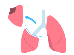
A pneumonectomy is a surgical procedure carried out to resect an entire lung. It is usually used to treat severe cases of lung diseases, including lung cancer, lung infections (in their advanced stages), or lung damage. The process is carried out when other treatments have been ineffective or are not viable.
Pneumonectomy
In pneumonectomy, the surgeon makes an incision in the chest from the side, usually on the affected lung, thereby gaining the required space. The bronchus, the embolized lung blood vessels, and other vessels are minutely disconnected and dissected. The entire lung is removed, together with all surrounding tissues and structures.
Indications
Lung Cancer: Pneumonectomy is advised in such cases if the damage has not metastasized beyond the lungs. It is sometimes considered when the piece of tissue involves any major lung area or is adjacent to the bronchus.
Severe Lung Infections: Sometimes, when a continuous, severe and repeated occurrence of respiratory infections is observed when antibiotics or any other ineffective treatment is being given, it is recommended to remove the affected lung to stop the spread of infection to other body parts.
Lung Trauma: Although targeted surgery may be applied in cases of trauma to a lung where only the damaged part of the lung is cut away, the entire lung or a portion of it may need to be removed due to extensive damage.
Lung Diseases: Bronchiectasis or emphysema, during the severe stages which lead to the impairment and non-functionality of the lung tissue, could need a pneumonectomy to be performed.
Contraindications
Poor Lung Function: A pneumonectomy cannot be performed if the remaining lung is not functioning correctly to provide the body with oxygen.
Advanced Heart Disease: Patients with severe heart conditions may not even withstand the hazard of the procedure and the altered circulation that follows pneumonectomy.
Severe Pulmonary Hypertension: Lung artery hypertension with pulmonary vascular pressures may make it hard or impossible to complete the procedure and may impair post-operative recovery.
Extensive Spread of Cancer: The larger lungs removal alone could not be enough for the cure if the cancer also is in the other organs or tissues in addition to the lungs is present.
Severe Chronic Obstructive Pulmonary Disease: COPD patients with severe respiratory insufficiency may not be able to sustain efficiency after the reduction of the lung function down to one lung.
Outcomes
Equipment
Operating Room Setup:
Operating table
Anesthesia machine
Monitoring device
surgical lights
Surgical Instruments:
Scalpel or electrocautery
Forceps
Scissors
Retractors
Hemostatic clamps or ligatures
Sutures and staplers
Thoracotomy Tray
Lung Isolation Devices
Chest Drains
Imaging Equipment
Suction Devices
Electrosurgical Unit
Sterile Dressings and Wound Care Supplies
Patient preparation
Medical Evaluation: The complete medical care is executed before the process for the purpose of deciding whether the patient is in good health, as for cardiovascular, pulmonary, kidney functions are concerned.
Pre-operative Optimization: Before surgery, it is necessary to take actions for the optimization of medical conditions, like smoking cessation, treatment of the coexistent medical disorders like diabetes and hypertension, and pulmonary rehabilitation or bronchodilator therapy optimization of respiratory function.
Medication Management: Any drugs the patient might be taking should be reviewed and the dosage is altered to moderate to blood pressure, sugar level in blood or any other relevant factors prior to surgery. Some medicines that must be stopped temporarily before the operation.
Nutritional Assessment: Malnutrition can enhance an individual’s chances of experiencing infections and retarding healing rates. As a result, if they are applicable, the assessment and the optimization of nutrition including the counselling about diet or supplementation will be needed.
Patient position
Usually, the patient is positioned in a lateral decubitus position.
Pneumonectomy
Step 1: The patient has given general anesthesia and a tube is placed into the trachea to ensure breathing by a pump.
Step 2: The surgeon begins to make an incision in the chest wall between the fourth or five ribs on that side of the lung being removed.
Step 3: The ribs are separated to allow the surgeon to access the lung.
The bronchus, or airway, and blood arteries supplying the lung are identified, clamped, and divided with utmost care.
Step 4: The lung is then taken outside the chest.
Step 5: To release any trapped air or fluid in the area where the lung was removed, the surgeon might choose to suture the remaining lung tissue and place a chest tube.
Step 6: Next, the chest wall is closed, while the incision is stitched.
Right-Sided Extrapleural Pneumonectomy
Step 1: Anesthesia and patient positioning
The patient is positioned appropriately, and general anesthesia is given.
Usually, a patient will be placed in his or her back with the arm resting above the head.
Step 2: Incision and exposure
Usually between the fifth and sixth ribs, a prolonged posterolateral thoracotomy incision is performed on the right side of the chest wall.
To give the surgeon more space, a portion of the rib may be excised.
Step 3: Dissection
The surgeon dissects the lungs and pleura conscientiously from the chest wall, diaphragm, and mediastinum (the middle portion of chest which contains important structures such as the heart, esophagus, and trachea).
This is done to reduce harm to the blood vessels and surrounding tissues.
Step 4: Division of Blood Vessels and Bronchus
The pulmonary artery, pulmonary veins and right bronchus are identified, dissected, and stapled or tied off, depending on the nature of the surgery, to minimize bleeding.
Lymph nodes are also dissected to remove any that may contain cancerous cells.
Step 5: Removal of lung and other tissues
The right lung is removed in its whole, including with the pericardium, diaphragm, and blood vessels and bronchus, in single piece.
Step 6: Reconstruction
The diaphragm or pericardium can be reconstructed using mesh or other material.
During the recovery process, chest tube is inserted for evacuation of any air or fluid that can develop in the chest cavity after the surgery.
Step 7: Postoperative Care
Postoperative care is followed, complications are minimised.
Laboratory tests
Arterial Blood Gas Analysis: This test is carried out to determine the blood’s pH, oxygen, and carbon dioxide concentrations. Moreover, it provides information on the efficiency of gas exchange that occurs at the lungs.
Pulmonary Function Tests: PFTs, for example, are used mainly to measure dysfunction of the lung before it is removed surgically. These tests assess lung capacity, airflow, and gas exchange.
Imaging Studies: A combination of respiratory scans such as chest X-rays, CT scans or PET scans are best suited for delineating the site, and stage of the identified lung mass. These imaging tests can show where tumors are located and help to determine whether segmentectomy can be used in the treatment.
Complications
Sepsis: It is a severe infection that starts when a person’s immune system works against the person and kills his own tissues and organs while fighting infection. It often starts at the time of an infection, but there is an increased risk after any type of surgery.
Pulmonary Edema: In the case of an immune response to transplanted organs, persistent fluid accumulation in the remaining lung may occur after surgery, resulting in pulmonary edema. This edema will hamper oxygen exchange with low atmospheric pressure and, hence, may lead to respiratory distress.
Infection: Surgical wounds, the remaining lung, or the area where surgery was performed could become infected post-pneumonectomy. This could lead to conditions such as a surgical site infection, pneumonia, or empyema.
Hemorrhage: Surgical intervention can cause hemorrhage, which is bleeding either during surgery or post-surgery. This might result in hypovolemic shock, a condition caused by excessive blood loss.
Bronchopleural Fistula: In this condition, a persistent state exists where the bronchial trees have an established unusual link with the pleural space. It may, therefore, cause various episodes of pneumonia and an increased chance for treating empyema.
Empyema: Intrapleural space contamination causes the formation of infected fluid, called empyema, which is treated by tube drainage and antibiotics.
A pneumonectomy is a surgical procedure carried out to resect an entire lung. It is usually used to treat severe cases of lung diseases, including lung cancer, lung infections (in their advanced stages), or lung damage. The process is carried out when other treatments have been ineffective or are not viable.
Pneumonectomy
In pneumonectomy, the surgeon makes an incision in the chest from the side, usually on the affected lung, thereby gaining the required space. The bronchus, the embolized lung blood vessels, and other vessels are minutely disconnected and dissected. The entire lung is removed, together with all surrounding tissues and structures.
Lung Cancer: Pneumonectomy is advised in such cases if the damage has not metastasized beyond the lungs. It is sometimes considered when the piece of tissue involves any major lung area or is adjacent to the bronchus.
Severe Lung Infections: Sometimes, when a continuous, severe and repeated occurrence of respiratory infections is observed when antibiotics or any other ineffective treatment is being given, it is recommended to remove the affected lung to stop the spread of infection to other body parts.
Lung Trauma: Although targeted surgery may be applied in cases of trauma to a lung where only the damaged part of the lung is cut away, the entire lung or a portion of it may need to be removed due to extensive damage.
Lung Diseases: Bronchiectasis or emphysema, during the severe stages which lead to the impairment and non-functionality of the lung tissue, could need a pneumonectomy to be performed.
Poor Lung Function: A pneumonectomy cannot be performed if the remaining lung is not functioning correctly to provide the body with oxygen.
Advanced Heart Disease: Patients with severe heart conditions may not even withstand the hazard of the procedure and the altered circulation that follows pneumonectomy.
Severe Pulmonary Hypertension: Lung artery hypertension with pulmonary vascular pressures may make it hard or impossible to complete the procedure and may impair post-operative recovery.
Extensive Spread of Cancer: The larger lungs removal alone could not be enough for the cure if the cancer also is in the other organs or tissues in addition to the lungs is present.
Severe Chronic Obstructive Pulmonary Disease: COPD patients with severe respiratory insufficiency may not be able to sustain efficiency after the reduction of the lung function down to one lung.
Operating Room Setup:
Operating table
Anesthesia machine
Monitoring device
surgical lights
Surgical Instruments:
Scalpel or electrocautery
Forceps
Scissors
Retractors
Hemostatic clamps or ligatures
Sutures and staplers
Thoracotomy Tray
Lung Isolation Devices
Chest Drains
Imaging Equipment
Suction Devices
Electrosurgical Unit
Sterile Dressings and Wound Care Supplies
Medical Evaluation: The complete medical care is executed before the process for the purpose of deciding whether the patient is in good health, as for cardiovascular, pulmonary, kidney functions are concerned.
Pre-operative Optimization: Before surgery, it is necessary to take actions for the optimization of medical conditions, like smoking cessation, treatment of the coexistent medical disorders like diabetes and hypertension, and pulmonary rehabilitation or bronchodilator therapy optimization of respiratory function.
Medication Management: Any drugs the patient might be taking should be reviewed and the dosage is altered to moderate to blood pressure, sugar level in blood or any other relevant factors prior to surgery. Some medicines that must be stopped temporarily before the operation.
Nutritional Assessment: Malnutrition can enhance an individual’s chances of experiencing infections and retarding healing rates. As a result, if they are applicable, the assessment and the optimization of nutrition including the counselling about diet or supplementation will be needed.
Patient position
Usually, the patient is positioned in a lateral decubitus position.
Step 1: The patient has given general anesthesia and a tube is placed into the trachea to ensure breathing by a pump.
Step 2: The surgeon begins to make an incision in the chest wall between the fourth or five ribs on that side of the lung being removed.
Step 3: The ribs are separated to allow the surgeon to access the lung.
The bronchus, or airway, and blood arteries supplying the lung are identified, clamped, and divided with utmost care.
Step 4: The lung is then taken outside the chest.
Step 5: To release any trapped air or fluid in the area where the lung was removed, the surgeon might choose to suture the remaining lung tissue and place a chest tube.
Step 6: Next, the chest wall is closed, while the incision is stitched.
Step 1: Anesthesia and patient positioning
The patient is positioned appropriately, and general anesthesia is given.
Usually, a patient will be placed in his or her back with the arm resting above the head.
Step 2: Incision and exposure
Usually between the fifth and sixth ribs, a prolonged posterolateral thoracotomy incision is performed on the right side of the chest wall.
To give the surgeon more space, a portion of the rib may be excised.
Step 3: Dissection
The surgeon dissects the lungs and pleura conscientiously from the chest wall, diaphragm, and mediastinum (the middle portion of chest which contains important structures such as the heart, esophagus, and trachea).
This is done to reduce harm to the blood vessels and surrounding tissues.
Step 4: Division of Blood Vessels and Bronchus
The pulmonary artery, pulmonary veins and right bronchus are identified, dissected, and stapled or tied off, depending on the nature of the surgery, to minimize bleeding.
Lymph nodes are also dissected to remove any that may contain cancerous cells.
Step 5: Removal of lung and other tissues
The right lung is removed in its whole, including with the pericardium, diaphragm, and blood vessels and bronchus, in single piece.
Step 6: Reconstruction
The diaphragm or pericardium can be reconstructed using mesh or other material.
During the recovery process, chest tube is inserted for evacuation of any air or fluid that can develop in the chest cavity after the surgery.
Step 7: Postoperative Care
Postoperative care is followed, complications are minimised.
Arterial Blood Gas Analysis: This test is carried out to determine the blood’s pH, oxygen, and carbon dioxide concentrations. Moreover, it provides information on the efficiency of gas exchange that occurs at the lungs.
Pulmonary Function Tests: PFTs, for example, are used mainly to measure dysfunction of the lung before it is removed surgically. These tests assess lung capacity, airflow, and gas exchange.
Imaging Studies: A combination of respiratory scans such as chest X-rays, CT scans or PET scans are best suited for delineating the site, and stage of the identified lung mass. These imaging tests can show where tumors are located and help to determine whether segmentectomy can be used in the treatment.
Sepsis: It is a severe infection that starts when a person’s immune system works against the person and kills his own tissues and organs while fighting infection. It often starts at the time of an infection, but there is an increased risk after any type of surgery.
Pulmonary Edema: In the case of an immune response to transplanted organs, persistent fluid accumulation in the remaining lung may occur after surgery, resulting in pulmonary edema. This edema will hamper oxygen exchange with low atmospheric pressure and, hence, may lead to respiratory distress.
Infection: Surgical wounds, the remaining lung, or the area where surgery was performed could become infected post-pneumonectomy. This could lead to conditions such as a surgical site infection, pneumonia, or empyema.
Hemorrhage: Surgical intervention can cause hemorrhage, which is bleeding either during surgery or post-surgery. This might result in hypovolemic shock, a condition caused by excessive blood loss.
Bronchopleural Fistula: In this condition, a persistent state exists where the bronchial trees have an established unusual link with the pleural space. It may, therefore, cause various episodes of pneumonia and an increased chance for treating empyema.
Empyema: Intrapleural space contamination causes the formation of infected fluid, called empyema, which is treated by tube drainage and antibiotics.
Both our subscription plans include Free CME/CPD AMA PRA Category 1 credits.
Digital Certificate PDF
On course completion, you will receive a full-sized presentation quality digital certificate.
medtigo Simulation
A dynamic medical simulation platform designed to train healthcare professionals and students to effectively run code situations through an immersive hands-on experience in a live, interactive 3D environment.
medtigo Points
medtigo points is our unique point redemption system created to award users for interacting on our site. These points can be redeemed for special discounts on the medtigo marketplace as well as towards the membership cost itself.
Community Forum post/reply = 5 points
*Redemption of points can occur only through the medtigo marketplace, courses, or simulation system. Money will not be credited to your bank account. 10 points = $1.
All Your Certificates in One Place
When you have your licenses, certificates and CMEs in one place, it's easier to track your career growth. You can easily share these with hospitals as well, using your medtigo app.