Neck disease crucial for the spread of squamous cell carcinoma from head sites.
Common sites of carcinoma spread mucosal areas including upper aerodigestive tract, especially larynx, oropharynx, and oral cavity.
Lymph node metastasis in squamous cell carcinoma cuts survival in half. Post-surgery patients with neck metastasis have less than 5% survival.
Radical neck dissection developed in 1906 to treat metastatic neck disease. It is a relatively simple surgery for trained head and neck surgeons.
Classic radical neck dissection remains standard to control neck metastasis.
Recent decades made progress in understanding of cervical fascial, lymphatic drainage, staging, and spread.
New findings show that nodal disease pattern varies with primary site and prompt modification of neck dissection to selectively preserve lymph nodes.
Indications
Lymph node metastases from head and neck cancers
In primary tumors of unknown origin with neck node metastasis
Nodal disease with extracapsular spread: Cancer that has breached the lymph node capsule and is invading adjacent tissues.
Recurrent or persistent nodal disease after non-surgical treatment: Patients with residual lymph node disease following radiation or chemotherapy.
Malignant melanoma with cervical node involvement: Melanoma with metastasis spread to the cervical nodes.
Advanced nodal involvement in N2 or N3 stage: Extensive nodal involvement requiring complete clearance for optimal control.
Contraindications
Widespread distant metastases
Involvement of unresectable structures
Poor general health or inability to tolerate surgery
Patient refusal of surgery
Radiation or chemotherapy as primary treatment
Involvement of bilateral neck nodes
Extensive local disease with poor prognosis
Previous surgery or radiation therapy to the neck
Outcomes
Patients may develop dysphagia or hoarseness after tissue removal and adjacent structure involvement when combined with radiation.
Reconstructive surgery can benefit some patients prioritizing function over appearance.
In thoracic duct injury and lymph fluid leakage cases proper treatment is required. Post-surgery radiation or chemotherapy are suggested to improved control and survival rate.
Equipment required
Instruments for neck surgery
Nerve and vessel management tools
Drain and wound closure materials
Anesthesia equipment
Patient Preparation:
Patients with malnourished are at risk of wound injuries thus they need nutrition supplements through feeding tube.
Preoperative consultation for anesthesia assesses airway risks from smoking, alcohol, or neck tumors to prevent complications.
Intraoperative nerve monitoring help to store spinal accessory nerve and other vital nerves during surgery.
Internal jugular vein management is important to prevent bleeding.
Patient Positioning:
The patient should be positioned in a supine position with the neck extension for easy access.
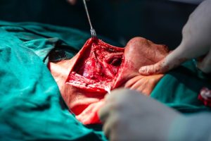
Figure. Radical Neck Dissection (left side)
Technique
Step 1: Incision
A long horizontal incision is made along with the lower margin of the mandible and extending towards the clavicle.
Step 2: Identification of Anatomic Structures
The sternocleidomastoid muscle is dissected and transected at both ends to allow exposure of underlying structures.
The spinal accessory nerve is identified to determine infiltration by tumor. If tumor infiltration is present, then IJV is ligated to ensure complete resection.
Step 3: Dissection of lymph node
The dissection proceeds systematically along with the carotid sheath and vascular structures to ensure safety of all lymphatic tissue.
Step 4: Management of the Thoracic Duct
The thoracic duct is identified and preserved on the left side of neck. In case of injury to the duct it should be ligated with non-absorbable sutures.
Step 5: Closure
Meticulous hemostasis is performed to prevent hematomas and excessive bleeding. Suction drains should be placed to prevent fluid accumulation.
Then the wound is closed with absorbable sutures for deeper tissues.
Complications:
Immediate complications are:
Hemorrhage/Hematoma: Injury to major vessels or inadequate hemostasis.
Nerve Injury: Early identification of injuries is referred to physiotherapy or nerve repair surgery if needed.
Chyle Leak: Damage to the thoracic duct during left-sided neck dissections.
Wound Infection: Contamination during surgery or poor postoperative care.
Seroma Formation: Accumulation of serous fluid due to incomplete lymphatic sealing.
Neck disease crucial for the spread of squamous cell carcinoma from head sites.
Common sites of carcinoma spread mucosal areas including upper aerodigestive tract, especially larynx, oropharynx, and oral cavity.
Lymph node metastasis in squamous cell carcinoma cuts survival in half. Post-surgery patients with neck metastasis have less than 5% survival.
Radical neck dissection developed in 1906 to treat metastatic neck disease. It is a relatively simple surgery for trained head and neck surgeons.
Classic radical neck dissection remains standard to control neck metastasis.
Recent decades made progress in understanding of cervical fascial, lymphatic drainage, staging, and spread.
New findings show that nodal disease pattern varies with primary site and prompt modification of neck dissection to selectively preserve lymph nodes.
Lymph node metastases from head and neck cancers
In primary tumors of unknown origin with neck node metastasis
Nodal disease with extracapsular spread: Cancer that has breached the lymph node capsule and is invading adjacent tissues.
Recurrent or persistent nodal disease after non-surgical treatment: Patients with residual lymph node disease following radiation or chemotherapy.
Malignant melanoma with cervical node involvement: Melanoma with metastasis spread to the cervical nodes.
Advanced nodal involvement in N2 or N3 stage: Extensive nodal involvement requiring complete clearance for optimal control.
Widespread distant metastases
Involvement of unresectable structures
Poor general health or inability to tolerate surgery
Patient refusal of surgery
Radiation or chemotherapy as primary treatment
Involvement of bilateral neck nodes
Extensive local disease with poor prognosis
Previous surgery or radiation therapy to the neck
Patients may develop dysphagia or hoarseness after tissue removal and adjacent structure involvement when combined with radiation.
Reconstructive surgery can benefit some patients prioritizing function over appearance.
In thoracic duct injury and lymph fluid leakage cases proper treatment is required. Post-surgery radiation or chemotherapy are suggested to improved control and survival rate.
Instruments for neck surgery
Nerve and vessel management tools
Drain and wound closure materials
Anesthesia equipment
Patient Preparation:
Patients with malnourished are at risk of wound injuries thus they need nutrition supplements through feeding tube.
Preoperative consultation for anesthesia assesses airway risks from smoking, alcohol, or neck tumors to prevent complications.
Intraoperative nerve monitoring help to store spinal accessory nerve and other vital nerves during surgery.
Internal jugular vein management is important to prevent bleeding.
Patient Positioning:
The patient should be positioned in a supine position with the neck extension for easy access.
Figure. Radical Neck Dissection (left side)
Step 1: Incision
A long horizontal incision is made along with the lower margin of the mandible and extending towards the clavicle.
Step 2: Identification of Anatomic Structures
The sternocleidomastoid muscle is dissected and transected at both ends to allow exposure of underlying structures.
The spinal accessory nerve is identified to determine infiltration by tumor. If tumor infiltration is present, then IJV is ligated to ensure complete resection.
Step 3: Dissection of lymph node
The dissection proceeds systematically along with the carotid sheath and vascular structures to ensure safety of all lymphatic tissue.
Step 4: Management of the Thoracic Duct
The thoracic duct is identified and preserved on the left side of neck. In case of injury to the duct it should be ligated with non-absorbable sutures.
Step 5: Closure
Meticulous hemostasis is performed to prevent hematomas and excessive bleeding. Suction drains should be placed to prevent fluid accumulation.
Then the wound is closed with absorbable sutures for deeper tissues.
Complications:
Immediate complications are:
Hemorrhage/Hematoma: Injury to major vessels or inadequate hemostasis.
Nerve Injury: Early identification of injuries is referred to physiotherapy or nerve repair surgery if needed.
Chyle Leak: Damage to the thoracic duct during left-sided neck dissections.
Wound Infection: Contamination during surgery or poor postoperative care.
Seroma Formation: Accumulation of serous fluid due to incomplete lymphatic sealing.
Both our subscription plans include Free CME/CPD AMA PRA Category 1 credits.
Digital Certificate PDF
On course completion, you will receive a full-sized presentation quality digital certificate.
medtigo Simulation
A dynamic medical simulation platform designed to train healthcare professionals and students to effectively run code situations through an immersive hands-on experience in a live, interactive 3D environment.
medtigo Points
medtigo points is our unique point redemption system created to award users for interacting on our site. These points can be redeemed for special discounts on the medtigo marketplace as well as towards the membership cost itself.
Community Forum post/reply = 5 points
*Redemption of points can occur only through the medtigo marketplace, courses, or simulation system. Money will not be credited to your bank account. 10 points = $1.
All Your Certificates in One Place
When you have your licenses, certificates and CMEs in one place, it's easier to track your career growth. You can easily share these with hospitals as well, using your medtigo app.